Reporting on a Study of One with Khavinson Peptides and Melatonin for Thymic Regrowth
This post reports on the outcome of a self-experiment with three of the Khavinson peptides (epitalon, thymogen, and vilon) and the supplement melatonin in an attempt to produce thymic regrowth. There is evidence in animals for some of these peptides to produce thymic regrowth, as well as evidence for some of these peptides (thymogen particularly) to reduce mortality in old human patients. All of this comes from the Russian research community, however, and original sources are not all that accessible. Certainly, no-one has checked to see whether the thymus is regrown in humans following treatment with these peptides.
Thymic regrowth is a desirable goal, a way to restore immune function in older people who have lost some, most, or all of the active thymic tissue needed for the production of naive T cells. The loss of this supply of new T cells is an important component in the age-related decline of the immune system. Thus it seems worth the effort to gather data on this front. Last year I posted a study outline for a self-experiment with Khavinson peptides, and this year I have a report from one adventurous self-experimenter, using a more aggressive version of that study protocol.
Study Outline
Based on the published outline, this was a nine-month study. On each of the first ten days of every month, a mix of 10mg epitalon, 10mg thymogen, and 10mg vilon in was injected subcutaneously. This was split between two injections 12 hours apart, morning and evening.
This study included the use of a high dose (20mg daily) of melatonin in addition to Khavinson peptides, on the basis that there is good safety data for melatonin, and a single study has shown an increase in thymic tissue resulting from supplementation with melatonin at the equivalent dose in mice. This is an entirely speculative addition, but years of data on melatonin use suggests no meaningful downside at this dose. The 20mg of melatonin were taken orally in the evening, which differs from the mouse study, in which melatonin was supplied in drinking water.
A CT scan of the thymus was taken before and after the study. A complete blood count assay was used to assess lymphocyte:monocyte ratio before and after the study. That is a number that becomes lower with age, and which should increase if the thymus is more rather than less active. Ideally an assay to measure recent thymic emigrants would have been included, but was not. Recent thymic emigrants are T cells recently emerged from the thymus, within the past few weeks.
Subject Details
The subject for the self-experiment was in the 45-55 age range, healthy and without chronic conditions, with a BMI of ~22 throughout the duration of the experiment. Diet and exercise were described as "relatively consistent" across the study duration. I feel that one should always be relatively skeptical of that sort of claim, however, no matter how formal or informal the study.
Results
CT images of the thymus showed a visible reduction in active tissue across the nine months of the study, the opposite of the hoped outcome. In the image below, paired cross-sections through the chest are shown, before on the left, after on the right. For guidance on reading CT scans of the thymus, refer to "Normal Thymus in Adults: Appearance on CT and Associations with Age, Sex, BMI and Smoking". In a cross-section of the chest, as below, and as in the examples given in that paper, the thymus is the triangular patchy grey structure closer to the top of the image, immediately below the sternum (white). Areas of fat will appear dark in the range chosen here, and thus a more atrophied thymus, in which more active tissue is replaced with fat, will appear darker. By late life, the thymus is entirely dark, fatty.
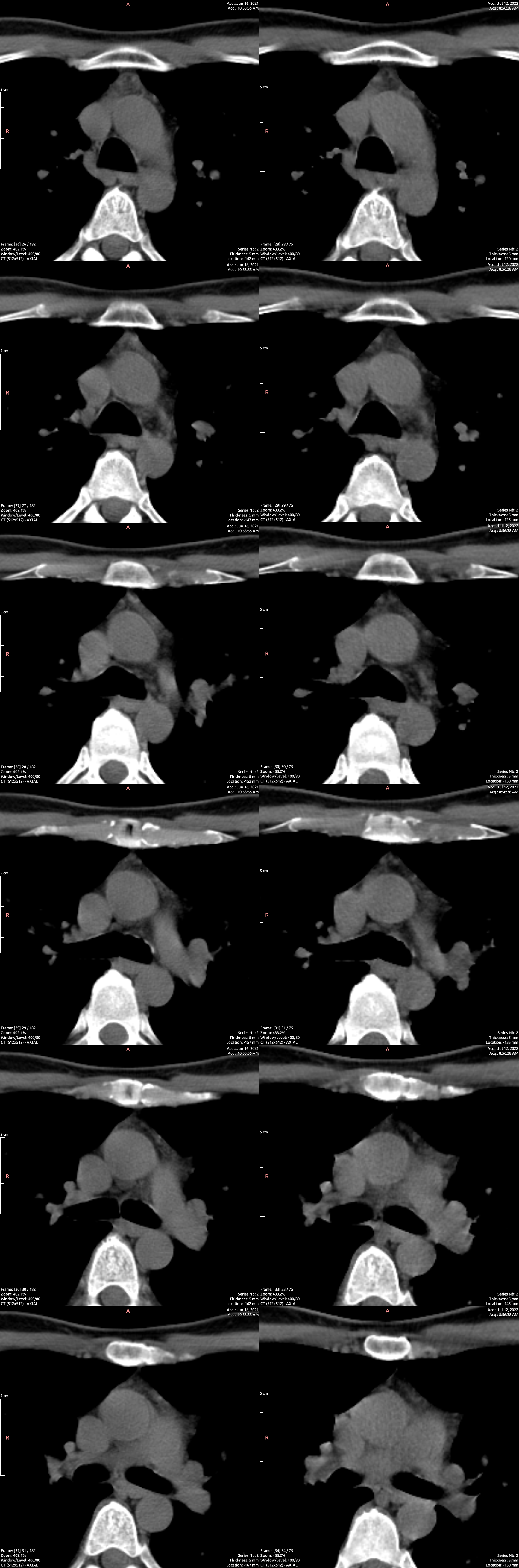
There was no meaningful change in lymphocyte:monocyte ratio. Over four years of complete blood count data prior to this study, leukocyte:monocyte ratio varied from 4.4 to 6.5, with no particular trend. In three measures after the study, leukocyte:monocyte ratio was 5.8, 6.0, and 4.0.
Conclusion
Use of the Khavinson peptides and melatonin in combination in this way, at this dose, negatively impacts the thymus, producing a reduction in active tissue and increase in atrophy to fatty tissue. The degree to which this atrophy occurred is greater than one would expect to take place over nine months of aging at this stage of life.
Why did this outcome occur, given the animal studies showing thymic regrowth, and the studies showing reduced later life mortality following use of thymogen? We can only speculate. Firstly, the dose makes the poison, and the dosing here may have been too high, too frequent. In one of the human studies, testing thymogen only, dosing for ten days occurred only one every six months, rather than monthly as here. Secondly, it may be that these peptides are pleiotropic in their effect on the thymus, and only beneficial after the thymus is very atrophied. Thirdly, it may be that in humans any benefit to the use of Khavinson peptides arises from increased peripheral T cell replication in useful populations, such as naive T cells. This could be beneficial on balance in late life, allowing greater resistance to infection, even if it pushes the patient further towards the accumulation of senescent and exhausted T cells. Lastly, the existing study data for Khavinson peptides relevant to this exercise may simply be dubious, wrong, or otherwise bad.
wow, awesome self experiment! Thankyou to the individual who did this.
Disappointing that things didn't work out. To my eye there's not really much of a difference between the left and right pictures (looking at the light grey areas in the center of the pictures).
Where did you find this experiment? do you have a link to the original post? and more importantly, is there a place on the internet where people share studies like this?
@GREGORY S SCHULTE: The original post describing the protocol is here: https://www.fightaging.org/archives/2021/11/how-to-plan-and-carry-out-a-simple-self-experiment-a-single-person-trial-of-khavinson-peptides-for-thymic-regrowth/
I don't think there's a reputable single place where people are posting good self-experiments, with measures.
could you provide the detailed analysis how the thymus shrinked? I analyse the picture(s) with the naked eye and in 2022 I see slightly bigger actives areas than in 2021.
@SilverSeeker: The analysis is of cellularity, not size. Look at the relative fraction of light pixels versus dark pixels within the thymus cross-section area.
@Reason Look at the whole pictures. The matching of left and right images is rather very approximate (none at all), but it's not the problem here. The main problem is that while characteristic curve of series 2021 is uniform (like they they were taken with same equipment and settings for the set), and series 2022 also uniform (like they they were taken with the same equipment for the set) these two light curves seem very different, from comparing darkest and lightest areas. Before comparing them by dark grey areas, the images would need to be transformed into equal curve. The proper setting would be to take both scans on the same equipment with the same settings. Judging from my experience with B&W photography it is not possible to tell that bigger gray areas are in the second series not the first, from such pictures.
If I had to guess: the studies are falsified, as are the peptides used (in this context, an indirect dosing problem).
@SilverSeeker: for what it is worth, those images were taken on the same machine (a year apart, obviously), with the same settings, and the same window/level settings in the image processing stage.
I'd like to know why an individual has to conduct such a study by themselves. The NIH, pharma, and the FDA are corrupt and less than useless (as proven on a national scale with horrific consequences since 2020). Reason: Please consider offering yourself as a schedule A appointee when sanity might return to Washington/Bethesda.
Does anyone know anything about that self-experiment re: thymus regeneration with Steve Horvath and Greg Fahy? Or is this the same one?
@Ann Cappola: This isn't the same one. The one you are thinking of is the clinical trial run by Intervene Immune, using a growth hormone based approach, which is in its second set of volunteers at the moment.
Thank you for this analysis. I'm afraid to say that the inadvertent dosing puts into question your results. If possible, please repeat the study with the recommended dosage and report the outcomes.
Furthermore, 10 mg is too high a dose for a synthetic peptide. The recommended dosages for synthetic peptides are around 200 micrograms per day.
It is not advisable to use several synthetic peptides at the same time, as this increases the load on the thymus.
As for natural peptides:
Orally: up to 20 milligrams.
Intramuscularly: 2-10 milligrams.
For artificial (synthetic) peptides, the dose should not exceed 200 micrograms (not milligrams!).
The issue might be the following:
All the peptides used in your experiment are synthetic.
In studies on elderly people where thymus and epiphysis peptides extended lifespan, natural peptides isolated from the thymus and epiphysis were used, not synthetic ones.
In animal experiments that successfully extended life, natural peptides, thymus and epiphysis extracts were also used, not artificial ones. Synthetic peptides yielded contradictory results and sometimes even reduced lifespan.
There is a study by Khavinson which shows that any peptides (even natural ones), if they are not isolated from the thymus, have a negative effect on it.
Natural thymus peptides contain a multitude of peptides (several dozen), unlike synthetic peptides.
Synthetic peptides often contain impurities, which can exacerbate the negative effect.
For the thymus restoration protocol, try using one natural thymus preparation instead of several synthetic ones.
Natural thymus peptides can be purchased in CIS countries such as Ukraine, Russia, etc. They are called "Тималин" in Russian.