New Insights into Liver Regeneration

The liver is the most regenerative of organs in mammals, capable of regrowing large amounts of lost tissue following injury. Its strategy for regrowth is somewhat different from that of other tissues, and somewhat different again from the mechanisms employed by species capable of proficient regeneration, such as salamanders. Evolution has produced many approaches to growth and regrowth, it seems. It may or may not be the case that researchers can find ways to make other organs behave more like the liver. I think it is far too early to say just how challenging a proposition this might be; even were there compelling mechanisms in hand and being worked on, that would be a tough prediction to make.

Meanwhile, investigative research continues. In the work noted here, researchers uncover a role for shifts in alternative splicing in liver regeneration. Alternative splicing allows for the production of different proteins from the same genetic blueprint, and is a complex enough epicycle atop all of the other complexity of cellular biochemistry to remain comparatively poorly explored in most specific cases. The researchers tie their findings to the Hippo signaling pathway, something that has attracted attention of late in the context of rejuvenation. A number of research groups are eyeing the Hippo pathway as a target for therapies that might enhance regeneration in various internal organs. This is all largely very early stage work, and it will likely be years before something emerges into the development pipeline.

Study: Damaged liver cells undergo reprogramming to regenerate

The liver is a resilient organ. It can restore up to 70 percent of lost mass and function after just a few weeks. We know that in a healthy adult liver, the cells are dormant and rarely undergo cell division. However, if the liver is damaged, the liver cells re-enter the cell cycle to divide and produce more of themselves. Using a mouse model of a liver severely damaged by toxins, researchers compared injured adult liver cells with healthy cells present during a stage of development just after birth. They found that injured cells undergo a partial reprogramming that returns them to a neonatal state of gene expression.

The team discovered that fragments of messenger RNA, the molecular blueprints for proteins, are rearranged and processed in regenerating liver cells in a manner reminiscent of the neonatal period of development. This phenomenon is regulated through alternative splicing, a process wherein exons (expressed regions of genes) are cut from introns (intervening regions) and stitched together in various combinations to direct the synthesis of many different proteins from a single gene.

"We found that the liver cells after birth use a specific RNA-binding protein called ESRP2 to generate the right assortment of alternatively spliced RNAs that can produce the protein products necessary for meeting the functional demands of the adult liver. When damaged, the liver cells lower the quantity of ESRP2 protein. This reactivates fetal RNA splicing in what is called the Hippo signaling pathway, giving it instructions about how to restore and repopulate the liver with new and healthy cells."

Alternative splicing rewires Hippo signaling pathway in hepatocytes to promote liver regeneration

During liver regeneration, most new hepatocytes arise via self-duplication; yet, the underlying mechanisms that drive hepatocyte proliferation following injury remain poorly defined. By combining high-resolution transcriptome and polysome profiling of hepatocytes purified from quiescent and toxin-injured mouse livers, we uncover pervasive alterations in messenger RNA translation of metabolic and RNA-processing factors, which modulate the protein levels of a set of splicing regulators.

Specifically, downregulation of the splicing regulator ESRP2 activates a neonatal alternative splicing program that rewires the Hippo signaling pathway in regenerating hepatocytes. We show that production of neonatal splice isoforms attenuates Hippo signaling, enables greater transcriptional activation of downstream target genes, and facilitates liver regeneration. We further demonstrate that ESRP2 deletion in mice causes excessive hepatocyte proliferation upon injury, whereas forced expression of ESRP2 inhibits proliferation by suppressing the expression of neonatal Hippo pathway isoforms. Thus, our findings reveal an alternative splicing axis that supports regeneration following chronic liver injury.

A Lower Estimate for the Degree to Which Cancer is Self-Inflicted

The consensus among researchers has long been that a sizable fraction of all cancers could be avoided, given more exercise, better diet, less excess fat tissue. This is even setting aside the matter of smoking and its significant relationship to cancer. The study here is notable for adopting a slightly different approach from most other analyses, and arriving at lower numbers when it comes to the risk of a poor lifestyle. Whether or not one concurs, it is worth bearing in mind that aging remains the greatest risk factor for cancer incidence.

Cancer is a numbers game, risk over time: the wrong mutation in the wrong place; a mutated cell failing to destroy itself; the immune system failing to save the day by destroying the errant cell; the local tissue environment dysfunctional enough to support cancerous growth of that cell. Live long enough and cancer will happen, even given rejuvenation therapies capable of restoring the immune system, damping down chronic inflammation, and addressing the other most important mechanisms relating to cancer. A comprehensive, robust cure for all cancer is a vital part of the planned future toolkit of longevity assurance treatments.

Excess weight, low physical activity, and unhealthy diet contribute substantially to the development of cancer. However, no information on the attributable cancer incidence is available for the general population in Germany. By applying the concept of population-attributable fractions, we estimated the incidence of cancers attributable to excess weight, low physical activity, and unhealthy diet. Our definitions of normal body weight, recommended level of physical activity and a healthy diet followed the cancer prevention guidelines of the World Cancer Research Fund. We considered all cancer types that have been shown to be related to those lifestyle factors in published meta-analyses of prospective studies comprising 5000 or more cancer cases.

Our study revealed a high prevalence of excess weight, low physical activity, and unhealthy diet among the population in Germany in the period 2008 to 2011. For the population aged 35 to 84 years in 2018 in Germany, we therefore estimated that 30,567 incident cancers will be attributable to excess weight and 27,081 to low physical activity in 2018, corresponding to 7% and 6%, respectively, of the expected total of 440,373 incident cancers in this population. 9,000 to 14,000 cancers (2-3%) will be attributable to low intakes of dietary fiber, fruit, and non-starchy vegetables and high consumption of processed meat, and some 1,000 to 2,000 cases (less than 1%) to high intakes of salt and red meat.

Link: https://doi.org/10.3238/arztebl.2018.0578

Older Mice Heal Skin Injuries More Slowly, but with Less Scarring

Researchers have recently provided evidence for regeneration of skin injuries in old mice to result in lesser degrees of scarring than is the case in young mice. The usual consideration of regeneration with age is that it is disrupted by rising levels of inflammation. Further, the same set of inflammatory mechanisms appear to cause the formation of inappropriate scar-like tissue in organs, the process of fibrosis that contributes to loss of function and organ failure. Finding a way to align those well established results with the data from this study should keep research groups busy for some years. Nothing is simple in mammalian biochemistry.

Organisms repair wounds using a combination of two biological processes: scar formation and tissue regeneration. Scar formation results in deposition of fibrous tissue that disrupts the original tissue architecture. Tissue regeneration results in reconstitution of the original and functional tissue architecture, including all cellular subtypes and absence of scar formation. Although amphibians regenerate lost limbs, mammals generally repair injured tissue with scar formation. However, limited examples of human tissue regeneration do exist, including adult liver regeneration, pediatric traumatic digit tip amputations, and fetal skin wounds. These examples suggest that the mechanisms mediating tissue regeneration remain conserved in mammals.

Human skin wounds invariably form scars. Aging slows the speed of skin re-epithelialization and the subsequent rate of wound repair, but the strength of re-epithelized skin remains roughly the same at any age. Researchers have observed that skin wounds in the elderly close with thinner scars. Indeed, the incidence of keloid and hypertrophic scar formation peaks in the second decade of life and decreases with age. These surprising and somewhat counterintuitive clinical observations suggest that the tissue-regenerative pathway in the skin, instead of being diminished, may be more effective in the elderly. Here we investigated the role of aging as a regulator of mammalian tissue regeneration.

We show that full-thickness skin wounds in aged but not young mice fully regenerate. This aging-induced switch between scar formation and tissue regeneration appears to be a gradual process rather than a binary decision. Exposure of aged animals to blood from young mice by parabiosis counteracts this regenerative capacity. The secreted factor, stromal-derived factor 1 (SDF1), is expressed at higher levels in wounded skin of young mice. Genetic deletion of SDF1 in young skin enhanced tissue regeneration. Our results counter the current dogma that tissue function inevitably worsens with age and uncovers potential mechanisms to explain the paradoxical effect of aging on skin tissue regeneration.

Link: https://doi.org/10.1016/j.celrep.2018.08.054

A Spotlight on Geroscience

It isn't entirely fair to categorize geroscience as the worse of the two serious and considered approaches to the treatment of aging as a medical condition, the one that isn't as good as the SENS methodology of rejuvenation through repair of molecular damage. Nor is it entirely the case that geroscience aims only to modestly slow aging to gain a few years while SENS aims at radical life extension and rejuvenation of the old. It is also inaccurate to say that geroscience is concerned only with calorie restriction mimetics and other ways to induce beneficial stress responses, the manipulation of metabolism to resist aging a little better without addressing its root causes.

Yet if you pick a random point in the SENS portfolio and a random point in the geroscience portfolio, the stereotypes above are what you'll likely land upon. Unless, of course, you happened to touch on some portion of the growing interest in senolytics, the selective destruction of senescent cells. This is the major area of overlap between the two at the present time, or - if you choose to look at things the way I do - the most prominent example of the way in which SENS will eventually take over the mainstream of research because it is demonstrably more effective. Senolytics has become a focus for an increasing fraction of the research community as the positive data continues to roll in, and justifiably so. Larger, more reliable effects are what is desired by everyone. Sadly it remains the case that most researchers and sources of funding still need to be persuaded to put aside their geroscience work in favor of the better SENS approach.

The S. Jay Olshansky article I noted a few days back is one of a few interesting position papers from a recent edition of the Journal of the American Medical Association focused on geroscience as an endeavor. The other two are noted below, and each is worth reading as a standalone piece. The bigger picture is that the tenor of the great cultural conversation about aging is changing, has changed significantly, is no longer what it was even a decade ago. The technologies that slow and reverse aging are starting to emerge and be demonstrated in ways that cannot be refuted. Treating aging as a medical condition is no longer mocked in the media - the serious people are convinced. The future is arriving.

Aging as a Biological Target for Prevention and Therapy

Chronic health problems related to the unprecedented aging of the human population in the 21st century threaten to disrupt economies and degrade the quality of later life throughout the developed world. Fortunately, research has shown that fundamental aging processes can be targeted by nutritional, genetic, and pharmacologic interventions to enhance and extend both health and longevity in experimental animal models. These findings clearly demonstrate that the biological rate of aging can be slowed.

The geroscience hypothesis, for which there is abundant evidence in animal models, links these biological discoveries to human health by proposing that targeting biological aging processes will prevent, or at a minimum delay, the onset and progression of multiple chronic diseases and debilities that are typically observed in older adults. For example, interventions that extend the life span of mice often also prevent or slow the progress of several types of cancer, reduce atherosclerotic lesions, improve heart function, alleviate normal age-related cognitive loss, and even improve vaccine response.

One of the main geroscience accomplishments is to highlight a small number of major "pillars," interacting molecular and physiological processes that underlie the biology of aging, for instance, metabolism, proteostasis, macromolecular damage, inflammation, adaptation to stress, epigenetics, and stem cells and their regeneration. The key feature of this conceptual framework is that these processes are understood to be tightly interrelated. These findings have emerged from the remarkable progress made in recent years in dissecting aging processes in model organisms. The discovery of cellular and molecular pathways that modulate healthy aging in diverse species across great evolutionary distances offers an unprecedented opportunity for intervention

Aging, Cell Senescence, and Chronic Disease: Emerging Therapeutic Strategies

Age is the leading predictive factor for most of the chronic diseases that account for the majority of morbidity, hospitalizations, health costs, and mortality worldwide. The fundamental aging processes that contribute to phenotypes characteristic of advanced old age, such as muscle weakness and loss of subcutaneous fat, also appear to underlie the major chronic diseases, geriatric syndromes, and loss of physical resilience. These aging processes can be broadly classified as follows: (1) chronic, low-grade inflammation that is "sterile" (occurring in the absence of known pathogens), together with fibrosis; (2) macromolecular and cell organelle dysfunction (such as DNA damage, dysfunctional telomeres, protein aggregation and misfolding, decreased removal of damaged proteins, or mitochondrial dysfunction); (3) changes in stem cells and progenitors that lead to reduced capacity to repair or replace tissues; and (4) cellular senescence.

Senescence involves essentially irreversible arrest of cell proliferation, increased protein production, resistance to programmed cell death (apoptosis), and altered metabolic activity. Senescent cells accumulate in multiple tissues as a result of chronological aging, especially after middle age, and in tissues central to the pathogenesis of chronic diseases. For example, senescent cells accumulate in and near bone in patients with age-related osteoporosis and in blood vessel walls in patients with vascular disease.

Some senescent cells develop a senescence-associated secretory phenotype (SASP) that entails release of proteins, bioactive lipids, nucleotides, extracellular vesicles, and other factors. The SASP contributes to inflammation and the breakdown of tissues, stem and progenitor cell dysfunction, and the spread of senescence to nonsenescent cells. The SASP, immune cells attracted and activated by the SASP, and spread of senescence contribute to profound local and systemic effects with even small numbers of senescent cells. For example, transplanting small numbers of senescent cells around knee joints in young mice leads to joint pain and pathologic changes closely resembling human osteoarthritis. Transplanting senescent cells into middle-aged mice so that only 1 in 10,000 cells in the recipients is a transplanted senescent cell is sufficient to cause profound physical dysfunction within 2 months, together with early death due to accelerated onset of age-related diseases as a group, compared with transplanting nonsenescent cells.

Intermittent Living as a Proposal for Enhanced Beneficial Cellular Stress Responses

Intermittent fasting, particularly in the form of fasting mimicking diets that enhance autophagy and last long enough to trigger significant reduction and replacement of immune cells, is growing in popularity as a way to activate the range of cellular stress responses known to modestly improve health. It isn't the only way to alter behavior and the environment to upregulate beneficial cellular stress responses such as autophagy, however. Thus the authors of this open access paper propose that a broader program of periodic challenges should be introduced as a best practice for human health, and be as strongly recommended as regular exercise. The health benefits may be in the same ballpark. They choose to call this "intermittent living." A great deal of gathering and analysis of data lies between here and the realization of their vision, of course.

The number of people with chronic diseases such as cardiovascular diseases (CVD), diabetes, respiratory diseases, mental disorders, autoimmune diseases and cancer has increased dramatically over the last three decades. The increasing rates of these chronic systemic illnesses suggest that inflammation, caused by excessive and inappropriate innate immune system activity, is unable to respond appropriately to danger signals that are new from the perspective of evolution.

These, mostly environment-driven, risk factors seem inevitable in current Western societies and their shares and intensities are most likely destined to further increase in the future. Importantly, many of these risk factors exhibit interaction, while contemporary humans are likely to suffer from these challenges in concert. This contrasts with the stress factors experienced by traditionally living populations who still live in the environment of our ancestors. In that environment, they had to cope with short-term mono-metabolic danger factors (e.g. hunger, thirst, cold, heat), whereas modern humans are exposed to multi-metabolic risk factors that stimulate an energy conflict between organs and major systems. The ensuing conflict between current experience and to what our genes and stress systems are adapted is the basis of the so-called 'mismatch hypothesis' of 'typically Western' diseases.

Mono-metabolic stress factors have shaped adaptive mechanisms for survival and reproduction, such as short-lasting inflammation, insulin resistance, activation of the sympathetic nervous system and others. Mild triggers might at least in part reset physiologic and metabolic dysfunctioning in patients with 'typically Western' diseases. In other words they may provide low-cost opportunities for secondary prevention. Conversely, the chronic absence of mild stress factors may have rendered modern 21st century humans less resistant to major toxic insults and susceptible to the development of many, 'typically Western', chronic diseases of affluence, including metabolic disorders.

Several of our studies showed that the combination of certain intermittent stress factors produce a hormetic early stress response with a compensatory improvement of multiple metabolic and immunological indices, and wellbeing. The employed hormetic triggers included: intermittent fasting, intermittent heat, intermittent cold, intermittent hypoxia, intermittent drinking, and the consumption of a great number of nutrients with hormetic effects. The use of intermittent challenges, combined in a homework-protocol, could serve as a vaccine against the deleterious effects of modern life. We named this concept "intermittent living", defined as the daily intermittent use of known ancient triggers for a period of seven days per month. We propose to use this concept as a basis for interventions for individuals with chronic disease and/or its prevention. Intermittent living is no more than the reintroduction of mild environmentally-based short lasting stress (including cold, heat, hunger, thirst).

Link: https://doi.org/10.1016/j.mehy.2018.08.002

Searching for Small Molecules that Can Break Down Protein Aggregates Involved in Neurodegenerative Disease

Considerable effort in the research community is devoted to the search for small molecule drugs that can break down or inhibit formation of the protein aggregates associated with various forms of neurodegenerative disease. One of these is α-synuclein, a prominent feature of Parkinson's disease. Potential treatments based on clearance of α-synuclein are at varying points in the development and regulatory approval pipeline. The materials here provide one of many examples of continued efforts to produce new drug candidates that can enter that pipeline. This is an uncertain process: scanning the compound libraries for new possibilities has unknown (but certainly low) odds of success in any given case. It is expensive and slow besides, and few sources of funding are willing to roll the dice given those points.

Parkinson's disease (PD) is characterized by a progressive loss of dopaminergic neurons, a process that current therapeutic approaches cannot prevent. In PD, the typical pathological hallmark is the accumulation of intracellular protein inclusions, known as Lewy bodies and Lewy neurites, which are mainly composed of α-synuclein. Recently, we have developed an accurate and robust high-throughput screening methodology to identify α-synuclein aggregation inhibitors. Here, we exploited this methodology to identify a small molecule (SynuClean-D) able to inhibit α-synuclein aggregation.

SynuClean-D significantly reduces the in vitro aggregation of wild-type α-synuclein and familiar variants in a substoichiometric molar ratio. This compound prevents fibril propagation in protein-misfolding cyclic amplification assays and decreases the number of α-synuclein inclusions in human neuroglioma cells. Computational analysis suggests that SynuClean-D can bind to cavities in mature α-synuclein fibrils and, indeed, it displays a strong fibril disaggregation activity. The treatment with SynuClean-D of two PD Caenorhabditis elegans models, expressing α-synuclein either in muscle or in dopaminergic neurons, significantly reduces the toxicity exerted by α-synuclein.

SynuClean-D-treated worms show decreased α-synuclein aggregation in muscle and a concomitant motility recovery. More importantly, this compound is able to rescue dopaminergic neurons from α-synuclein-induced degeneration. Overall, SynuClean-D appears to be a promising molecule for therapeutic intervention in Parkinson's disease.

Link: https://doi.org/10.1073/pnas.1804198115

Recent Papers Illustrative of Present Efforts to Quantify the Benefits of Exercise

It is fairly settled in the scientific community, barring the odd few objections here and there, that regular moderate exercise improves health in the long term, relative to a sedentary lifestyle. When it comes to the details of the dose-response curve for exercise, however, the scientists of the field are still somewhere in the midst of a slow and grand debate that has lasted decades and seems likely to last for decades more. Extracting solid conclusions from human epidemiological data is a challenging endeavor at the best of times. The papers noted below are illustrative of a score or more similar efforts published every year, as researchers add ever more analysis to the existing mountain of thought on exercise and health.

Present evidence is leaning in the direction of a big leap in benefits in the transition from no exercise and minimal physical activity. Benefits increase thereafter up to the point of an hour or so a day, and then may or may not decline with further increases. Clearly there is a point at which too much exertion is harmful, but does that occur prior to the level of exercise undertaken by profession athletes? If so, how to account for their longevity compared to the rest of the population? It may be that people who can become professional athletes are just more robust than everyone else to start with, or alternatively it has to do with social status, wealth, and other confounding factors.

Further, even if there ever comes to be solid agreement on how much exercise is best, what about the different forms of exercise? Are repeated short bursts better or worse than extended effort? Can short term effects be separated from long term effects? Is strength training so important as to be worth sacrificing time on aerobic exercise to undertake it? Is cycling better or worse than rowing? Or swimming? Or moving plants around the garden? An endless series of questions might be posed. Few of them will be definitively answered before we find ourselves in an era in which optimizing the effects of exercise is an amusing hobby and little more, because rejuvenation biotechnologies exist. Their effects on health and life span will far exceed anything that might be produced by finding a way to do a little better than the currently recommended level of exercise.

The Goldilocks Zone for Exercise: Not Too Little, Not Too Much

Homo sapiens are evolutionarily adapted to be very physically active throughout life, and thus habitual physical activity (PA) is essential for well-being and longevity. Never the less, middle-aged and older individuals engaging in excessive strenuous endurance exercise appear to be at increased risk for a variety of adverse cardiovascular effects including atrial fibrillation, myocardial fibrosis, and coronary atherosclerosis. An emerging body of evidence indicates U-shaped or reverse J-shaped curves whereby low doses and moderate doses of PA significantly reduce long-term risks for both total mortality and cardiovascular mortality, however, at very high doses of chronic strenuous exercise much of the protection against early mortality and cardiovascular disease is lost.

The optimal dose, or what we term 'Goldilocks Zone,' of PA may be: at least 150 minutes per week of moderate-intensity aerobic exercise or 75 minutes per week of vigorous-intensity aerobic activity, but not more than four to five cumulative hours per week of vigorous (heart-pounding, sweatproducing) exercise, especially for those over 45 years of age. It is also important to take at least one day per week off from vigorous exercise. There appears to be no concerns about an upper threshold for safety for leisure-time low-to-moderate intensity activities such as walking at a comfortable pace, housework, gardening, etc. After every 30 consecutive minutes spent sitting, stand up and move, ideally walking briskly for about five minutes.

Now you just need to remember to exercise!

In a study of 36 healthy young adults, the researchers discovered that a single 10-minute period of mild exertion can yield considerable cognitive benefits. Using high-resolution functional magnetic resonance imaging, the team examined subjects' brains shortly after exercise sessions and saw better connectivity between the hippocampal dentate gyrus and cortical areas linked to detailed memory processing. "The hippocampus is critical for the creation of new memories; it's one of the first regions of the brain to deteriorate as we get older - and much more severely in Alzheimer's disease. Improving the function of the hippocampus holds much promise for improving memory in everyday settings."

While prior research has centered on the way exercise promotes the generation of new brain cells in memory regions, this new study demonstrates a more immediate impact: strengthened communication between memory-focused parts of the brain. "We don't discount the possibility that new cells are being born, but that's a process that takes a bit longer to unfold. What we observed is that these 10-minute periods of exercise showed results immediately afterward."

Mitochondrial changes similar in short sprint exercise versus longer moderate-intensity workouts

A team of researchers studied eight young adult volunteers as they participated in cycling workouts of varying intensity: (a) moderate intensity consisted of 30 minutes of continuous exercise at 50 percent peak effort; (b) high-intensity interval exercise consisted of five four-minute cycling sessions at 75 percent peak effort, each separated by one minute of rest; (c) sprint cycling consisted of four 30-second sessions at maximum effort, each separated by 4.5 minutes of recovery time.

The research team measured the amount of energy the volunteers spent on each workout and compared mitochondrial changes in the participants' thigh muscles before and after each exercise session. The researchers found that fewer minutes of higher-intensity exercise produced similar mitochondrial responses compared to a longer moderate-intensity activity. "A total of only two minutes of sprint interval exercise was sufficient to elicit similar responses as 30 minutes of continuous moderate-intensity aerobic exercise. This suggests that exercise may be prescribed according to individual preferences while still generating similar signals known to confer beneficial metabolic adaptions."

Lifespan.io is Crowdfunding a Nicotinamide Mononucleotide Mouse Lifespan Study

Lifespan.io has launched their latest crowdfunded study, and seeks more donors to join the more than 100 philanthropists of our community who have pledged already in the first few days. It is an assessment of the capacity of nicotinamide mononucleotide (NMN) to slow aging in mice, treating both normal mice and a lineage exhibiting accelerated aging. The final stretch goal in this fundraiser will provide enough funding to kick off a full life span study. This work is carried out in partnership with David Sinclair's lab, home of the past fifteen years of work on calorie restriction mimetics associated with sirtuins, a line of research that has evolved to these days to focus instead on nicotinamide adenine dinucleotide, NAD+.

NMN is one of a number of options that can be used to treat the loss of NAD+ in older individuals. While this approach doesn't address any of the causes of aging, meaning the rising levels of molecular damage that take throughout the body, it does serve to boost mitochondrial function. Mitochondrial function is well known to falter with age, and researchers consider this important in a range of age-related conditions. The evidence to date suggests that enhancing NAD+ levels may to some degree diminish measures of aging. To pick one recent example from the data, a small human trial of nicotinamide riboside, another of the options to raise NAD+ levels, demonstrated a reduction in blood pressure in hypertensive older patients.

One of the best studied anti-aging treatments is a diet reduced in calories, yet high enough in nutrients to avoid malnutrition. Known as calorie restriction (CR), this dietary regimen provides irrefutable evidence of the importance of metabolism in the aging process. While CR has been studied extensively and even tested in human trials, long term adherence to a CR dietary regimen is extremely difficult for most individuals to maintain.

One method to achieve the benefits of CR for everyone would be to administer compounds which act as "CR mimetics". A major metabolic signaling molecule that we and others have shown to exhibit significant declines with increasing age is NAD+. Importantly, CR reverses the age-related decline of bioavailable NAD+. This key metabolite plays a crucial role in regulating the activity of many important signaling molecules involved in age-related diseases.

However, feeding or administering NAD+ directly to organisms is not a practical option. The NAD+ molecule cannot readily cross cell membranes to enter cells, and therefore would be unavailable to positively affect metabolism. Instead, precursor molecules to NAD+ must be used to increase bioavailable levels of NAD+. Recently, we have shown that by administering the NAD+ precursor NMN (Nicotinamide Mononucleotide) in drinking water to older mice, NAD+ levels were restored to those normally associated with younger healthy animals. By administering NMN to mice for just one week, our lab demonstrated a robust correction in age-associated metabolic dysfunction and restored muscle mitochondrial function in old mice to levels seen in younger control mice.

Although the restorative properties of NMN treatment drive many of the same cellular signaling pathways which underlie CR, trials of greater than one week or two months are needed to properly evaluate whether NMN can reverse the aging process. Starting with mice that are 20 months old (roughly equivalent to a 50 year old human), longer-term NMN treatments will be applied in order to restore levels of cellular NAD+ to those found in youthful mice. Along with a large cohort of normal mice, we will also use a cohort of our novel genetically engineered mouse, termed the ICE mouse (Induced Changes In Epigenome). These ICE mice manifest an accelerated aging phenotype.

Your donations will not only allow us to purchase the materials necessary to perform this experiment, but also pave the way for human clinical trials aimed at showing, for the first time, that we can actually slow down human aging. We find ourselves at a turning point in history, and together we have the chance to accelerate technologies that will allow us to live healthfully at any age. This is a future that is coming, and whether it arrives in our lifespan or only for future generations is up to us.

Link: https://www.lifespan.io/campaigns/can-nmn-increase-longevity/

Destroying Mutant Mitochondrial DNA as a Strategy to Treat Mitochondrial Disease

The herd of bacteria-like mitochondria in each of our cells are vital cellular components, and come equipped with their own small genome, the mitochondrial DNA, one or more copies in each mitochondrion. If that DNA is broken, then harm results. Mitochondrial diseases bear only superficial similarities to the mitochondrial DNA damage that is a root cause of degenerative aging; while it is the case that mitochondrial DNA is mutated in both cases, the distribution of those mutations in cells and tissues is quite different. Nonetheless, it seems a reasonable proposition that a strategy of selectively destroying damaged mitochondrial DNA may work in each situation, though for different reasons.

In inherited mitochondrial disease, there is some split between healthy and damaged copies of mitochondrial DNA in cells. Destroy the bad genome copies and the good ones will hopefully replicate to make up that loss. In aging, just a few cells become entirely overtaken by clones of mitochondria with damaged genomes, but they exert a sizable negative influence on health via generation of oxidative molecules. Destroying all of the mitochondrial DNA in those cells might be expected to kill them, for all of the obvious reasons. Since there are few of them, destroying them is probably the most expedient approach to dealing with the issue. In both cases, effectiveness would be determined by how clean a sweep is made, though one might imagine temporary or partial benefits resulting from removing even half of the damaged mitochondrial genomes.

"Mitochondrial replacement therapy is a promising approach to prevent transmission of mitochondrial diseases, however, as the vast majority of mitochondrial diseases have no family history, this approach might not actually reduce the proportion of mitochondrial disease in the population. One idea for treating these devastating diseases is to reduce the amount of mutated mitochondrial DNA by selectively destroying the mutated DNA, and allowing healthy DNA to take its place."

To test an experimental gene therapy treatment, which has so far only been tested in human cells grown in petri dishes in a lab, the researchers used a mouse model of mitochondrial disease that has the same mutation as some human patients. The gene therapy treatment, known as the mitochondrially targeted zinc finger-nuclease, or mtZFN, recognises and then eliminates the mutant mitochondrial DNA, based on the DNA sequence differences between healthy and mutant mitochondrial DNA. As cells generally maintain a stable number of mitochondrial DNA copies, the mutated copies that are eliminated are replaced with healthy copies, leading to a decrease in the mitochondrial mutation burden that results in improved mitochondrial function.

The treatment was delivered into the bloodstream of the mouse using a modified virus, which is then mostly taken up by heart cells. The researchers found that the treatment specifically eliminates the mutated mitochondrial DNA, and resulted in measures of heart metabolism improving. Following on from these results, the researchers hope to take this gene therapy approach through clinical trials, in the hope of producing an effective treatment for mitochondrial diseases.

Link: https://www.cam.ac.uk/research/news/mitochondrial-diseases-could-be-treated-with-gene-therapy-study-suggests

Thoughts on Attending RAADfest 2018 in San Diego

I spent an interesting few days last week attending RAADfest, and came away somewhat optimistic that this strange collision of subcultures may herald an acceleration in the adoption of solid science and working therapies on the part of the anti-aging marketplace, accompanied by a driving out of the ineffective nonsense and fraud of past decades. This sea change is very much a work in process, and there is plenty of that nonsense still to be found. Yet the advent of senolytic therapies to clear senescent cells has clearly invigorated certain groups, who have now turned a sizable amount of their advocacy and attention to the adoption of this first legitimate rejuvenation therapy, an implementation of the SENS model of damage repair.

RAADfest is a stage show of presentations and a floor show of company booths. The core of RAADfest, the presentations on stage, might be best understood as almost a secular church, an adoption of the methods of American revivalism when it comes to firing up an audience, presenting a message, and encouraging people to set forth and tell their friends the good news. The presenters are effusive and largely very charismatic characters, the audience leaps up to applaud and shout encouragement. There are few other places you will see scientists gain a standing ovation and rousing cheers on presenting patient benefits, or explaining how their work will help in the future.

The hosts, the leaders of the Coalition for Radical Life Extension represent a community of long-standing longevity advocates and their followers, associated for decades with the anti-aging marketplace, the original iconoclasts from an era in which the scientific community was much more hostile to the message of longevity. Among them are some of those who built the original businesses such as the Life Extension Foundation because they believed that more could be accomplished than was possible at the time. Where businesses succeeded, they ended up selling at best marginal and ineffective products, hoping for a better future, and coming to be surrounded by an industry in which the fraud of false promises became a way of life. So what happens when real, working rejuvenation therapies start to show up? I hope that the good drives out the bad, and that process seems to be underway. The past generation of longevity advocates, those who built an audience and a logistics pipeline, now have a chance to redeem themselves.

Still, there is an element of the tragic here, in that these people have adopted a mode of advocacy that does not allow them to admit that there is the slightest chance that they will not make it, that the pace of process will be too slow. Technology cannot advance fast enough to save today's oldest demographic, and their only realistic option is cryopreservation (Alcor had a booth, and good for them). There is a certain pathos in folk in late life standing on stage to call for radical life extension, to say that they are fully engaged with living a great life, that they wish to live forever and have fun doing it. In the process they are doing good in the sense of aiding the advance of rejuvenation therapies in their own way, but it is clear that they will be not be around for long enough to see the true flowering of this field of medicine.

On the topic of senolytics, I took a sizable number of handouts to the conference and distributed them all. I feel quite strongly that it is crazy that so little progress is being made in getting the senolytic dasatinib to the tens of millions of older people who might benefit significantly from even a single dose, given that it is a cheap, easily available, FDA-approved generic drug with well characterized pharmacology that can be used off-label. Millions are suffering needlessly, and they and their physicians just need to be told of the opportunity in order to assess it and take advantage of it.

As it turned out, no such effort on my part was actually needed, as the formal conference materials contained a fairly comprehensive section covering senolytics, featuring the dasatinib and quercetin combination front and center. The position put forward by Bill Faloon of the Life Extension Foundation at the conference is that older people should absolutely be taking advantage of NAD+ boosting therapies such as nicotinamide riboside, then senolytics, then mesenchymal stem cell therapies, in that order, envisaged as an ascending stairway of benefits. There is good evidence to suggest that positive results for older individuals will result from all of these, though senolytics are by far the most impressive in animal studies. I'm omitting the presence of basic good health practices and supplements in this stairway picture, but they are still there and being emphasized. That said, I have to think that they will drift away from being as central to the anti-aging message as they have been in the past, as the biotechnologies improve.

I had said this was a collision of cultures. The older longevity advocates of past decades are the organizers, and then into the big tent are invited scientists developing the foundations of real, working rejuvenation therapies; investors funding the latest startups; entrepreneurs commercializing the range of new approaches to treat aging; the transhumanist community with its focus on transcending human limits; health advocates of all varieties; stem cell physicians of the proven and unproven variety, and of course the strange detritus of the anti-aging marketplace, a mix of cynics and true believers offering little more than hope and strangeness, ineffective products wrapped in the veneer of science. It is quite the mix. One might hope that out of this cross-pollination perhaps an acceleration will emerge, a speeding of the otherwise slow process of outreach and awareness in the matter of longevity science. It was certainly the case that I met a couple of interesting new faces, people undertaking or considering what I see to be worthwhile endeavors.

Among the presentations of note, Aubrey de Grey gave an update on the growing number of companies now emerged from or associated with the SENS Research Foundation community (a list that includes the startup Repair Biotechnologies, founded by Bill Cherman and myself). The audience here might find it very interesting to note that Revel Pharmaceuticals, the glucosepane cross-link breaking company in the lengthy process of being founded by David Spiegel is apparently now funded by Juvenescence and Kizoo Technology Ventures. So it looks like we'll be hearing more on that front soon, and a good thing too, as this part of the SENS rejuvenation research agenda is likely to be just as big as senolytics once realized.

Bill Andrews gave an interesting update on progress towards human trials of telomerase gene therapy at Libella Gene Therapeutics; the technology is coming together. Liz Parrish of BioViva was also present at the event to discuss her company and present efforts. Gene therapies are still a pain to develop, to optimize sufficiently to gain high levels of transduction of target cells. This will change in the years ahead, but for now it is still an expensive and complicated business to deal with all of the issues that arise, and every therapy is significantly different in its needs, a hand-crafted product unlike any of its peers. I remain in the camp of wait and see on telomerase therapies; my concern is the sizable difference between mouse and human telomere dynamics. Were I older, the risk/reward calculus would be different, but I have to admire the brave souls who have undergone or will in the near future undergo these therapies. Their risk-taking benefits all of us.

Vocal investor Jim Mellon, who at times appears to be more or less single-handedly driving the funding of our nascent rejuvenation biotechnology industry, gave a rousing presentation. Longevity is a market that will make every past growth trend and bubble seem tiny. Mellon doesn't care about the money, save as a means to an end - "I have enough money" - but rather he wants to see the job accomplished, rejuvenation realized, life spans extended by decades and more. The best way to do that is to build an industry large enough and vibrant enough and sufficiently well known to draw in a flood of capital, packed with enough companies to develop every possible portion of and take on the SENS rejuvenation research portfolio that we can come up with. As he points out, the anti-aging market, selling things that don't work, is worth $140 billion today. How much will it be worth when the products do work? Over the timescale of decades, it is large and healthy industries that truly change the world.

These are interesting times that we live in.

Researchers Generate Esophagus Organoids from Pluripotent Stem Cells

Every tissue requires a different recipe for the production of a functioning structure from the starting point of a few cells: different signals, different environment, different timing. Researchers in the heavily funded tissue engineering community are working their way through the lengthy and costly task of establishing those recipes for every type of organ that might be replaced or repaired. At the same time, there is at present no reliable way to produce the capillary networks needed to support tissues thicker than a few millimeters. The result is an era of organoids, tiny functional sections of organ tissue that are primarily used to accelerate research rather than for the production of therapies. Therapies are nonetheless possible in some cases: sheets of functional tissue can in principle be transplanted onto or into an organ, or even used to generate free-standing tiny assistive organs elsewhere in the body, as demonstrated by the ongoing work at Lygenesis. The set of organoid recipies is expanding and the quality of the end results are improving, with the report here being a representative example of the state of progress in this field.

Scientists working to bioengineer the entire human gastrointestinal system in a laboratory now report using pluripotent stem cells to grow human esophageal organoids. The newly published research is the first time scientists have been able to grow human esophageal tissue entirely from pluripotent stem cells (PSCs), which can form any tissue type in the body. Scientists have already used PSCs to bioengineer human intestine, stomach, colon, and liver. "Disorders of the esophagus and trachea are prevalent enough in people that organoid models of human esophagus could be greatly beneficial. In addition to being a new model to study birth defects like esophageal atresia, the organoids can be used to study diseases like eosinophilic esophagitis and Barrett's metaplasia, or to bioengineer genetically matched esophageal tissue for individual patients."

The scientists based their new method for using human PSCs to general esophageal organoids on precisely timed, step-by-step manipulations of genetic and biochemical signals that pattern and form embryonic endoderm and foregut tissues. They focused in part on the gene Sox2 and its associated protein - which are already known to trigger esophageal conditions when their function is disrupted. The scientists used mice, frogs, and human tissue cultures to identify other genes and molecular pathways regulated by Sox2 during esophagus formation. The scientists report that during critical stages of embryonic development, the Sox2 gene blocks the programming and action of genetic pathways that direct cells to become respiratory instead of esophageal. In particular, the Sox2 protein inhibits the signaling of a molecule called Wnt and promotes the formation and survival of esophageal tissues.

After successfully generating fully formed human esophageal organoids - which grew to a length of about 300-800 micrometers in about two months - the bioengineered tissues were compared biochemically to esophageal tissues from patient biopsies. Those tests showed the bioengineered and biopsies tissues were strikingly similar in composition. The research team is continuing its studies into the bioengineering process for esophageal organoids and identifying future projects to advance the technology's eventual therapeutic potential.

Link: https://www.cincinnatichildrens.org/news/release/2018/human-esophageal-organoids

An Update on Dkk1 as a Therapeutic Target in Alzheimer's Disease

Six years ago, researchers reported that dkk1 appears to be involved in the destruction of synapses in Alzheimer's disease. More recent work expands the understanding of dkk1 in this context, placing it in a positive feedback loop related to amyloid-β, in which synaptic damage drives more synaptic damage. The researchers provide evidence to suggest that dkk1 protein can be a therapeutic target for treatments that slow the progression of Alzheimer's disease. Fortunately, there is an existing approved drug that might be used to produce a proof of concept in human patients, so it seems likely that we will be hearing more from this line of research in the years ahead.

Overproduction of the protein beta-amyloid is strongly linked to development of Alzheimer's disease but many drugs targeting beta-amyloid have failed in clinical trials. Beta-amyloid attacks and destroys synapses - the connections between nerve cells in the brain - resulting in memory problems, dementia, and ultimately death. In a new study researchers found that when beta-amyloid destroys a synapse, the nerve cells make more beta-amyloid driving yet more synapses to be destroyed. "We show that a vicious positive feedback loop exists in which beta-amyloid drives its own production. We think that once this feedback loop gets out of control it is too late for drugs which target beta-amyloid to be effective, and this could explain why so many Alzheimer's drug trials have failed."

The researchers also found that a protein called Dkk1, which potently stimulates production of beta-amyloid, is central to the positive feedback loop. Previous research identified Dkk1 as a central player in Alzheimer's, and while Dkk1 is barely detectable in the brains of young adults its production increases as we age. Instead of targeting beta-amyloid itself, the researchers believe targeting Dkk1 could be a better way to halt the progress of Alzheimer's disease by disrupting the vicious cycle of beta-amyloid production and synapse loss. "Importantly, our work has shown that we may already be in a position to block the feedback loop with a drug called fasudil which is already used in Japan and China for stroke. We have convincingly shown that fasudil can protect synapses and memory in animal models of Alzheimer's, and at the same time reduces the amount of beta-amyloid in the brain."

Link: https://www.eurekalert.org/pub_releases/2018-09/kcl-dce091818.php

Clearing Senescent Cells Reduces Tau Aggregation and Cognitive Loss in Mouse Models of Alzheimer's Disease

Researchers have once again demonstrated that a senolytic therapy capable of selectively destroying senescent cells can reduce tau aggregation and consequent loss of cognitive function in a mouse model of Alzheimer's disease. The research materials noted below report on the use of navitoclax, also known as ABT-263, in mice and follow very closely on the heels of two other studies that produced very similar results using different senolytic treatments, piperlongumine in one study and the dasatinib / quercetin combination in the other. This is very characteristic of research into the removal of senescent cells: any approach that succeeds in destroying a significant fraction of senescent cells produces significant gains in health; there is no shortage of different approaches; the treatments employed cost very little and are easily purchased in the global marketplace; and researchers can readily replicate the findings of other groups. This a very robust intervention in the aging process, producing data that is far more consistent than any other approach I am aware of.

Why does the removal of senescent cells work so well? Firstly, there are few of these cells, perhaps a few percent by number in aged tissues, so selective destruction is not particularly disruptive. It leaves little debris to clean up, and the few lost cells can be rapidly replaced. Secondly, these few senescent cells produce produce sizable harm through the continuous secretion of inflammatory and other harmful signals. A large fraction of that harm is in effect an altered, degraded state of tissue function that is actively maintained via signaling. The moment that this unwanted signaling is cut back, the environment shifts back to a more youthful, less inflammatory, less disrupted state. Regenerative capacity picks up, and many other forms of cellular function improve. This change is rapid. Near any age-related inflammatory condition is mostly likely significantly driven by the accumulation of senescent cells, whether that is arthritis, fibrosis, or Alzheimer's disease. In animal models, removal of senescent cells has been demonstrated to reverse measures of aging in numerous diseases and near all major organs.

It is interesting to compare dosing strategies between the three studies that demonstrated reductions in tau aggregation and cognitive decline. They are illustrative of the behavior of different classes of senolytic drugs, as each of navitoclax, piperlongumine, and the dasatinib / quercetin combination use different mechanisms to kill senescent cells. The navitoclax study here used long-term intermittent administration, five days on and sixteen days off over the full lifetime of the mice. The piperlongumine study used a daily dose over eight weeks. The dastinib study used a bi-weekly schedule of administration over twelve weeks. This is consistent with other data from studies using these compounds, in which dasatinib appears to require far less frequent dosing to achieve more or less the same outcome.

Zombie cells found in brains of mice prior to cognitive loss

Zombie cells are the ones that can't die but are equally unable to perform the functions of a normal cell. These zombie, or senescent, cells are implicated in a number of age-related diseases. In a mouse model of brain disease, scientists report that senescent cells accumulate in certain brain cells prior to cognitive loss. By preventing the accumulation of these cells, they were able to diminish tau protein aggregation, neuronal death, and memory loss.

In the current study, the team used a model that imitates aspects of Alzheimer's disease. "We used a mouse model that produces sticky, cobweb like tangles of tau protein in neurons and has genetic modifications to allow for senescent cell elimination. When senescent cells were removed, we found that the diseased animals retained the ability to form memories, eliminated signs of inflammation, did not develop neurofibrillary tangles, and had maintained normal brain mass." They also report that pharmacological intervention to remove senescent cells modulated the clumping of tau proteins.

"Two different brain cell types called microglia and astrocytes were found to be senescent when we looked at brain tissue under the microscope. These cells are important supporters of neuronal health and signaling, so it makes sense that senescence in either would negatively impact neuron health. We had no idea whether senescent cells actively contributed to disease pathology in the brain, and to find that it's the astrocytes and microglia that are prone to senescence is somewhat of a surprise."

Clearance of senescent glial cells prevents tau-dependent pathology and cognitive decline

Cellular senescence, which is characterized by an irreversible cell-cycle arrest accompanied by a distinctive secretory phenotype, can be induced through various intracellular and extracellular factors. Senescent cells that express the cell cycle inhibitory protein p16INK4A have been found to actively drive naturally occurring age-related tissue deterioration and contribute to several diseases associated with ageing, including atherosclerosis and osteoarthritis. Various markers of senescence have been observed in patients with neurodegenerative diseases; however, a role for senescent cells in the aetiology of these pathologies is unknown.

Here we show a causal link between the accumulation of senescent cells and cognition-associated neuronal loss. We found that the a mouse model of tau-dependent neurodegenerative disease accumulates p16INK4A-positive senescent astrocytes and microglia. Clearance of these cells as they arise using INK-ATTAC transgenic mice prevents gliosis, hyperphosphorylation of both soluble and insoluble tau leading to neurofibrillary tangle deposition, and degeneration of cortical and hippocampal neurons, thus preserving cognitive function. Pharmacological intervention with a first-generation senolytic modulates tau aggregation. Collectively, these results show that senescent cells have a role in the initiation and progression of tau-mediated disease, and suggest that targeting senescent cells may provide a therapeutic avenue for the treatment of these pathologies.

Humanin Improves Cognition in Aged Mice

Researchers here demonstrate that delivery of humanin to aged mice can improve cognitive function. Humanin appears to trigger increased levels of autophagy, the collection of processes responsible for recycling damaged proteins and cell structures. Increased autophagy is associated with many of the approaches shown to modestly slow aging in short-lived species such as nematodes, flies, and mice. These approaches largely involve applying mild stress to cells, such as via heat, lack of nutrients, or other methods, or directly triggering the signals that normally result from such stress. Increased autophagy for some period of time is the primary outcome.

One of the more important mechanisms by which autophagy influences aging may be the removal of damaged mitochondria. Swarms of mitochondria act as the power plants of the cell, producing chemical energy store molecules, but their function degrades with age. This is in part a reaction to rising levels of cellular damage, but it is also the case that a tiny fraction of mitochondria can malfunction dramatically due to DNA damage. This leads to errant behavior in cells that can damage the tissue environment. Greater recycling of mitochondria appears to reduce their dysfunction in aging, though nowhere near as much as we'd like it to. We have a good guide as to what happens when autophagy is upregulated in humans, namely the practice of calorie restriction. Far greater benefits than those realized via calorie restriction seem unlikely to arise through this set of mechanisms.

Humanin is the first member of a new class of peptides originating from small, alternative open reading frames within the mitochondrial genome. Since it was discovered, humanin has been shown to be neuroprotective in multiple in vitro and animal studies. The importance of the mitochondria in the etiology of Alzheimer's disease is becoming more apparent and evidence suggests that humanin protects from various insults both in cellular models and in vivo models of Alzheimer's disease. Because circulating humanin levels have been shown to decrease in humans as they age, humanin could also play a role in age-related cognitive decline although this has not been investigated.

Because humanin is encoded within the mitochondrial genome, mitochondrial genetics may influence the expression of humanin, which in turn may directly influence cognition during aging. In fact, many of the differences in disease incidence between haplogroups and ethnicities are in diseases that have been linked to humanin in animal models such as Alzheimer's, diabetes, and cardiovascular disease. Thus, in this study we examined the role of humanin in cognition and its use as a possible intervention across several experimental models and paradigms.

We show that humanin has neuroprotective effects both in vitro and in vivo. We further show that humanin administration is sufficient to prevent some of the normal behavioral and cognitive deficits that occur with age in common laboratory mice. This suggests that the decline in humanin seen with increasing age may be one of the reasons for the age-related decline in cognition and related physiological parameters.

Link: https://doi.org/10.1038/s41598-018-32616-7

$1.5 Million Raised for Ichor Therapeutics Portfolio Company Auctus Biologics

The folk at Ichor Therapeutics are spinning off subsidiary companies at a fair pace, each devoted to either SENS rejuvenation biotechnology or life science infrastructure technologies: LysoClear; Antoxerene; Auctus Biologics; RecombiPure. At this point, the staff at Ichor Therapeutics are quietly turning their portion of Upstate New York into a biotech hub pretty much all by themselves. I'm doing my small part to help by situating Repair Biotechnologies right next door. This growth seems likely to continue, as there is no shortage of work to be done when it comes to realizing the SENS portfolio of rejuvenation therapies.

The latest funding round for this collection of companies, just closed, puts $1.5 million into Auctus Biologics, which is for now starting out with the aim of commercializing a most interesting infrastructure technology. But then Antoxerene also started out this way, and that company is now using their infrastructure technology to produce new senolytic drugs, a way to selectively remove senescent cells and thus turn back that contribution to aging. The Auctus Biologics platform initially offers the promise of being able to replace injections with pills for a range of therapies; if realized it will be a big deal for everyone in the industry who cares deeply about the logistics and cost of delivery therapies to patients.

Auctus Biologics, Inc., a new portfolio company of Ichor Therapeutics, Inc., announced today the closure of $1.5 million in seed funding. The company will develop RPtag, a hyper-stable antibody mimetic scaffold published earlier this year, to take on conventional clinical antibody therapy as an orally bioavailable formulation. New high priority immunosenescence and gastrointestinal targets will also be pursued. "Although there may be significant opportunities to develop this platform as an oral formulation to replace the need for conventional intravenous infusions, we are also very excited about the prospect of deploying this technology to modulate gut micro-flora and to go after other gut targets that may drive age-associated disease and related processes."

"This program is a testament to the excellence of our research teams and their ability to identify unique value in all its manifestations. This technology was originally developed for protein expression applications. By following the data and affording the team an appropriate level of scientific freedom, we have created a robust therapeutic platform that can operate in environments where biologics are traditionally limited."

Link: https://www.businesswire.com/news/home/20180921005524/en/Ichors-Auctus-Biologics-Closes-Quick-1.5MM

SENS Research Foundation Scientists Plan their First Mouse Studies for Allotopic Expression of Mitochondrial Genes

I notice that the lead researcher on the MitoSENS program at the SENS Research Foundation recently gave an interview at Longecity. This work is focused on the prevention of the mitochondrial contribution to degenerative aging, and has been underway for some years. A separate update at the Life Extension Advocacy Foundation, where a crowdfunding event for MitoSENS was organized back in 2015, notes that progress in this work has continued quietly since the last big announcement, and a transition from cell studies to the first mouse studies for that team lies ahead.

Mitochondria are the power plants of the cell, the descendants of ancient symbiotic bacteria. They retain a tiny remnant of the original bacterial genome, encoding thirteen genes essential to mitochondrial function. Most of the other genes moved to the cell nucleus over the course of evolutionary time, as mitochondria became ever more integrated as cellular components. Unfortunately mitochondrial DNA is more vulnerable to damage than nuclear DNA, and some forms of damage can produce malfunctioning mitochondria, faulty because they lack the essential proteins produced by now broken genes. These errant mitochondria can quickly overtake a cell, crowding out their undamaged peers. That cell then becomes a dysfunctional exporter of harmful oxidative molecules, an outcome that contributes to a range of age-related conditions. Oxidized lipids, for example, contribute to the progression of atherosclerosis.

The goal of the MitoSENS research program is to generate backup copies of all mitochondrial genes in the cell nucleus via gene therapy, a process known as allotopic expression. This will in principle prevent damage to mitochondrial DNA from contributing to aging, by providing an additional source of the proteins necessary for correct mitochondrial function. Of course, this is easier said than done, always true in biotechnology. The genes must be altered in ways that allow the proteins to migrate back to mitochondria, and optimizing the insert and the migration so as to produce an acceptable result is an enormous task. There is a huge configuration space of options to explore, with little guidance from historical studies as to where the best results are to be found in that space. This and the low levels of funding for this part of the field are why work has progressed so slowly over the years since allotopic expression was first demonstrated.

MitoSENS Update September 2018

Hi, everyone! Time for another exciting mitochondrial update. This time, we've got 2 teasers for you. The first is that we're preparing a story about a new trick that we've discovered to improve the allotopic expression of mitochondrial genes. We're still confirming that we're 100% sure that we're right before writing up the manuscript and making an announcement, but we're very close. Yes, that means we're getting it to work on more genes. Stay tuned!

The second is that we're in the late stages of planning our first mitochondrial mouse, and we're going to ask for your help in getting it started! This will involve combining two technologies that SENS Research Foundation helped invent: an advanced transgenic mouse technology and applying what we learned from the 2016 paper to a mouse model that we think will prove to the world that mitochondrial gene therapy is the future. You should see an announcement about the new mouse project soon.

Interview with Dr. Matthew O'Connor

This week we are once again profiling the work at one of Longecity's affiliate labs, the SENS Research Center is leading the charge in the damage theory of aging. One part of the human system that is damaged and declines in function as we age is the cellular mitochondria. The SENS idea to fix this problem, termed MitoSENS, is one of the more ambitious and technically difficult fixes for damaged mitochondria. There have been some significant developments lately and you'll hear about them in this interview with the leader of MitoSENS, Dr. Matthew O'Connor.

I: Welcome to the program!

M: Hi, it is a pleasure to be with you. Thank you.

I: For any first time listeners, could you provide the digest version of MitoSENS?

M: Sure. We have been working on technologies to try to develop a gene therapy for mitochondrial mutations, the idea being that the mitochondria has its own DNA, its own genes, very few of them, only 13 protein coding genes, but they are all important, essential genes. They are a problem when they get mutated, either through an inherited mutation from your mother, or if you develop a mutation with age.

I: And those mutations that develop with age affect pretty much everyone, correct?

M: Exactly. We don't understand it perfectly yet, but all indications are that mitochondrial function decreases with age, and that this is an important aspect of aging that everyone feels and experiences, for example in their muscles, as they get weaker with age.

I: When we last spoke, you were just in a proof of concept stage, trying to move mitochondrial genes in to the nucleus. That's what we're talking about here, the first concept of MitoSENS was to move some of those protein coding genes into the cell nucleus, where they could be protected, and continue to do good work instead of bad work. What is the latest? Have you moved on from the original two genes you were targeting?

M: Yes, so as you point out the mitochondrial DNA is more susceptible to damage because the mitochondria is specialized into making energy, not for protecting and housing DNA. That is the job of the nucleus, which is where all of our chromosomes live. The mitochondria produce energy and the byproduct of energy production is free radicals, which DNA is pretty sensitive to. So we've been working on trying to create a backup copy for any of these thirteen protein coding genes in the nucleus. You raised the two that we had been working on and talking about for a while. We published something on those two at the end of 2016, and so that went very well, we were able to show clearly that we could take the cell that was taken from a patient who had a mutation in two of the thirteen genes, and rescue that mutation by performing our gene therapy in a petri dish of these cells. We could make them behave and survive more like normal cells.

I: So you were able to fix some mitochondrial mutation, rescue some mitochondrial function in those cells. That sounds pretty big.

M: Yes, so it was very clear that by a number of measures. We could show the mitochondrial energy production, we could show the mitochondrial oxygen consumption - the reason that we breathe, that we consume oxygen, is that our mitochondria need it for energy production. We could show that their survival improved. We could grow the cells under two different conditions: the conditions in which the cells could survive without oxygen, growing anaerobically, the way cancer cells usually do, or the way bacteria grow, or we grew them under conditions where they could only survive aerobically, if they could consume oxygen using the mitochondria. Under those conditions only the rescued cells survived, and the mutant cells all died.

I: So you had some success with those first two genes that you were focusing on. What about the other 11 genes? Any plans to work on any of those any time soon?

M: Yes, in fact we are already working on all of them to various extents, and I can tell you a little bit about that. We've made constructs for all of the, meaning we've designed DNA targeting vectors for all 13 of the protein coding genes, and we've tested them to various extents for their ability to produce the gene products to send them to the mitochondria. We've had varying levels of success, so they are not all working sufficiently well yet that we can declare victory and go home. But we will have some new progress to report soon on all of them, I think. We'll show which ones are working best and which ones are working less well. We'll be able to talk about strategies that we are working on to improve the continuous process of engineering these genes for targeting to the mitochondria.

I: Now I'm not a bioengineer, so could you explain the mechanism by which genes that might be sent to the nucleus and encode mitochondrial proteins, how do those proteins end up back at the mitochondria? How does that happen?

M: So the mitochondria only codes 13 proteins, the nucleus codes over a thousand proteins that go to the mitochondria. That is more the normal course of events, whereas the weird part is making proteins in the mitochondria. What we've done is we've studied the way that the nucleus does the job normally, and are trying to adapt the mitochondrial proteins to act more like the nuclear ones. The two simplest components of that are that, for one, the mitochondrial DNA is written in a slightly different language than the nuclear DNA. They still use the same four bases, A, T, C, and G, but the way you read that string of letters is slightly different. The first thing we need to do is to translate that into a language that the nucleus understands. The second thing that we have to do is put a targeting sequence on the front of the gene, and this is called a mitochondrial targeting sequence. We pick one or more to test, and we've tested many in our lab, of these sequences, and move them from a different gene to be in front of any of these 13 mitochondrial genes, and use that to target the product to the mitochondria.

I: That sounds pretty difficult, technically speaking. You've been working on it for a few years now, what is the biggest challenge in terms of speeding along this potential therapy to rejuvenate the human body?

M: Actually, the two things I just laid out are relative the easy part, and the hard part is optimizing the way the code works with the targeting sequence, and then other kinds of regulatory sequences that surround the gene, upstream and downstream of the gene, where the gene goes into the genome, how many times it is inserted. There are a lot of different aspects to this that we are playing with that end up being the difficult part, and understanding how evolution has created this system, and figuring out how we can adapt it to the mitochondrial genes. We are constantly engineering and reengineering, trying different little tweaks to the sequence of these genes, in order to to try to figure out how to improve the production of the gene product, the targeting to the mitochondria, and then the import into the mitochondria, and then measuring whether or not it is behaving functionally.

I: People who follow rejuvenation research, such as the stuff that you are doing, know that it is slow, it is tedious, and this kind of work is very complicated. Are there any new tools that you see arriving on the scene that might help produce results more efficiently?

M: There are two tools that are helping us right now. One is that in the current era of synthetic biology, when we have more and more tools to create new DNA sequences, such that today, it is relatively affordable, in the hundreds to low thousands of dollars, to have a company synthesize any DNA sequence that we want to test just from scratch. So these days, as opposed to when I was in graduate school, we can just type on a computer the code that we want to create, and have it synthesized. In the old days, we had to use a lot of fancy tricks that would take up weeks and months of a scientist's time, to create a new version, but these days it is becoming more and more affordable just to type it out and send it off. That has been a huge boon to us, and our ability to test new ideas. A second one is CRISPR, and this is something new, not to molecular biology, but new to this project, that is allowing us to control where in the nuclear genome we are inserting our sequences. That takes out a variability that traditionally scientists have had to content with, where when you are trying to insert your gene of interest into the genome, usually it goes in randomly, anywhere, and that is an aspect that can complicate things. We are now starting to control this by inserting genes more specifically using CRISPR.

I: Anyone who begins any kind of research project into rejuvenation, there are a lot of companies out there nowadays, they look at one aspect of aging, and it seems all of a sudden there crop up a few roadblocks or unexpected things along the way. I know you've been very careful in planning out how MitoSENS is going to progress. Over the past few years, what has been the most surprising thing, or roadblock that you didn't anticipate?

M: One problem that we have is that the models that are available to study mitochondrial mutations and mitochondrial disease are quite limited. For example, I was just talking about using CRISPR to target nuclear DNA specifically. Now for inserting our sequences, that's great, but if you wanted to target something into the mitochondria, you can't use CRISPR, it doesn't work there, or at least no-one has figured out how to make it work there. So there's no way to manipulate the mitochondrial genome, and that means that no-one can create custom mutations in mitochondrial DNA. We are left with random mutations that occur naturally. Furthermore, there aren't very many models of these mutations in model systems that are usually studied in the labs, like mice. There are very few mouse models of mitochondrial disease available, and so most of us actually use humans. That doesn't mean that we're experimenting on people, but we do use human cells. We are restricted to cells that are collected from patients who have these very rare mitochondrial mutations, and to make that even a little bit more rare, our group is picky about the kind of mutations that we want to study, because we want constrained mutations that only affect one, maybe two genes at a time, so that we can ask and answer simple questions. Trying to do everything all at once turns out to be a messy and careless way of doing things, and doesn't produce results very quickly. I'd say that has been one of the biggest roadblocks slowing us down, a lack of good cell lines to work with. We're always on the lookout in the literature and at conferences for the right kind of cells to work on.

I: That does give me a followup question: when do you anticipate that you will be working with whole organisms rather than just with cells in a petri dish?

M: Great question, and I have an encouraging answer for you. We are planning on launching some fundraising for a mouse project in the coming months. We are writing up funding proposals for this as we speak. We have quotes from a transgenic mouse facility that could produce the mice for us. We have fully designed the mice we want to make. What I said before, that it is rare to find mice that have these mutations, we have found one. It is not as dramatic of a mutation as the ones that we usually work on in the cell lines, but if it was then the mouse probably wouldn't be around to be talking about it, because mitochondrial mutations are so damaging to health. But we have one that does have a mild mutation, and we've already done the experiments on the cells from this mouse, and they are deemed to be working. So I think we're going to have mice fairly soon, but it will be a couple of years before we have progress to report in terms of figuring out whether we've actually rescued the mutation. Nonetheless, we should have mice with our gene in maybe less than a year.

I: That sounds great. A final question here: you work with damaged mitochondria and the SENS theory of aging says, hey, let's just fix the damage and things are going to get a lot better. Do you have any thoughts on a lot of the current products that are out there that people take, supplements that supposedly target mitochondrial function? Antioxidants like MitoQ, or NAD precursors - what do you think about them? Do you think there is much efficacy with some of these supplements?

M: It is a difficult question for me, as it is not my main area of expertise, but I can opine on it a bit. I would say that there is some tentative, encouraging research suggesting that boosting your NAD levels through one or more of these supplements that are available might actually be having some beneficial effects on your mitochondrial function. Whether or not that is going to help you to stay healthy longer or live longer, I think is far from a settled question yet. But they might be modestly boosting mitochondrial energy production. Then the mitochondrially targeted antioxidants I would also say are tentatively encouraging, but I don't want to recommend that people run out and start dosing themselves with it, but I do think it is an area of research worth keeping your eyes on. A generation past, an era in which everyone was talking about taking megadoses of vitamin C and vitamin E to try to soak up all the free radicals being produced by mitochondria, it turns out that those don't get into your mitochondria efficiently, but some of these targeted ones do seem to get into your mitochondria. The hesitation is that this is a sensitive system, that you don't want to mess around with too much. There have been experiments that have shown that some of these targeted antioxidants can do too good of a job and actually end up damaging the function of mitochondria in some of these cases. So I'm going to sit tight before I start taking a lot of these supplements, but I am keeping my eye on the research.

An Investigation of Gene Expression Changes in the Aging of B Cells

The immune system ages in a variety of ways. It might be considered several overlapping systems that all interact closely with one another, while each component has its own distinct forms of dysfunction that arise in later life. Researchers here report on their investigation of aging in B cells, responsible for the generation of antibodies. They link aging to altered expression of genes related to IGF-1, an area of biochemistry long known to be influential in determining the pace of aging in mammals. Like most such well known aging-related regions of mammalian biochemistry, this touches on stress response and nutrient sensing, and is involved in the mechanisms by which calorie restriction extends life span.

Antibodies are generated in specialized cells called B cells. The pathway that generates these cells is highly complex, encompassing many precursor cell types. All of the early steps of this process occur in the bone marrow where dedicated precursor B cells are generated from hematopoietic stem cells. The aging process strongly impacts these early steps, with reduced numbers of precursor B cells and a decline in the developmental flow of these cells towards mature B cells that secrete antibodies. Importantly, this reduces the diversity of the antibody repertoire.

Since each B cell produces a different antibody, this is essentially a numbers game - the fewer B cell precursors you have, the less chance you have of producing a mature B cell with a good antibody match for any infection you may encounter. Precisely why the numbers of these precursors decline in the aged is not known. One theory is that this is linked to how genes are affected upon aging. Many genes code for proteins, the tools cells use for their function. Others code for regulatory molecules that control these proteins. The way genes are packaged and organized in the nucleus has a major impact on their expression, for example if they are switched on or off.

To test this theory, we decided to explore whether changes in the gene expression apparatus and genome organization in B cell precursors contribute to this decline. When we compared the expression of genes in B cell precursors from young and old mice, we found that aging affected only a relatively narrow set of genes. Significantly, several of these genes, including long, non-protein coding transcripts and small regulatory transcripts called microRNAs, participate in pathways that respond to nutritional status and link to growth and proliferation.

In particular, several key genes in the insulin-like growth factor (IGF) signaling pathway, a highly conserved regulatory pathway that is initiated by growth hormones in many cell types, were downregulated in the aged B cell precursor cells. We identified changes in genome organization that are linked to this downregulation. Our study suggests that relocation of genes between active and repressive nuclear environments might contribute to changes in gene expression upon aging. This is an unusual method of downregulation, since normally signalling pathways are modulated by fine tuning of cytoplasmic events, such as phosphorylation.

Link: https://blogs.biomedcentral.com/on-biology/2018/09/05/aging-and-the-intricacies-of-the-immune-system/

Considering the Potential Benefits of Photobiomodulation

It is possible to beneficially influence the behavior of cells with suitable wavelengths and intensities of laser light, and for some narrow uses this may be roughly analogous to a limited form of small molecule drug development. Light can provoke cells into changing their internal operations, just like small molecules, and no doubt has side-effects, just like small molecules. The open access commentary here makes for interesting reading, though it seems that the marketplace for low level laser light treatments is somewhat ahead of the scientific understanding of the basis for benefits.

Mitochondria play key roles in regulating the ageing process. When their membrane potential and function declines, their production of adenosine triphosphate (ATP) reduces and they can signal cell death. This is particularly marked in the energy demanding central nervous system, where the neurons and glia, undergo some key structural and functional changes during ageing.

Recently, photobiomodulation, the application of red to infrared light on body tissues has been reported to alter the course of aged decline. These wavelengths are absorbed by cytochrome c oxidase, the rate limiting enzyme in mitochondrial respiration, increasing its activity along with mitochondrial membrane potential and ATP production. For the neurons, photobiomodulation improves function, as measured by electroretinograms, in the retina of aged mice, together with reducing cell death in a range of experimental pathologies in the brain.

The precise mechanisms used by photobiomodulation are unclear. Mitochondrial and physiological functions are improved, but increased ATP production alone is unlikely to underpin the physiological improvement, as this is relatively temporary. Hence, there are likely to be cascades of signalling between mitochondria and other structures including the nucleus and endoplasmic reticulum that have a wide ranging impact on metabolism that sustain longer term positive changes. For the neurons, several studies have reported that photobiomodulation activates various transcription factors leading to the expression of stimulatory and protective genes related to beneficial cellular features, for example neurogenesis, synaptogenesis, and an increase in neurotrophic growth factors. For the glial cells, the mechanisms are less clear.

A key issue for consideration at this point is whether the photobiomodulation-induced benefits seen in the animal models of ageing can be translated to humans. One problem would be method of application, given the large size of the human brain. Photobiomodulation has been reported to penetrate 20-30mm through a range of body tissues, from bone to brain. Hence, from a transcranial approach, photobiomodulation would only reach cortical layers of the brain (less than 10mm), but it would penetrate the retina.

Link: https://doi.org/10.18632/aging.101556

A Call to Focus on Extension of Healthspan, Not Lifespan

S. Jay Olshansky is one of the researchers behind the Longevity Dividend initiative, a long-standing and fairly conservative academic initiative aimed at producing far greater funding for research to slow aging. It is one of a number of groups attempting to change the present academic and public research edifice from the inside. Olshansky recently issued a call to action, arguing for the research community to focus on increased healthspan rather than increased lifespan. From my perspective he makes this argument for all the wrong reasons, based on an expectation that it will prove impossible to produce large gains, say two decades or more, in either life span or health span in our lifetimes. This is actually a fairly common viewpoint among researchers, many of whom believe (a) that the only viable way forward is through incremental alteration of the processes of metabolism in later life, an enormously slow and expensive proposition with a limited potential to produce benefits, or (b) that biology is too complex for the existence of any simple strategy to produce sizable improvements in life span.

For my part, I'm not sure that it much matters whether the focus is on healthy lifespan or overall lifespan, as a comparatively simple strategy that should produce large gains in life span does in fact exist, and is described in detail in the SENS rejuvenation research proposals. The strategy is to identify and repair the known forms of cell and tissue damage that cause aging, that arise as a side-effect of the normal operation of metabolism and have no deeper cause themselves. The best analogy is rust in a complex metal structure; rust is very simple, but the progression of decay as the structure falls apart will be as complicated as its shape. Aging has simple causes, it is exactly an accumulation of damage, but it appears complex in its progression because cellular biology and its reactions to damage are complex.

Since aging and age-related ill health have the same cause, are in fact the same phenomenon, it is the case that repair is the best approach whether targeting either healthspan or lifespan. Competition between researchers and developers will lead to the rapid spread of repair-based therapies once any such treatments start to be tested in earnest. The current enthusiasm and increased funding for clearance of senescent cells serves to illustrate this point. Clearance of senescent cells as a method of rejuvenation was a part of the SENS program from day one, but was ignored by near all of the research community until the first demonstrations were carried out. Now in a few short years, numerous approaches are showing far more robust and sizable effects on inflammatory age-related diseases than have yet been achieved via other methods.

Extension of healthy life span is inextricably linked with extension of overall life span when following a repair strategy. Health persists until unrepaired damage reaches critical levels. To the extent that damage can be repaired, health will last longer. To the extent that health lasts longer, life will last longer. So I think the present challenge is less a matter of where the focus on aging falls, but more a matter of obtaining that focus in the first place. It remains the case that work on therapies to treat aging as a medical condition is a minority concern, with minimal funding in comparison to research programs that only investigate aging. In turn, aging research as a whole has minimal funding in comparison to other fields of medical research. Given that aging is the majority cause of death in our species, and the cause of death of 90% of all people in the wealthier regions of the world that fund most life science research, this is a strange and unfortunate state of affairs. It isn't helped by researchers who declare that only minor gains are there to be had in our lifetimes, not exactly a way to fire up enthusiasm for the cause of human rejuvenation.

Shifting focus from life extension to 'healthspan' extension

Olshansky discusses how human longevity has reached into its upper limits and has little room for further gains. He notes that at the turn of the 20th century, life expectancy at birth in most developed nations ranged from 45 to 50 years. With the emergence of major public health initiatives in the late 19th century - including sanitation and the public provision of clean water - mortality rates dropped, and life expectancy increased rapidly. The rise in longevity has slowed considerably in recent decades, and maximum lifespan has never changed much throughout human history.

"There's been a lot of focus in the news lately about what is the maximum human lifespan, with some researchers claiming that it has the potential to be infinite, but there is a biologically based limit imposed largely by the way in which our bodies are designed, and it can be expressed mathematically." Based on the science and medicine available today, he contends that the probability of any significant increase in maximum lifespan in this century is remote.

"There is reason to be optimistic that future breakthroughs in aging biology, if pursued, could allow humanity to live healthier longer. You don't want to live to be over 100 years old if the last 20 years of your life are spent in pain and sickness. Ideally, you want to compress the years of decay and disease - what I call the 'red zone' - into as few as possible at the very end of life. We should not continue to pursue life extension without considering the health consequences of living longer lives. This will be the only way science can push through the biological barriers to life extension that exist today. Life extension should no longer be the primary goal of medicine when applied to people over age 65 - the principal outcome and most important metric of success should be extension of the healthspan."

From Lifespan to Healthspan

Over the past century, the relatively easy gains in life expectancy have been achieved by reducing mortality of younger people; more recently, scientists have focused on how much higher life expectancy can increase and what the maximum lifespan is for humans. The former is a population-based metric that involves national vital statistics for groups of people; the latter is the world record for longevity held by 1 person. Regarding maximum lifespan, only a small proportion of all humans are capable of living to 115 years of age. As such, the probability of any substantial increase in maximum lifespan in this century is remote.

Regarding life expectancy, one view developed in 1990 suggested that the increase in life expectancy would soon decelerate because the easy gains had already been achieved. Any substantive future increases require improvements in mortality at older ages, although components of the human body (e.g., brain, heart, knees) are not designed for long-term use. Others suggested that historical trends in the increase in life expectancy will continue indefinitely into the future due to yet-to-be-developed medical advances and improved lifestyles. Not one of the anticipated high-life-expectancy scenarios is remotely plausible today. In fact, a new trend in the opposite direction has emerged in much of the developed world, indicating that death rates for many major causes of death have either leveled off, experienced declining improvement, or increased since 2008.

Reductions in childhood diseases can occur only once for a population; once such gains are achieved, the only outlets for further gains in life expectancy must come from extending the lives of older people. Given that multiple fatal conditions accrue in older people because of biological aging. Once survival past age 65 years becomes common in a country, life expectancy gains will decelerate, even with medical advances and improved lifestyles. Because the point of diminishing returns on life expectancy and the longevity limit for the species has been approached in many parts of the world, there is good reason to conclude that the goal of life extension has largely been achieved.

The conventional approaches used to counteract the diseases of older age have been to improve behavioral risk factors, find ways to detect them earlier, and use medical technology to extend survival for those who already have diseases. The more important goal of public health, medicine, biotechnology, and the health sciences should now shift toward delaying and compressing the period of the lifespan when frailty and disability increase substantially. Referred to as the first health revolution, this new approach for public health (which is to target aging) is seen as a highly effective method of primary prevention.

A consortium of scientists as well as public health experts and organizations has formed with the purpose of developing this new approach to extend healthspan, address the diseases of aging, and help to ameliorate the economic challenges of an anticipated rising prevalence of late-onset diseases. This effort is called the Longevity Dividend Initiative or geroscience. Clinical trials designed to target aging have been approved by the US Food and Drug Administration, with the first trial set to begin in 2019. Large investments in aging biology have already begun through Google's Calico and Human Longevity Inc. The National Institute on Aging has established the Interventions Testing Program to rigorously and quickly test prospective aging interventions for free. The National Institutes of Health has reduced the barriers between its disease-oriented research silos, and the American Federation for Aging Research is spearheading a global effort to secure funds to launch the Longevity Dividend Initiative in 2019. The time has come to recognize the achievement of life extension. Efforts should be focused on achieving the goals of extending and improving the healthspan.

There is no real difference between modest aspirations and a determination to fail. Aim low, and the results will definitely be a disappointment. To pick one example, there is good evidence to suggest that the present outer limit on human life span is determined by accumulation of transthyretin amyloid in the cardiovascular system, leading to heart failure. This is what kills the majority of supercentenarians, based on autopsy data established after David Gobel of the Methuselah Foundation thought to ask Steven Coles of the Gerontology Research Group to check on cause of death. A number of companies are presently working on ways to clear transthyretin amyloid from the body, and there has been one quite successful clinical trial of such a methodology. What then happens to this vaunted limit on human lifespan once it is possible to remove this form of metabolic waste, a form of damage, that degrades cardiovascular function and kills the oldest people? All of aging is this way, all just damage that is amenable to repair.

Salicylates as an Autophagy Based Approach to Modestly Slow Aging in Nematodes

A sizable fraction of the many methods demonstrated to slow aging and increase longevity in nematode worms involve increased levels of autophagy. This collection of cellular maintenance and recycling mechanisms becomes more active following any sort of cellular stress, from heat to toxicity to lack of nutrients. Life span in short lived species is highly plastic in response to environmental circumstances; any minor stress can produce a net benefit. This can make it somewhat challenging to determine whether any particular approach shown to slow aging is in fact acting directly or indirectly via the controlling mechanisms of autophagy, or just stressing cells in some novel way. In the case of salicylates, a category that includes acetylsalicylic acid, better known as aspirin, there is by now enough data to be more certain about what is going on under the hood, however.

It is known that salicylates have beneficial activity on several pathways implicated in inflammation. For example, acetylsalicylic acid (ASA) is known to act as an anti-inflammatory. Interestingly, salicylates and other nonsteroidal anti-inflammatory drugs were also shown to extend lifespan of yeast and fly through inhibition of tryptophan uptake. Salicylates have also been shown to activate the adenosine monophosphate-activated protein kinase (AMPK) pathway, which has been suggested to control the aging process in general. Targeting AMPK has been discussed as a potential strategy to slow down aging in humans.

Interestingly, ASA has recently been revealed as a lifespan-extending treatment in both mice and nematodes. Salicylic acid also extends lifespan of C. elegans, albeit with a less pronounced effect than ASA. Work on the molecular mechanism in C. elegans has shown that activation of AAK-2/AMPK and DAF-16/FOXO was required for the lifespan-extending activity of ASA. These results led us to investigate in the present work another salicylic acid derivate, 5-octanoyl salicylic acid (referred to as C8-SA).

Unlike for ASA or salicylic acid, no anti-inflammatory activity has been detected for C8-SA. However, we were able to show that C8-SA displays a similar activity to ASA with regard to lifespan in the roundworm Caenorhabditis elegans. C8-SA activates AMPK and inhibits TOR both in nematodes and in primary human keratinocytes. We also show that C8-SA can induce both autophagy and the mitochondrial unfolded protein response (UPRmit) in nematodes. This induction of both processes is fully required for lifespan extension in the worm. In addition, we found that the activation of autophagy by C8-SA fails to occur in worms with compromised UPRmit, suggesting a mechanistic link between these two processes.

Link: https://doi.org/10.1111/acel.12830

Dietary Fiber in the Context of Gut Bacteria, Inflammation, and Aging

Gut microbes have some level of influence over the pace of natural aging. It isn't yet clear as to how large this influence might be, but it may well turn out to be of a similar magnitude to that of exercise. Identifying the most important mechanisms by which the microbiota of the gut affect aging is an ongoing process, still in its comparatively early stages. Many researchers are, quite reasonably, focused on inflammation as a primary concern. Inflammation rises with age, and accelerates the development of all of the common age-related conditions. Scientists are thus attempting to trace back the ways in which different bacterial populations and byproducts can spur the immune system into inappropriate chronic inflammation, and link those mechanisms with known dietary changes and bacterial population changes that take place in later life.

As mammals age, immune cells in the brain known as microglia become chronically inflamed. In this state, they produce chemicals known to impair cognitive and motor function. That's one explanation for why memory fades and other brain functions decline during old age. Dietary fiber promotes the growth of good bacteria in the gut. When these bacteria digest fiber, they produce short-chain fatty acids (SCFAs), including butyrate, as byproducts. "Butyrate is of interest because it has been shown to have anti-inflammatory properties on microglia and improve memory in mice when administered pharmacologically."

Although positive outcomes of sodium butyrate - the drug form - were seen in previous studies, the mechanism wasn't clear. A new study reveals, in old mice, that butyrate inhibits production of damaging chemicals by inflamed microglia. One of those chemicals is interleukin-1β, which has been associated with Alzheimer's disease in humans. Understanding how sodium butyrate works is a step forward, but the researchers were more interested in knowing whether the same effects could be obtained simply by feeding the mice more fiber.

The concept takes advantage of the fact that gut bacteria convert fiber into butyrate naturally. Butyrate derived from dietary fiber should have the same benefits in the brain as the drug form, but no one had tested it before. The researchers fed low- and high-fiber diets to groups of young and old mice, then measured the levels of butyrate and other SCFAs in the blood, as well as inflammatory chemicals in the intestine. "The high-fiber diet elevated butyrate and other SCFAs in the blood both for young and old mice. But only the old mice showed intestinal inflammation on the low-fiber diet. It's interesting that young adults didn't have that inflammatory response on the same diet. It clearly highlights the vulnerability of being old." On the other hand, when old mice consumed the high-fiber diet, their intestinal inflammation was reduced dramatically, showing no difference between the age groups. The researchers examined about 50 unique genes in microglia and found the high-fiber diet reduced the inflammatory profile in aged animals.

Link: http://news.aces.illinois.edu/news/dietary-fiber-reduces-brain-inflammation-during-aging

A Primitive Form of Memory Exhibited by the Innate Immune System may Contribute to the Overall State of Immune Aging

The innate immune system evolved long before the adaptive immune system arose as a more sophisticated layer atop it. It is generally considered that only jawed vertebrates have an adaptive immune system, but there are interesting examples of stranger, adaptive-like innate immune systems in some of the more ancient jawless vertebrate lineages, such as lampreys. An overly simplistic view of the difference between innate and adaptive immunity is that the innate immune response is always the same, that for a given stimulus it will respond in the same way tomorrow as it does today. The adaptive immune system, on the other hand, maintains a memory. It will respond far more quickly and efficiently to any future incidence of a stimulus that it has encountered in the past.

Nothing in biology is simple, however. Researchers have become aware that the innate immune response in mammals can in fact change over time in response to stimuli, a phenomenon termed trained immunity. This appears to be an epigenetic process, and thus may or may not be truly lasting for any given individual - it may fade over time, if the stimulus is removed. Nonetheless, in the open access commentary noted below, researchers suggest that trained immunity may contribute to the age-related decline of the immune system into chronic inflammation and incapacity.

The impact of persistent infection or overall lifetime burden of infection on immune aging is more usually considered in terms of its effects on the adaptive immune system. Since the supply of new T cells declines with age as the thymus atrophies, the adaptive immune system behaves ever more like a resource-limited system. Only so many T cells that can become devoted to memory or to specific threats before there are too few naive T cells remaining to effectively tackle new pathogens. Prevalent and persistent herpesviruses such as cytomegalovirus are considered to be the most important burden in this sense, and the immune system uselessly devotes ever more resources to futile attempts to remove these viruses. It is interesting to consider that an analogous harmful reaction to persistent infection may be taking place in the innate immune system as well.

Be aware, innate immune cells remember

Aging is one of the most powerful independent risk factors for the development of atherosclerosis. Among many other explanations, this could be driven by age-related changes in the immune system. Systemic inflammation contributes to atherogenesis and an increased low-level inflammation during the aging process ("inflammaging") has been proposed as a culprit for many age-related diseases. Monocyte-derived macrophages are the most abundant immune cells in atherosclerotic plaques, and are key to the formation, growth, and rupture of these lesions. Monocyte production capacity for several pro-atherogenic inflammatory cytokines was higher with increasing age.

In the past few years, three novel mechanisms have been proposed to contribute to this age-related activation of the innate immune system. First, cellular senescence, a permanent arrest of cell growth, is associated with an enhanced secretion of pro-inflammatory mediators, e.g. cytokines. Secondly, due to an accumulation of acquired mutations in hematopoietic stem cells that confer a competitive advantage, more than 10% of subjects aged over 70 years have significant amounts of mutant clones in peripheral leukocytes, which is called clonal hematopoiesis of indeterminate potential (CHIP). CHIP is associated with an increased risk for cardiovascular disease because these clonal leukocytes have an increased NLRP3 inflammasome-mediated interleukin-1β secretion. Thirdly, we and others have described that innate immune cells can effectively build a non-specific immunological memory that results in an increased proinflammatory phenotype, a process which is termed trained immunity.

Recent studies have shown that circulating monocytes and myeloid progenitor cells in the bone marrow have the intriguing capacity to reprogram towards a long-term non-specific pro-inflammatory phenotype following initial exposure to microorganisms or microbial products. Although beneficial in the context of resistance against reinfections, this mechanism might be detrimental in non-infectious chronic inflammatory conditions in which myeloid cells contribute to disease progression, such as atherosclerosis. We have recently proposed this mechanism to contribute to the well-known association between acute and chronic infections and atherosclerosis. Interestingly, trained immunity is not only induced by microbial products, but also by endogenous sterile atherogenic stimuli such as oxidized low-density lipoprotein (oxLDL) or lipoprotein(a).

Osteoporosis Linked with Increased Risk of Later Development of Dementia

The pace of aging varies to some degree between individuals, largely a result of differences in lifestyle and choice. Genetics only begins to significantly influence the outcome at a very late age, and by that time it becomes a question of resilience to high levels of molecular damage. Between 60 and 80, the span of time in which age-related diseases become very prevalent given today's state of medical science, it is the case that very few people can claim genetics to have a significant contribution to their present state of health.

Of the unifying mechanisms one can invoke to explain links between lifestyle and pace of aging, chronic inflammation and raised blood pressure are two of the obvious choices. These two contribute in some way to all of the common age-related conditions, directly or indirectly. So when faced with an epidemiological study that shows a broad correlation between existing osteoporosis and risk of later dementia, chronic inflammation is the obvious candidate. There is plenty of evidence for it to contribute to both disruption of bone maintenance and the progression of neurodegeneration, and lifestyle choices such as exercise and weight gain both strongly influence the state of chronic inflammation in later life.

"There is big interest in the relationship between osteoporosis and dementia. This study is the first to address this question in a very large database enabling the case-control-comparison between patients with and without osteoporosis." This retrospective cohort study used data from the Disease Analyzer database (IQVIA), which compiles information on drug prescriptions, diagnoses, and demographic data obtained directly and in anonymous format from computer systems used by general practitioners and specialists. This database has already been used in several studies focusing on osteoporosis and dementia in recent years.

The study included patients diagnosed with osteoporosis between January 1993 and December 2012 (index date) and were followed for up to 20 years. After applying similar inclusion criteria, controls were matched (1:1) to osteoporosis patients using propensity scores based on age, gender, index year, several comorbidities, and co-therapies. The main outcome of the study was to determine the proportion of patients with all-cause-dementia diagnoses within 20 years of the index date.

The study included 29,983 patients with osteoporosis and 29,983 controls without osteoporosis. After 20 years of follow-up, 20.5% of women with osteoporosis and 16.4% of controls had been diagnosed with dementia. At the end of the follow-up period, dementia was found in 22.0% of men previously diagnosed with osteoporosis and 14.9% of men without this chronic condition. Osteoporosis was associated with a 1.2-fold increase in the risk of being diagnosed with dementia in women and a 1.3-fold increase in the risk of being diagnosed with dementia in men.

"The major hypothesis to explain the association between osteoporosis and dementia is that these two conditions have similar risk factors. These factors include APOE4 allele of the apolipoprotein E, a major cholesterol carrier, lower vitamin K levels, vitamin D deficiency, but also androgens and estrogens." The main limitations of the study are missing data on bone mineral density and on lifestyle-related risk factors (e.g., smoking, alcohol, and physical activity).

Link: https://www.iospress.nl/ios_news/impact-of-osteoporosis-on-the-risk-of-dementia-in-almost-60000-patients-followed-in-general-practices-in-germany/

An Independent Group Working on a LysoSENS Medical Bioremediation Program

This is an interesting and welcome development; a group independent of the SENS Research Foundation and its scientific network has chosen of their own accord to work on one of the LysoSENS rejuvenation research programs. This sort of thing is a sign of progress, a point at which newcomers turn up out of the blue and pitch in with no prompting required. The team is in the early stages of assessing bacterial species for their ability to break down 7-ketocholesterol, a form of metabolic waste important in aging. Cells struggle to degrade this and similar forms of oxidized lipids, and a faster progression of atherosclerosis is one of the numerous consequences. The next step for the team is to identify the specific enzymes employed by promising bacterial species, and assess them for potential use as the basis for a therapy.

Intrinsic insufficiencies in cellular catabolism and transport, particularly in post-mitotic and senile cells, lead to the build up of specific compounds that exert deleterious effects on cellular function and viability. One example of accumulation of pathogenic compounds is the formation of transformed oxysterols that exhibit cytotoxicity towards mammalian cells and are shown to participate in the pathogenesis of several age-related diseases. The major intracellular cholesterol oxide, 7-ketocholesterol, has been involved in pathogenesis of age-related diseases such as atherosclerosis, Alzheimer's disease, Parkinson's disease, and cancer. This compound is a natural oxysterol produced via autooxidation of cholesterol and cholesterol-fatty acid esters and mainly found in oxidized lipoprotein deposits associated with atheromatous plaques.

Therefore, the delivery of microbial sterol-catabolizing enzymes into affected cell types may be advantageous for controlling elevated 7-ketocholesterol levels, and consequently help to reduce the severity of the diseases associated with the accumulation of this oxysterol. Several human enzymes are capable of metabolizing 7-ketocholesterol, but the main limitation is their localization in cellular compartments other than the lysosomes that makes them not very efficient at preventing lysosomal membrane permeabilization as well as resulting death-signalling cascade. The goal of this study was to isolate the microorganisms with high catabolic activity towards 7-ketocholesterol from diverse environmental samples (sea water sediment, soil, manure piles).

Four bacterial isolates, showing high catabolic activity towards 7-ketocholesterol were isolated: Alcanivorax jadensis IP4 (sea water sediment), Streptomyces auratus IP2 (soil), Serratia marcescens IP3 (soil) and Thermobifida fusca IP1 (manure piles). All the isolates were capable of utilizing 7-ketocholesterol as the sole organic substrate, resulting in its mineralisation. Overall, these results support the notion that oxysterol levels might be controlled by biodegradation processes, and further investigation of specific microbial enzymes involved in catabolism as well as the specific pathways involved in microbial 7-ketocholesterol degradation can be the next goals leading to come up with identifying enzymes capable of transforming oxysterols for potential environmental, industrial, pharmaceutical, and medical applications.

Link: https://doi.org/10.1134/S0003683818030110

Methuselah Fund Closes Initial Fundraising, Reports on Some Early Investments

I'm pleased to note that the Methuselah Fund has closed its first fundraising effort after hitting the target amount, obtaining the support of many long-standing members of our community. The fund is a mixed for-profit/non-profit vehicle that is intended to expand the investment efforts undertaken by the Methuselah Foundation in past years, helping promising lines of rejuvenation research to make the leap from laboratory to commercial development. At the present point in time there are few enough rejuvenation focused companies that doing this well requires extensive connections within the research community, and a willingness to step in and help specific teams and lines of work to crystallize into startup companies sooner than might otherwise have been the case. Traditional venture capital tends to do more sitting on the sidelines, waiting for opportunities to arise. That works, more or less, in a more mature field, but not here, not yet.

Given the rise of senolytics startups and the notable financial success of Unity Biotechnology, an increasing number of venture funds are starting to pay attention and take the treatment of aging seriously. Their principals should take notes regarding the the activities of the highly connected early participants - such as the Methuselah Fund, Longevity Fund, Kizoo Technology Ventures, and so forth - as following the standard biotechnology venture playbook probably won't work all that well for another few years at least. This is a field in the early stages of a sweeping transition and what will ultimately be enormous growth, in which one really has to dive in and get to know the researchers and research programs. Success comes from reaching into academia and helping companies to form; backing specific models of intervention and specific researchers, not the offerings of specific companies and entrepreneurs.

The Methuselah Foundation successfully closes its boutique venture fund, the Methuselah Fund, focused on companies that can extend the healthy human lifespan.

The Methuselah Foundation, promoting the extension of the healthy human lifespan for 17 years, announces the successful closing of fundraising for its boutique venture fund, Methuselah Funds LLC (M Fund). The M Fund is mission-oriented and focused on seed-stage companies that have technology to increase the healthy human lifespan in multiple ways. The investment thesis of the M Fund is based around six pathfinding strategies that provide a structure to how the Fund believes healthy longevity can be achieved. These strategies have been purposefully named in a non-academic way in order to explain the thought process via analogies and recognizable ideas. These strategies and the details are:

1) New Parts for People - As we age, the wear and tear we put on our bodies begins to take a toll. As one body component begins to weaken, this leads to an exponential strain on the body that stresses remaining parts, leading to failure and eventual death. This strategy focuses on technologies that create replacement parts of our bodies, such as organs, cartilage, bones, and vasculature.

2) Get the Crud Out - Cellular processes of life result in by-products that are harmful if not cleared by the cell. As we age, there is an increasing amount of DNA damage and accumulation of wastes, which negatively affect cellular and organ function. This strategy focuses on technologies that clear harmful substances from the body at both the microscopic (cellular), and macroscopic (organ) level.

3) Restore the Rivers - As an individual ages, the vascular system becomes less effective due to vessel stiffening, less effective pumping, insufficient waste clearance, poor oxygen exchange, and inadequate angiogenesis. This affects every process of the body, down to the sub-cellular level. This strategy addresses the need to restore the circulatory system to youthful competence.

4) Debug the Code - The code includes DNA, and also the "action code", RNA, and proteins that actually do the work of the cells, which become damaged and altered with age. This strategy deals with the informational life of the cell and its expression.

5) Restock the Shelves - As we age, stem cells become fewer and less effective, senescent cells become more prolific, and the immune system becomes weakened. This strategy addresses the need to provide the aged body with the tools required to rebuild and protect itself.

6) Lust for Life - Among the aged, depression, loss of purpose, loss of senses, loss of independence, and social isolation are serious problems. This strategy addresses the need to help elderly patients want to increase their longevity, and to empower them to make the most of longer life.

The M Fund was conceived after successful angel mission-focused investments by the Methuselah Foundation. These include being the lead investor in the seed-stage round of Organovo Holdings, Inc, a medical laboratory and research company which designs and develops functional, three-dimensional human tissue for medical research and therapeutic applications. Investment in the longevity field is heating up significantly and the M Fund anticipates that investments will begin pouring into the area over the next 18 months. The M Fund's current portfolio companies include:

OncoSenX - A pre-clinical cancer company that targets solid tumors based on transcriptional activity using a unique lipid nanoparticle and plasmid DNA. OncoSenX is working on the next generation in cancer therapy that will be more targeted and with fewer side effects. Their treatment delivers a simple program that induces apoptosis in cancerous cells.

Leucadia - Has a unique and compelling approach on how to potentially predict, halt, and cure early stage Alzheimer's disease. 25 years of research have focused on plaques and tangles as the cause of AD. At Leucadia, it is known that those are previously undiscovered pathological effects of a more serious underlying condition. Leucadia's technology may allow for the creation of a simple, yet sophisticated surgical procedure bypassing the unsuccessful small-molecule approach.

Oisin - Their research and platform technology demonstrate that one of the solutions to mitigating the effects of age-related diseases is to address the damage resulting from the aging process itself. Oisín is developing a highly precise, DNA-targeting platform to clear senescent cells. Oisín's platform has shown as much as an 80% reduction in senescent cells in cell culture and significant reductions of senescent cell burden in naturally aged mice.

Revercell - Is developing global and transformational epigenetic solutions, moving past the single gene/pathway manipulations of traditional approaches, to address the multifaceted manifestation of cellular age, with tissue and organ level benefit. The company is developing the technology to effectively turn mature differentiated cells to a dramatically younger state, without first turning them into totipotent or pluripotent cells.

A Possible Role for Sirtuin 7 in Osteoporosis

Researchers recently provided evidence to suggest that sirtuin 7 is involved in the imbalance between bone creation and and bone destruction that arises in old age, leading to osteoporosis. The extracellular matrix of bone tissue is constantly remodeled, with osteoclast cells breaking it down and osteoblast cells building it up. In older people the activity of osteoclasts begins to outweigh the activity of osteoblasts, weakening bones. There are many possible contributing causes, from the effects of inflammation on the generation of these cells to altered signaling environments in aged tissue affecting the pace at which the cells undertake work. Overall it has the look of a condition in which the proximate cellular cause of imbalanced bone remodeling is a fair way downstream from the roots of aging.

Bone is a living tissue that is repeatedly broken down (bone resorption) and remade (bone formation) little by little every day. If this balance collapses and bone resorption exceeds bone formation, bone density decreases and can lead to osteoporosis. Sirtuins are enzymes that play important roles in controlling aging, stress responses, various areas of the metabolism, and several other body functions. In mammals, there are seven types of sirtuins, SIRT1 to SIRT7. Although SIRT7 has been reported to be involved in cancer and lipid metabolism, its role in bone tissue and bone aging was unknown.

Recent experiments showed that mice lacking the SIRT7 gene had reduced bone mass. Analysis showed that bone formation and the number of osteoblasts (bone-building cells) had been reduced. Furthermore, the researchers obtained similar results using osteoblast-specific SIRT7 deficient mice, thereby showing that osteoblast-specific SIRT7 is important for bone formation. To clarify the mechanism, the researchers compared sirtuin (SIRT1, 6, and 7) expression in the skeletal tissue of young and old mice, and found that SIRT7 decreased with age. Additionally, the expression of genes indicating osteoblast differentiation was also decreased, thereby revealing that SIRT7 controls the differentiation of osteoblasts.

Researchers found that the transcription activity of SP7 (also known as Osterix), a protein known to induce differentiation of pre-osteoblasts into mature osteoblasts and osteocytes, was markedly decreased in osteoblasts that lacked the SIRT7 gene. "In situations where SIRT7 does not work sufficiently, such as in an older individual, osteoblast formation is impaired due to low SP7/Osterix transcriptional activity. We believe that this decreased osteogenesis is associated with osteoporosis. Our results show that the regulatory pathway of SIRT7 - SP7 / Osterix is a promising target for new therapeutic agents to treat decreased osteogenesis and osteoporosis."

Link: https://www.eurekalert.org/pub_releases/2018-09/ku-bon090318.php

Daily Low Dose Aspirin Fails to Extend Healthy Life Spans in Older Patients

Aspirin is arguably a calorie restriction mimetic, able to spur some of the same beneficial cellular stress responses that are activated by low nutrient levels. Calorie restriction itself, practiced over the long term, does not have a very large effect on human life span. Given the existing demographic data, a gain of even five years of life would be very surprising. Further, it is well established that the life extension resulting from calorie restriction scales down as species life span scales up. Mice live up to 40% longer on calorie restricted diets, but we humans certainly don't.

Aspirin has other effects besides increasing cellular stress responses, some good and some bad. Either no effect, a very small reduction, or a very small gain in life span are all plausible predictions for the outcome of a study on use of aspirin in older patients. The initial results from this study of aspirin cannot be used to discuss overall life span, but the data does show no gain in a common measure of healthy life span, free from disability. Nonetheless, this is a result that can be compared to studies in short-lived species in which it does modestly extend healthy life. This is more or less exactly what we should expect to see from most of the current crop of calorie restriction mimetic drugs. It would be surprising to see large effects on life span in humans, given what is known of the underlying mechanisms, and given that most of these compounds are only mildly mimetic of the actual calorie restriction response.

The large ASPirin in Reducing Events in the Elderly (ASPREE) trial is intended to determine the risks and benefits of daily low-dose aspirin in healthy older adults without previous cardiovascular events. Initial results show that aspirin did not prolong healthy, independent living (life free of dementia or persistent physical disability). Risk of dying from a range of causes, including cancer and heart disease, varied and will require further analysis and additional follow-up of study participants.

ASPREE is an international, randomized, double-blind, placebo-controlled trial that enrolled 19,114 older people (16,703 in Australia and 2,411 in the United States). The study began in 2010 and enrolled participants aged 70 and older; 65 was the minimum age of entry for African-American and Hispanic individuals in the United States because of their higher risk for dementia and cardiovascular disease. At study enrollment, ASPREE participants could not have dementia or a physical disability and had to be free of medical conditions requiring aspirin use. They were followed for an average of 4.7 years to determine outcomes.

In the total study population, treatment with 100 mg of low-dose aspirin per day did not affect survival free of dementia or disability. Among the people randomly assigned to take aspirin, 90.3 percent remained alive at the end of the treatment without persistent physical disability or dementia, compared with 90.5 percent of those taking a placebo. Rates of physical disability were similar, and rates of dementia were almost identical in both groups.

The group taking aspirin had an increased risk of death compared to the placebo group: 5.9 percent of participants taking aspirin and 5.2 percent taking placebo died during the study. This effect of aspirin has not been noted in previous studies; and caution is needed in interpreting this finding. The higher death rate in the aspirin-treated group was due primarily to a higher rate of cancer deaths. A small increase in new cancer cases was reported in the group taking aspirin but the difference could have been due to chance. As would be expected in an older adult population, cancer was a common cause of death, and 50 percent of the people who died in the trial had some type of cancer.

The researchers also analyzed the ASPREE results to determine whether cardiovascular events took place. They found that the rates for major cardiovascular events - including coronary heart disease, nonfatal heart attacks, and fatal and nonfatal ischemic stroke - were similar in the aspirin and the placebo groups. In the aspirin group, 448 people experienced cardiovascular events, compared with 474 people in the placebo group.

Link: https://www.nia.nih.gov/news/daily-low-dose-aspirin-found-have-no-effect-healthy-life-span-older-people

The Conservatism Inherent to Human Nature Strives to Kill Us All

All people are conservative, their first impulse being to preserve the status quo. There are few examples of day to day life that is so terrible it will not be defended against change. Near all change is resisted, viewed with suspicion, and rouses resentment against the effort of will and thought required. This is the case whether or not the change is positive. The greater the change, the more that people dig in their heels. These highly conservative urges are set deep within the core of the human condition, a part of the primate evolutionary heritage of hierarchy and state of mind.

Now consider that we are proposing to up-end everything to do with aging, to change everything in the trajectory of a life through the introduction of rejuvenation therapies. To change the view of parents and grandparents, to change relationships with all older people, to throw out all long term plans for the future and replace them with different ones. The result will be a world made enormously better, in the sense that the disease, suffering, and slow death of later life will diminish rapidly and eventually go away entirely. Yet people are genuinely slow to buy in to this vision: it is a struggle to discard an accepted, known certainty (even if it is of aging, pain, and death) in order to take on the unwanted effort of engaging with future change (even if it is an end to that aging, pain, and death). So people stick with what they know.

This is a deep and serious flaw in our species. Our inherent conservatism strives to kill us in this era of rapid progress in biotechnology, by encouraging us to reject the greatest and most beneficial applications of new life science technologies. It is possible to bring an end to aging in the decades ahead, but that will require the sort of massive funding and widespread support that attends efforts to treat cancer. It requires an acceptance that the new status quo - for now - is to live in a world that strives for healthy longevity, in which the future of a life has an uncertain and unbounded upside. Everything changes for the better, but all planning and assumptions must be reworked. This sweeping change in the public view of aging has yet to happen, and as a consequence funding for rejuvenation research remains anemic.

The Status Quo of Aging

One of the reasons why the idea of rejuvenating people isn't all that easy to sell is that it challenges the status quo. For good or bad, we're used to the fact that our health goes south on us as time goes by, ultimately killing us if nothing else does. That's not a nice certainty to have, but our species is one of planners; we tend to prefer bad certainties to uncertainty. For example, some people want to be certain that, at some point, they won't be fit for work anymore and will need to retire; they prefer this over the uncertainty of not knowing how they'd make a living at age 150.

That's not the only reason. Radical change requires radical rethinking of anything affected by the change itself; as rejuvenation would affect our social contracts, the job market, future planning, our idea of life milestones, of family, what it means to be old, and many other things, it would take a lot of rethinking - which is something humanity generally does only grudgingly and on its own sweet time.

Think about it: "Granny" is more likely to make you think of a sweet, gray-haired lady with large glasses on her nose baking a cake than of an attractive girl out one late night with friends. Yet, in a world in which comprehensive rejuvenation is common, the granny and grandpa that inhabit our collective imagination would simply not exist; rather, you'd find that grannies and grandpas in their late 80s can't be told apart from people in their 20s; elderly would look just as young as "truly young" people, would be just as healthy, and would be engaged in the activities they prefer rather than having their activities limited by their declining health.

This is only one example out of many more new situations that we, having grown up in a world plagued by aging, would have to get used to; newer generations born in a post-aging world would hardly have any problem with it and would probably end up wondering how anyone could possibly have opposed it in the past. Examples like this are different from concerns such as overpopulation in that they don't represent a potential but tangible issue that might arise as a consequence of rejuvenation; people may have problems with biologically young elderly people simply because they're new and unfamiliar ideas, not because they pose any actual problem.

The Chronic Inflammation of Aging Impairs Nerve Maintenance and Regeneration

Chronic inflammation arises in aging for a variety of reasons. Researchers focused on immune system dysfunction refer to inflammaging, a state in which the immune system is both roused and ineffective. This is in part a result of the burden of persistent infection gained across a lifetime, but also a consequence of growing numbers of senescent cells. The immune system should be removing these cells, but progressively fails at that task also. Thus immune system failure feeds upon itself, accelerating like all aspects of age-related decline. Damage causes damage.

A more subtle consequence of continual inflammation is disruption of the normal processes of tissue maintenance and regeneration. Brief and localized inflammatory signaling is a necessary part of the normal operation of regenerative processes in youthful tissues, helping to guide the intricate interactions between stem cells, immune cells, and somatic cells that is required to rebuild and repair tissue structures. Constant inflammation runs roughshod over the delicate relationships at the heart of regeneration.

The regenerative capacity of peripheral nerves declines during aging, contributing to the development of neuropathies, limiting organism function. Changes in Schwann cells prompt failures in instructing maintenance and regeneration of aging nerves; molecular mechanisms of which have yet to be delineated. Here, we identified an altered inflammatory environment leading to a defective Schwann cell response, as an underlying mechanism of impaired nerve regeneration during aging.

Chronic inflammation was detected in intact uninjured old nerves, characterized by increased macrophage infiltration and raised levels of monocyte chemoattractant protein 1 (MCP1) and CC chemokine ligand 11 (CCL11). Schwann cells in the old nerves appeared partially dedifferentiated, accompanied by an activated repair program independent of injury. Upon sciatic nerve injury, an initial delayed immune response was followed by a persistent hyperinflammatory state accompanied by a diminished repair process. As a contributing factor to nerve aging, we showed that CCL11 interfered with Schwann cell differentiation in vitro and in vivo.

Our results indicate that increased infiltration of macrophages and inflammatory signals diminish regenerative capacity of aging nerves by altering Schwann cell behavior. The study identifies CCL11 as a promising target for anti-inflammatory therapies aiming to improve nerve regeneration in old age.

Link: https://doi.org/10.1111/acel.12833

Linking Altered Signaling to Splicing Factors and Cellular Senescence in Aging

Alterations in the levels and behaviors of splicing factors have gained more attention of late in the study of aging, particularly in the context of the increased numbers of senescent cells present in aged tissues. Researchers here report on an exploration of some of the connections that exist between splicing factors, cellular senescence, and a number of proteins already known to undergo age-associated changes in their gene expression.

Aging is at root the consequence of numerous forms of molecular damage, but every tissue is a dynamic system in which any given change leads to countless chains of consequences: altered signaling, altered mechanisms, a complex dance of interlocking feedback loops. Tracing these paths is an enormous task, and building a full map is far beyond the present capacity of the research community. It will require decades to make even modest inroads into thin slices of cellular biochemistry - just look at the history of sirtuin research for an example of such a lengthy and narrowly focused research effort.

Waiting for full understanding before taking action is not the right strategy in the matter of aging. We do not have the luxury of time. Given that the molecular damage that causes aging has been identified with a high degree of confidence, the right path is to repair this damage and then see whether benefits result. As efforts related to the selective destruction of senescent cells have demonstrated in recent years, the beneficial outcomes will be very clear and the effect sizes large and reliable if the target is in fact a significant cause of aging.

A study has found that certain genes and pathways that regulate splicing factors - a group of proteins in our body that tell our genes how to behave - play a key role in the ageing process. Significantly, the team found that disrupting these genetic processes could reverse signs of ageing in cells. Aged, or senescent, cells are thought to represent a driver of the ageing process and other groups have shown that if such cells are removed in animal models, many features of ageing can be corrected. This new work found that stopping the activity of the pathways ERK and AKT, which communicate signals from outside the cell to the genes, reduced the number of senescent cells in in cultures grown in the laboratory. Furthermore, they found the same effects from knocking out the activity of just two genes controlled by these pathways - FOXO1 and ETV6.

The ERK and AKT pathways are repeatedly activated throughout life, through aspects of ageing including DNA damage and the chronic inflammation of ageing. The research suggests that this activation may hinder the activity of splicing factors that tell genes how to behave. This, in turn, could lead to a build-up of senescent cells - those which have deteriorated or stopped dividing as they age. To stop the activity of the ERK and AKT pathways, the study used inhibitors which are already used as cancer drugs in clinics. When the pathways were disrupted, the team observed an increase in splicing factors, meaning better communication between protein and genes. They also noted a reduction in the number of senescent cells. Researchers saw a reversal of many of the features of senescent cells that have been linked to the ageing process.

Link: http://www.exeter.ac.uk/news/featurednews/title_681386_en.html

A View of Advanced Glycation End-Products that is Primarily Inflammatory

In the materials noted here, a Buck Institute researcher puts forward a view of just one side of the science of advanced glycation end-products (AGEs) and their role in degenerative aging. AGEs are sugary metabolic byproducts of many different varieties, both present in the diet and generated in the body. In the view of AGEs and aging expounded here, near all of the many types of AGE are important, most are transient and levels will vary in response to day to day circumstances, dietary intake of AGEs probably has a significant negative influence on long-term health, and AGEs present in tissues disrupt metabolism by hammering on a set of receptors that trigger chronic inflammatory signaling and a range of other inappropriate cellular behavior.

This leads to proposals for interventions that run along the lines of eating a better diet, finding ways to block the interaction between AGEs and receptors such as RAGE and RANKL, and so forth. If successful, these approaches could be expected to slightly slow the pace of aging, largely via reduced levels of chronic inflammation. It isn't an unreasonable viewpoint: the evidence for AGEs to cause inflammation is fairly robust; the involvement of RAGE is well demonstrated; inflammation does indeed accelerate the progression of all of the common age-related diseases. The question of whether or not dietary intake of AGEs is important in comparison to the creation of AGEs in the body can be debated. It is hard to separate this one potentially negative contribution to health from the many others associated with the sort of sugary, fatty diet that is high in AGEs.

All of this, however, is just the one side of considerations of AGEs and aging. In the materials here there is no mention of the other side, which is that in humans, the overwhelming majority of persistent cross-links formed by AGEs involve glucosepane rather than any of the other varieties of AGE. So when it comes to damage to the material properties of the extracellular matrix, leading to structural change in skin and blood vessels due to loss of elasticity, or structural change in bone and cartilage due to loss of tensile strength, only one type of AGE really counts. In this view of AGEs and aging, the vast majority of short-lived AGEs ebb and flow, while age-related degeneration is driven by the glucosepane AGEs that persist to shackle molecules of the extracellular matrix to one another, weakening and stiffening tissues.

A key challenge in this area of research is that the important classes of persistent AGEs and cross-links are completely different between mammalian species, and hence (a) past attempts to remove cross-links failed to translate from mice to humans, while (b) the ability to work with glucosepane at all was only developed comparatively recently, as this compound isn't a focus for groups working primarily in mice, and (c) ongoing work on AGEs in short-lived species is of little relevance to cross-links and aging in humans. That said, give it another five to ten years or so and I'd imagine we'll have solid evidence to back a declaration regarding which of these views of AGEs is the more important in aging. Glucosepane cross-link breaker development at the Spiegel Lab and elsewhere has been nearing the leap from laboratory to startup company for a few years now. If the Buck Institute is signaling interest in the other side of the AGE field, then approaches on that side of the house may also start to emerge in the near future.

Advanced Glycation End Products As Drivers of Age-Related Disease

An inevitable by-product of metabolism, advanced glycation end products (AGEs) are toxic molecules formed when proteins, DNA, and fats become bound after exposure to sugar. They are also in some of the foods we eat. Some Buck Institute researchers think the research community has neglected the importance of AGEs because they are challenging to study. Now they are on a mission to get scientists to focus on them as a driver of many age-related diseases. AGEs affect nearly every cell type and our bodies have inherent defense mechanisms that can clear them. But the production of AGEs really ramps up when blood sugar is high, and eating a typical high-carbohydrate, highly processed Western diet can overwhelm those natural defenses. Further, some of us are likely to be genetically prone to develop more of them, no matter what we eat.

AGEs make our cells old before their time, and over time the molecules accumulate in our tissues. The AGEs cause chronic inflammation, make proteins lose their shape, and send our metabolism into a sugar burning state, making it hard to lose weight. To make matters worse, the molecular damage from AGEs is irreversible. AGEs contribute to obesity and metabolic syndrome. They've long been implicated in insulin-resistant type 2 diabetes and are linked to its complications. In addition, AGEs are now seen as potential players in neurodegeneration. Recent findings associate AGEs with familial, early-onset and sporadic forms of Parkinson's disease, and with proteins linked to Alzheimer's disease. In one study, plaques extracted (post-mortem) from brains of patients with Alzheimer's show a 3-fold increase in AGEs content compared to age-matched individuals who died from other causes. AGEs are even found in cataracts.

The chemistry behind the formation of AGES was discovered in 1912 and an AGEs-based theory of aging was proposed more than three decades ago. Interest in the then red-hot field flagged when a drug designed to clear AGEs in diabetic kidney disease failed in clinical trials in 1998. But it's nearly impossible to study the biological development of AGEs and their implications in humans because they take decades to accumulate and there are obvious ethical concerns in encouraging the development of the toxic molecules in test subjects. So how to get researchers excited about understanding and exploiting the biology of AGEs?

The Buck Impact Circle, a donor group that pools its resources to support collaborative early-stage research at the Institute, has chosen to fund many projects involving AGEs. In addition to supporting research on the complications of diabetes and the link between AGEs and Parkinson's disease, the group has also funded projects aimed at determining if a ketogenic diet can protect against the complications of diabetes. This year they put their money toward research that tests compounds that show promise in lowering AGES associated with Alzheimer's disease pathology.

The Role of Advanced Glycation End Products in Aging and Metabolic Diseases: Bridging Association and Causality

Accumulation of advanced glycation end products (AGEs) on nucleotides, lipids, and peptides/proteins are an inevitable component of the aging process in all eukaryotic organisms, including humans. To date, a substantial body of evidence shows that AGEs and their functionally compromised adducts are linked to and perhaps responsible for changes seen during aging and for the development of many age-related morbidities. However, much remains to be learned about the biology of AGE formation, causal nature of these associations, and whether new interventions might be developed that will prevent or reduce the negative impact of AGEs-related damage. To facilitate achieving these latter ends, we show how invertebrate models, notably Drosophila melanogaster and Caenorhabditis elegans, can be used to explore AGE-related pathways in depth and to identify and assess drugs that will mitigate against the detrimental effects of AGE-adduct development.

An Update from the CellAge Team

The Life Extension Advocacy Foundation staff note a recent update from the CellAge team. That company was partially funded by a crowdfunding event, held at Lifespan.io, that completed in early 2017. The founders are now moving forward with their work on synthetic promoters as a way to identify senescent cells and quantify the burden of senescence in specific tissues. The senolytics development community has spent the past few years forging ahead with ways to destroy senescent cells, but improvements in the state of assays for senescence has lagged behind.

Staining a tissue sample for simple markers of senescence, such as expression of p16, is the present standard procedure. It is good enough for development, but really not acceptable for either commercial use or more sophisticated research in the years ahead. If someone wants to assess on a month to month or year to year basis just how many senescent cells are in specific tissues, a much better approach will be needed. That demand will arise rapidly enough once human data starts to arrive from trials of early senolytic therapies. The microfluidics approach to counting senescent cells by size that was published last year is a step in the right direction, and hopefully the CellAge work will in the fullness of time lead to still better options.

We have been quiet for a while so we thought it was time for a small update about the Cellage project. We are working with Circularis to screen for new senescent cell promoters using a unique technological platform never used before with human or senescent cells. A promoter is a region of DNA that initiates the expression of a particular gene. Promoters are located near the transcription start sites of genes, on the same strand and upstream on the DNA. In this case, we are searching for gene expression relating to cellular senescence and using p16 and CMV promoters as our positive controls.

If this is successful we will then move onto screening for synthetic promoters from a library of over 100,000 novel synthetic promoters. The objective being to identify suitable promoters so we can develop a highly accurate way to detect the presence of senescent cells that surpasses the current state of the art methods such as p16.

Link: https://www.leafscience.org/cellage-september-2018/

Exosomes from Young Mice are Shown to Reverse Changes in Expression of Aging-Associated Genes in Old Mice

Work on heterochronic parabiosis, in which an old and young mouse have their circulatory systems joined, has led to a wide variety of investigations into which signal molecules present in the bloodstream might be important in aging. The signaling environment changes in response to rising levels of molecular damage with age, leading to alterations in cellular behavior, some of which help to compensate, and some of which cause further harm.

At the same time, there is a rising level of interest in the roles played by various forms of extracellular vesicle in intracellular communication. These membrane-wrapped packages contain a diverse set of signal molecules, and are passed promiscuously back and forth between cells. That vesicles are conveniently packaged and distinguishable by size makes it comparatively easy to harvest them from cell cultures or blood samples, and from there they can be analyzed, or perhaps used as the basis for a therapy to change the behavior of cells in old tissues.

Changing the signaling environment may produce benefits large enough to be worth chasing, as the stem cell research community has demonstrated over the past twenty years. Most first generation cell therapies work because of the signals generated by transplanted cells, not because the cells manage to survive and integrate into tissue. Unfortunately, this approach doesn't target the underlying damage that causes aging, and thus will always be limited as to how great the benefits can be at the end of the day. If the molecular damage of aging remains unrepaired, it will continue to cause pathology.

Understanding the regulatory mechanisms and the involved molecules underlying aging has aroused interest to prevent or delay aging or aging-associated diseases. It has been reported that the upregulated or downregulated miRNAs induce cellular senescence. In cell-to-cell signaling in systemic aging, miRNAs are reported to be released in circulation and transferred to remote tissues. The released miRNAs can affect their levels in circulation in aged individuals, and in a recent study, they served as regulatory molecules to control aging speed. Therefore, they are strongly considered as aging-associated biomarkers, possibly determined by minimally invasive or noninvasive methods. So far, several studies comparing miRNA expression profiles from the blood of young and old animals have revealed differences in the expression levels of several miRNAs with aging.

One of the ways by which miRNAs are released in circulation is via vesicles blebbed out from cellular membranes. A representative type of these vesicles is exosomes, which are tens to hundreds of nanometers in diameter. The exosomes released from parent cells enter systemic circulation, which thus explains the signaling process among remote tissues. Cells under stress would release more exosomes in vitro to dispose unnecessary molecules or communicate their signals to the surrounding cells. Actually, aging is a type of cellular stress; thus, exosomes are secreted at higher levels from senescent cells than from normal cells. However, limited information is available on changes in the miRNA contents of exosomes in naturally aged individuals and their effects in the aging process. Therefore, the identification of miRNA molecules deregulated in exosomes in the aging process would be required to understand the mechanisms underlying aging and may have potential applications in evaluating or reversing the aging status of an individual.

In this study, we primarily identified differentially expressed miRNAs in exosomes from aged mice and compared them with those from young mice. If the miRNAs in exosomes have regulatory capability in systemic aging, their increased levels in young exosomes were expected to exert a reversing effect on tissues of old mice. Therefore, after intravenously injecting exosomes from young mice to aged mice, changes in aging-associated molecule levels were analyzed in aged mice. In the aged tissues injected with young exosomes, mmu-miR-126b-5p levels were reversed in the lungs and liver. Expression changes in aging-associated molecules in young exosome-injected mice were obvious: p16Ink4A, mTOR, and IGF1R were significantly downregulated in the lungs and/or liver of old mice. In addition, telomerase-related genes such as Men1, Mre11a, Tep1, Terf2, Tert, and Tnks were significantly upregulated in the liver of old mice after injection of young exosomes. These results indicate that exosomes from young mice could reverse the expression pattern of aging-associated molecules in aged mice. Eventually, exosomes may be used as a novel approach for the treatment and diagnosis of aging animals.

Link: https://doi.org/10.2147/IJN.S170680

Tau Aggregation in the Aging Brain Disrupts Nuclear Pores, Possibly Explaining Loss of Function in Alzheimer's Disease

As the amyloid cascade hypothesis of Alzheimer's disease has it, the condition begins with growing levels of amyloid-β in the brain. The amyloid forms solid deposits with a surrounding halo of harmful biochemistry, degrading the function of nearby cells. Perhaps this is caused by failing drainage of cerebrospinal fluid, perhaps by the innate immune response to persistent infections, perhaps by other mechanisms such as the age-related failure of the immune system to clear up molecular waste as aggressively as it should. The amyloid sets the stage for mild cognitive impairment and the later deposition of altered forms of tau protein into neurofibrillary tangles. It is the tau aggregation that is associated with the real damage of Alzheimer's disease: the inflammation, the major dysfunction, the death of neurons in large numbers.

How exactly is tau wreaking such havoc, however? This is an open question, still awaiting a definitive collection of evidence and consensus. There is the hope that, given a good answer to this question, some form of molecular sabotage could prove to be the basis for a therapy to rescue patients who are far along in the progression of Alzheimer's disease. This would be an alternative to the more mainstream strategy of building ways to clear tau from the brain, analogous to the existing lines of work on anti-amyloid immunotherapies. Could this work? I'm not sure, and my feeling is that it is unlikely to be more cost effective than attempts to remove tau aggregates. Finding and blocking any one mode of damage without removing the neurofibrillary tangles will still leave all of the other modes of damage - and there will always be more than one path to harm. Biochemistry is nothing if not exceedingly complex. This is, more generally, the usual objection to adjusting the state of a diseased metabolism rather than removing the cause of disease.

The research here reports on an association between nuclear pore dysfunction and tau aggregration, and this may prove to be a significant contribution to neuronal dysfunction in tauopathies such as Alzheimer's disease. It is interesting to consider that nuclear pores in neurons contain some of the longest-lived proteins in the body. The very same molecule, the same atoms in the same configuration, might accompany you throughout life from birth to death. There is some speculation regarding these and other extremely long lived proteins as the next frontier of longevity science, the challenge that arises after all of the SENS rejuvenation programs are somewhere near completion, and we can largely repair all of the common forms of damage that cause aging. How to deal with potentially damaged molecules deep within countless vital brain cells that our biochemistry will never replace if left to its own devices? Perhaps there will be good answers to that question sooner rather than later, but it is beyond current capabilities, if not beyond present vision.

Tau interferes with nuclear transport in Alzheimer's disease

Researchers have long known that tau accumulates in the brains of individuals with Alzheimer's disease (AD), a major component of AD's hallmark neurofibrillary tangles. Precisely how tau contributes to the disease has remained a mystery. Now scientists have found that the nuclear pore complex, which controls the transport of molecules into the cell nucleus, is defective in animal and human AD cells and that the defect is associated with tau aggregation inside neurons. In a cell, the nucleus is surrounded by a membrane separating contents inside the nucleus from everything else within the cell. The nucleus communicates with the cell through the protein-rich structures known as nuclear pores. Defects in these pores have been suggested in other causes of dementia, particularly frontotemporal dementia, and in amyotrophic lateral sclerosis (ALS).

The nuclear pore complex includes more than 400 different proteins. Researchers focused on one of the major structural proteins of the pore, Nup98. In the presence of tau, the Nup98 nuclear pores are not evenly spaced throughout the structure as expected. Instead, they were physically disrupted, fewer in number, and coalesced with each other. Nup98 seemed to leak or be mislocalized in the cytoplasm of AD brain cells rather than remaining in the nuclear pore. Whenever it was mislocalized, those same cells tended to have aggregates of tau. The team found that the more extreme the AD disease was while patients were alive, at autopsy they had worse pathology related to Nup98 mislocalization with tau. In mice models, when human tau was added to cultures of living rodent neurons, Nup98 was mislocalized in the cytoplasm and functional nuclear import was disrupted.

Tau Protein Disrupts Nucleocytoplasmic Transport in Alzheimer's Disease

Here, we show that phospho-tau-positive cells in human AD and tau transgenic mouse brains, as well as in cellular models of tau-related AD neuropathology, have an impaired nuclear transport. Indeed, we found that tau can interfere with nuclear pore complex (NPC) integrity in different ways. Tau directly interacts with the nucleoporin Nup98 in vitro, leads to cytoplasmic mislocalization of Nup98 in neurofibrillary tangles (NFTs) and in neurons with phospho-tau in vivo, and induces a disruption of the NPC distribution in the nuclear membrane.

Consequently, we observe failure of nuclear pore transport and diffusion-barrier properties, with changes in pore permeability to inert test molecules (dextrans) of various sizes, as well as alterations in active protein import and export, including Ran, an endogenous protein whose localization is known to be sensitive to NPC dysfunction. We further show that tau and Nup98 directly interact as assessed by co-immunoprecipitation from human AD brain tissue and surface plasmon resonance (SPR) of recombinant proteins. In addition, in vitro experiments show that Nup98 triggers tau aggregation and accelerates tau fibrilization and thereby possibly contributes to tau aggregation and tangle formation or stabilization in neuronal somata in AD and tauopathy brains.

In summary, we provide in vivo and in vitro evidence for a pathogenic model in which accumulation of tau in the somatodendritic compartment, as occurs in AD and tauopathies, increases the tau concentration in the perinuclear space and enables abnormal interaction of tau with Nups, which in turn leads to impairment in nuclear transport. These tau:Nup interactions may induce a pathological disruption of NPC function and contribute to tau-induced neurotoxicity. Targeting this pathway could provide a new therapeutic strategy for AD and similar neurodegenerative diseases.

Identifying Genes Responsible for Human Longevity Relative to Other Primates

We humans are unusually long-lived for our size, as compared to other mammals. This is particularly noticeable in comparison to our nearest primate relatives. Since our exceptional longevity among primates arose only comparatively recently in evolutionary time, coincident with intelligence, culture, and modernity, it is thought feasible to identify genetic changes likely involved in this process. That effort proceeds in tandem with more theoretical considerations regarding how it is that natural selection produced this gain in species life span, such as the Grandmother hypothesis, and the two lines of work can inform one another as they progress.

Aging is a complex process affecting different species and individuals in different ways. Comparing genetic variation across species with their aging phenotypes will help understanding the molecular basis of aging and longevity. Although most studies on aging have so far focused on short-lived model organisms, recent comparisons of genomic, transcriptomic, and metabolomic data across lineages with different lifespans are unveiling molecular signatures associated with longevity. Here, we examine the relationship between genomic variation and maximum lifespan across primate species.

We used two different approaches. First, we searched for parallel amino-acid mutations that co-occur with increases in longevity across the primate linage. Twenty-five such amino-acid variants were identified, several of which have been previously reported by studies with different experimental setups and in different model organisms. The genes harboring these mutations are mainly enriched in functional categories such as wound healing, blood coagulation, and cardiovascular disorders. We demonstrate that these pathways are highly enriched for pleiotropic effects, as predicted by the antagonistic pleiotropy theory of aging.

A second approach was focused on changes in rates of protein evolution across the primate phylogeny. We show that some genes exhibit strong correlations between their evolutionary rates and longevity-associated traits. These include genes in the Sphingosine 1-phosphate pathway, PI3K signaling, and the Thrombin/protease-activated receptor pathway, among other cardiovascular processes.

To our knowledge, this is the first systematic report providing direct evidence of gene-phenotype evolution of aging-related traits in primates. Genes and biological processes reported in this study could be added to the list of genes that increase lifespan when overexpressed or mutated (gerontogenes) and represent a valuable resource for examination of new candidate interventions that mimic gene evolution associated with natural changes in lifespan. Although our results may reflect local adaptive responses of species to their environment, we observed nonrandom association of gene evolution with pathways mainly related to wound healing, coagulation, and many cardiovascular processes. This would make sense from a biological perspective, since flexible and adjustable control of coagulation mechanisms is required for species that live longer.

Link: https://doi.org/10.1093/molbev/msy105

Removing Inflammatory Regulatory T Cells Reverses Aspects of Heart Failure in Mice

The progression of heart failure following a heart attack is driven by sustained levels of chronic inflammation. Researchers have now demonstrated the importance of this inflammation through the targeted removal of a critical population of T cells in mice, cells that become inappropriately inflammatory after injury to heart tissue. This selective destruction of immune cells produces a reversal of detrimental remodeling of heart muscle, as well as improvement in other inflammation-linked aspects of heart failure, such as fibrosis in heart tissue. Interestingly, this approach seems to result in lasting effects, as the replacement T cells, newly generated by the body, do not provoke further inflammation. All in all, this is a very promising set of data.

A heart attack triggers an acute inflammatory response, followed by resolution of inflammation and wound healing. A severe heart attack, however, can cause chronic and sustained inflammation that leads to heart failure and death. Researchers have found that a group of immune cells called regulatory T-lymphocyte cells, or T-regs, appear to go rogue in heart failure. Instead of their normal job to resolve inflammation, the dysfunctional T-reg cells become pro-inflammatory and prevent the growth of new capillaries. Experimental removal of those dysfunctional T-reg cells from heart-failure mice acted as a reset button to reverse heart failure, and the replacement T-regs that the mice produced resolved inflammation.

In a previous study, researchers had seen that CD4+ T-cells - which include T-regs - were globally expanded and activated in mouse heart failure, and there was persistent inflammation and activation of effector T cells, despite the increased numbers of T-reg cells that normally should help resolve inflammation. This led to the hypothesis for the present work - that the T-reg cells in heart failure themselves become dysfunctional, pro-inflammatory and tissue-injurious, and that that altered phenotype contributes to sustained inflammation and the pathologic enlargement of the heart's main pumping chamber. Such enlargement is known as left-ventricular remodeling.

The current study shows that dysfunctional T-reg cells are essential for adverse left-ventricular remodeling. Researchers selectively ablated the dysfunctional T-reg cells four weeks after heart failure. Ablation was accomplished by giving diphtheria toxin to genetically engineered mice that have the diphtheria toxin receptor inserted into T cells at the Foxp3 gene site, or by giving the mice anti-CD25 antibodies. T-reg ablation reversed left-ventricular remodeling over the next four weeks. Also, ablation with antibody halted further increase in left-ventricular remodeling, while remodeling in the heart failure mice given a non-specific antibody continued to worsen. Ablation alleviated fibrosis and systemic inflammation in the heart, and it enhanced growth of new capillaries.

Importantly, the new T-reg cells produced by the mice after an ablation pulse were no longer pro-inflammatory - instead, they showed restoration of normal T-reg immunosuppressive capacity. Thus, ablation of the pathogenic and dysfunctional T-reg cells acted, in effect, as a reset that restored the mouse T-reg cells back to their normal immunomodulatory function.

Link: https://www.uab.edu/news/research/item/9733-a-reset-of-regulatory-t-cells-reverses-chronic-heart-failure-in-mouse-model

Reporting on the 2018 International Cellular Senescence Association Meeting

Research into cellular senescence is at present one of the most exciting areas of the science of aging, as it is in this part of the scientific community that the first real, actual, legitimate rejuvenation therapies were discovered. These senolytic treatments, capable of selectively destroying senescent cells, are now in the process of verification in human trials. They offer the possibility of significant reversal of all inflammatory age-related disease, to a far greater degree than can be offered by any past therapy: osteoporosis; the fibrosis that drives dysfunction of the lung, heart, and kidney; neurodegenerative conditions such as Alzheimer's; atherosclerosis; and more. All of these conditions are either largely or partly caused by the accumulation of senescent cells that takes place in later life.

In the community of self-experimenters, many have chosen not to wait for the results of formal human trials. The evidence in mice from the past five years is robust and compelling; researchers have found it easy to reproduce benefits resulting from the removal of senescent cells, and have used a variety of small molecule drug families and other classes of therapy to achieve this goal. To the extent that an approach can destroy senescent cells, it works. The first generation of senolytic pharmaceuticals are both cheap and readily available, and tens of millions of older people in the US alone could benefit, given only the understanding and the proof of the first formal human data. A sweeping change is coming in what it means to be old, a great improvement in health across the board, at a very low cost per patient.

Meanwhile, the scientific community is forging ahead, building the foundations for the next generation of improved senolytic therapies, capable of removing a greater fraction of senescent cells with fewer accompanying side-effects. The near future of this field is bright, as is the future of our health in later life. We are now truly entering the era of human rejuvenation, a milestone in our technological progress as a species that will not soon be forgotten.

Cellular senescence, geroscience, cancer and beyond

More than two hundred scientists gathered in Montreal in July 2018 for the International Cellular Senescence Association (ICSA) Meeting to discuss the biological and medical impact of cellular senescence. In his welcoming speech, Dr. Ferbeyre summarized the key aspects that have attracted so much interest in cellular senescence including its ability to act as a tumor suppressor mechanism but also to promote aging and age-linked diseases.

One of the most exiting trends in senescence research is the concept of senolysis or the specific elimination of senescent cells. Jan Van Deursen (Mayo Clinic, USA) presented recent evidence that the elimination of senescent cells can induce regression of advanced atherosclerosis without any detectable side effects. Jennifer Hartt Elisseeff (Johns Hopkins, USA) showed that clearance of senescent cells using senolytics attenuates osteoarthritis development.

The connection between senescent cells and immune responses to injury and repair was presented. Darren Baker (Mayo Clinic, USA) presented experimental evidence that senescent cells promote neurodegeneration in mutant tau mice and their elimination attenuates disease. James Kirkland (Mayo Clinic, USA), showed that transplanting senescent cells to young mice caused frailty, diabetes, and osteoporosis, accelerating death from all causes. A cocktail of quercetin and dasatinib, a SRC-family kinase inhibitor, can kill senescent cells and revert their pathological effects both in senescent-cells transplanted young mice or in naturally aged mice, extending median life span up to 36%.

Salvador Macip (University of Leicester, UK) found another kinase, BTK, which activates the tumor suppressor p53 inducing senescence. Ibrutinib, a clinically approved inhibitor for this kinase increased life span in flies and in a mouse model of progeria. Irina Conboy (UC Berkeley, USA) used parabiosis to demonstrate the presence of factors in the serum of old mice that can induce senescence in young mice suggesting that some senescent cells in vivo may originate from extrinsic factors. She also presented interesting data on enhanced myogenesis and reduced liver adiposity, but no improvement in hippocampal neurogenesis in the old 3MR mice, when p16-high cells were experimentally ablated.

Myriam Gorospe, (NIH, USA) identified proteins expressed at the surface of senescent cells. SCAMP4 was found to favor the senescence-associated secretory phenotype (SASP) and DPP4 was found to allow the selective elimination of senescent cells using anti-DPP4 antibodies. Maria Almeida (University of Arkansas for Medical Sciences, USA) discussed the role of senescent osteocytes in age-related bone loss via production of increased levels of RANKL and the therapeutic potential of senolytic agents in preventing and treating osteoporosis by targeting senescent cells in the bones.

The promise that clearance of senescent cells with a therapeutic agent may prolong the health span and treat age-related diseases stimulates the research in finding new senolytic agents, therapeutic strategies, and delivery methods. Daohong Zhou (University of Florida, USA) presented some new development of Bcl-xl-targeted senolytic agents using proteolysis targeting chimera (PROTAC) technology. These Bcl-xl PROTACs that target Bcl-xl to an E3 ligase for ubiquitination and degradation exhibit an improved potency against senescent cells but reduced toxicity to normal cells and platelets compared to navitoclax and thus have the potential to be developed as a safer senolytic agent.

John Lewis (Oisin Biotechnologies, USA) described a clinically viable gene therapy consisting of a suicide gene under a senescent cell promoter delivered in vivo with fusogenic lipid nanoparticles (LNPs) to deplete senescent cells. This approach represents a first-in-class therapeutic that targets cells based on transcriptional activity, rather than surface markers or metabolism. Guangrong Zheng (University of Florida, USA) identified a dietary natural product, piperlongumine, as a novel senolytic agent. It can selectively kill senescent cells by targeting oxidation resistance 1 (OXR1), an important oxidative stress sensor that regulates the expression of a variety of antioxidant enzymes. His finding may lead to the development of better senolytic agents.

Daniel Munoz-Espin (University of Cambridge, UK) described the design of a new targeted-drug delivery system to senescent cells using the technology of the encapsulation of drugs with galacto-oligosaccharides because of the high lysosomal β-galactosidase activity of senescent cells. He showed that gal-encapsulated cytotoxic drugs can selectively target senescent cells in a tumor xenograft mouse model to improve tumor regression and toxicity. At the end of the meeting Ned David (Unity Biotechnology, USA) delivered a talk summarizing how his company is translating basic research on senescence into clinical trials using several senolytics. Senescence is undoubtedly at the forefront of biomedical research.

Scheduled Feeding Shown to Slow Aging in Mice

The metabolism of mice and rats is very sensitive to the stress of hunger; cells dial up their recycling and maintenance activities in response, and over time this adds up to a significant benefit to health and longevity. Calorie restriction has a sizable effect on longevity in short-lived rodents, but then so does intermittent fasting, even if the overall calorie intake is kept to the same level. Researchers here explore the lower end of this effect, using scheduled feeding to create comparatively short daily fasts between meals. This still produces health benefits.

Increasing time between meals made male mice healthier overall and live longer compared to mice who ate more frequently, according to a new study. Health and longevity improved with increased fasting time, regardless of what the mice ate or how many calories they consumed. "This study showed that mice who ate one meal per day, and thus had the longest fasting period, seemed to have a longer lifespan and better outcomes for common age-related liver disease and metabolic disorders. These intriguing results in an animal model show that the interplay of total caloric intake and the length of feeding and fasting periods deserves a closer look."

The scientists randomly divided 292 male mice into two diet groups. One group received a naturally sourced diet that was lower in purified sugars and fat, and higher in protein and fiber than the other diet. The mice in each diet group were then divided into three sub-groups based on how often they had access to food. The first group of mice had access to food around the clock. A second group of mice was fed 30 percent less calories per day than the first group. The third group was meal fed, getting a single meal that added up to the exact number of calories as the round-the-clock group. Both the meal-fed and calorie-restricted mice learned to eat quickly when food was available, resulting in longer daily fasting periods for both groups.

The scientists tracked the mice's metabolic health through their lifespans until their natural deaths and examined them post-mortem. Meal-fed and calorie-restricted mice showed improvements in overall health, as evidenced by delays in common age-related damage to the liver and other organs, and extended longevity. The calorie-restricted mice also showed significant improvement in fasting glucose and insulin levels compared to the other groups. Interestingly, the researchers found that diet composition had no significant impact on lifespan in the meal fed and calorie restricted groups.

Link: https://www.nia.nih.gov/news/longer-daily-fasting-times-improve-health-and-longevity-mice

Evidence for a Ketone Body Produced During Calorie Restriction to Reduce the Creation of Senescent Cells

Researchers here report on the identification of a fairly direct link between the biochemistry of calorie restriction and a reduced accumulation of senescent cells, one of the root causes of aging. All aspects of aging are slowed somewhat by the practice of calorie restriction, though far less so in humans than is the case in short-lived mammals such as mice. Since calorie restriction changes near everything in the operation of cellular metabolism, finding the few important links between those changes and the mechanisms of aging has proven to be a slow and expensive task. Still, as this example demonstrates, evidence emerges eventually.

"As people become older, they are more susceptible to disease, like cancer, cardiovascular disease and Alzheimer's disease. Age is the most important so-called risk factor for human disease. How to actually delay aging is a major pathway to reducing the incident and severity of human disease. The most important part of aging is vascular aging. When people become older, the vessels that supply different organs are the most sensitive and more subject to aging damage, so studying vascular aging is very important. This study is focused on vascular aging, and in old age, what kind of changes happen and how to prevent vascular aging."

Researchers identified an important small molecule that is produced during fasting or calorie restriction conditions. The molecule, β-Hydroxybutyrate, is one type of a ketone body, or a water-soluble molecule that contains a ketone group and is produced by the liver from fatty acids during periods of low food intake, carbohydrate restrictive diets, starvation, and prolonged intense exercise. "We found this compound, β-Hydroxybutyrate, can delay vascular aging. That's actually providing a chemical link between calorie restriction and fasting and the anti-aging effect. This compound can delay vascular aging through endothelial cells, which line the interior surface of blood vessels and lymphatic vessels. It can prevent one type of cell aging called senescence, or cellular aging."

Senescent cells can no longer multiple and divide. The researchers found β-Hydroxybutyrate can promote cell division and prevent cells from becoming senescent. Because this molecule is produced during calorie restriction or fasting, when people overeat or become obese this molecule is possibly suppressed, which would accelerate aging. In addition, the researchers found when β-Hydroxybutyrate binds to a certain RNA-binding protein, this increases activity of a stem cell factor called Octamer-binding transcriptional factor (Oct4) in vascular smooth muscle and endothelial cells in mice. Oct4 increases a key factor against DNA damage-induced senescence. "We think this is a very important discovery, and we are working on finding a new chemical that can mimic the effect of this ketone body's function."

Link: https://news.gsu.edu/2018/09/10/researchers-identify-molecule-with-anti-aging-effects-on-vascular-system-study-finds/

For Senolytics Companies, an Effective Piperlongumine would be a Greater Competitive Threat than Dasatinib

Senolytic treatments selectively destroy senescent cells, and several different approaches have been shown to produce some degree of rejuvenation in mice: reversal of measures of aging; reversal of the progression of specific age-related conditions; extension of life span. Most of these initial senolytics are repurposed pharmaceuticals drawn from cancer research databases, with the exceptions being the engineered peptide FOXO4-DRI, the suicide gene therapy developed by Oisin Biotechnologies, and SIWA Therapeutics' immunotherapy. Where animal study data has been published, the results produced by these varied senolytics are remarkably similar: up to 50% clearance of senescent cells from old tissues in mice, varying widely from tissue to tissue.

One of the repurposed pharmaceuticals is dasatinib, a drug already approved by the FDA for cancer treatment, with a sizable amount of human data by which we can judge side-effects and safety. Dasatinib is a generic drug that is mass produced by numerous manufacturers worldwide, whether with or without approval from the US government, and as a consequence it costs very little. This presents an interesting challenge for those companies attempting to produce senolytic therapies, as new treatments must run through clinical trials at enormous expense. In addition to proving new drug candidates or other classes of treatment, these trials will also provide supporting evidence that will allow physicians to prescribe off-label use of dasatinib at a tiny fraction of the cost that must be charged for new therapies in order to recoup development expenditure.

The principals of senolytic development companies will thus find themselves needing to produce treatments that can clear senescent cells far more effectively than the dasatinib and quercetin combination therapy. Even given the choice between a $100 drug that can clear 50% of senescent cells versus a $20,000 drug that can clear 80% of senescent cells, a company might struggle to obtain the desired level of sales over the long term. Though in fact the situation is more complex than this overly simplistic example, given the variability of results tissue by tissue, and there will be room for senolytics that turn out to be better for the heart, or lungs, or specific other organ than the competitors. But still, you see the challenge. This is particularly problematic for small molecule development, in which it is very expensive, uncertain, and time-consuming to attempt to improve specific aspects of an existing family of drugs. It is by no means certain that small molecule developers such as Unity Biotechnology will be able to produce drugs that are better enough to justify the price premium over dasatinib.

Dasatinib provides a certain degree of sink or swim encouragement to do better, but this pales before the state of affairs that will result should piperlongumine turn out to be senolytic to much the same level in mammals. Which may well be the case, given recent data, but nothing is yet proven in certainty. If piperlongumine is in fact approximately as good at removing senescent cells as the dasatinib and quercetin combination, then this discovery will unleash the dietary supplement industry and in short order allow them to become the major players in the senolytic marketplace, rather than merely a gaggle of hopeful onlookers. Piperlongumine is a plant extract, a natural product that is regulated in a completely different way from small molecule drugs and other medical biotechnologies. It costs far less in time and funding to bring a new natural product to the marketplace, and the resulting supplements are as a result far cheaper than medicine. Given effectiveness for piperlongumine, established dietary product concerns will be selling low-cost senolytics to much of the world well prior to the point at which the first expensive senolytic therapies emerge from the FDA regulatory process.

One could argue that this particular vision is unlikely to come to pass on the basis that the other potentially senolytic categories of natural product are not in fact capable of killing enough senescent cells to be worthy of the name. The flavonoid quercetin, for example, doesn't do much on its own. Certainly not enough to be an alternative to a real senolytic, no matter how cheap it might be. Is this a valid argument to direct at piperlongumine? Maybe so, maybe not. We shall see when the data arrives. Anyone with a few hundred thousand dollars to invest could run the necessary mouse studies to prove or disprove the senolytic capacity of piperlongumine, and that is not a large number in comparison to what it requires to build a new supplement manufacturing and distribution business. Given this, one might wonder whether or not anyone in the industry is already working on this project.

Might Means of Reversing Atherosclerosis also Prevent Macular Degeneration?

Atherosclerosis is characterized by growing lipid deposits that weaken and narrow blood vessels. Age-related macular degeneration is also characterized by a deposition of lipids in and around the retina in its early stage. One might therefore speculate as to whether age-related problems with the mechanisms responsible for clearing lipids might be at the root of both conditions. Macrophages are the cell responsible for gathering up unwanted lipids, which they then hand off to HDL particles for the journey back to the liver and consequent excretion, a process known as reverse cholesterol transport. This system works well in enough in youth, but falters with age. Macrophages become dysfunctional, with one theory being that this is due to increasing levels of oxidized lipids that cannot easily be broken down, and thus clog up the vital functions of macrophage cells.

A sizable amount of research into reverse cholesterol transport has taken place in the context of atherosclerosis, and this has given rise to a varied set of attempts to increase the flow of cholesterol through macrophages. So far this has resulted in failed clinical trials and limited benefits to patients, but efforts continue on the next generation of potential therapies. Less work has taken place in the context of macular degeneration. The authors of the open access paper here disable reverse cholesterol efflux in mice and observe the resulting deposition of lipids in the retina, making the argument that the results indicate that the situation is much the same as in atherosclerosis. Thus methods of treating atherosclerosis that are based on improved rates of reverse cholesterol transport may also turn out to prevent macular degeneration.

Advanced age-related macular degeneration (AMD), the leading cause of blindness among people over 50 years of age, is characterized by atrophic neurodegeneration or pathologic angiogenesis. Early AMD is characterized by extracellular cholesterol-rich deposits underneath the retinal pigment epithelium (RPE) called drusen or in the subretinal space called subretinal drusenoid deposits (SDD) that drive disease progression. However, mechanisms of drusen and SDD biogenesis remain poorly understood. Although human AMD is characterized by abnormalities in cholesterol homeostasis and shares phenotypic features with atherosclerosis, it is unclear whether systemic immunity or local tissue metabolism regulates this homeostasis.

Here, we demonstrate that targeted deletion of macrophage cholesterol transporters ABCA1 and ABCG1 leads to age-associated extracellular cholesterol-rich deposits underneath the neurosensory retina similar to SDD seen in early human AMD. These mice also develop impaired dark adaptation, a cardinal feature of RPE cell dysfunction seen in human AMD patients even before central vision is affected. Subretinal deposits in these mice progressively worsen with age, with concomitant accumulation of cholesterol metabolites including several oxysterols and cholesterol esters causing lipotoxicity that manifests as photoreceptor dysfunction and neurodegeneration.

These findings suggest that impaired macrophage cholesterol transport initiates several key elements of early human AMD, demonstrating the importance of systemic immunity and aging in promoting disease manifestation. Polymorphisms in genes involved with cholesterol transport and homeostasis are associated with a significantly higher risk of developing AMD, thus making these studies translationally relevant by identifying potential targets for therapy.

Link: https://doi.org/10.1172/jci.insight.120824

TXNIP, Associated with Aging in Flies, Shown to Influence Cellular Senescence in Mice

Now that the accumulation of senescent cells is broadly accepted to be one of the fundamental causes of aging, ever greater funding is flowing into this part of the scientific ecosystem. Many research groups are investigating aspects of the biochemistry of cellular senescence: how cells become senescent; the harmful signaling they produce; ways to prompt them to self-destruct, thereby removing their contribution to the aging process. One of the results of this expansion of effort is that some proteins previously known to be associated with aging are now being found to either influence or act through cellular senescence. The research here is an interesting example of the type, in which TXNIP, a protein associated with oxidative stress and aging in flies, is now implicated in cellular senescence in mice.

Cells are constantly exposed to metabolic stress, a major cause of cellular senescence. Recent reports have shown that metabolic changes influence aging in model systems, from the budding yeast to mouse models. One of the prominent cellular senescence markers is the accumulation of reactive molecules, such as reactive oxygen species (ROS), a product of an essential energy production. Glucose serves as an energy source in virtually all eukaryotic cells. A high concentration of glucose increases the metabolic input into cells and consequently induces oxidative stress via ROS production, thereby inducing DNA, protein, and lipid damage, causing premature senescence.

Thioredoxin-interacting protein (TXNIP) is an α-arrestin family protein that is induced by a rise in glucose and oxidative stress and is known to be a tumor suppressor and inhibit thioredoxin (TRX), an antioxidant protein, via a direct interaction. Many studies have examined the role of TXNIP in glucose uptake and metabolism. TXNIP expression is related to mitochondrial fuel switching under conditions of starvation, diabetes, and exercise in skeletal muscle. Previously, we suggested that TXNIP is highly expressed and acts as an antioxidant protein to regulate cellular ROS by activating p53 activity or by inhibiting p38 mitogen-activated protein kinase (MAPK) activity.

AKT is a serine-threonine kinase that is involved in a variety of cellular processes including cell survival, proliferation, and metabolism. AKT plays an essential role in the insulin-regulated transport of glucose and in whole-body glucose homeostasis. Activation of the AKT pathway is directly correlated with increased rates of glucose metabolism. The activation of AKT induces intracellular ROS by inducing oxygen consumption or inhibiting the forkhead box O (FOXO) family of transcription factors, in turn, promoting cellular senescence and apoptosis. AKT also activates the mechanistic target of rapamycin (mTOR) and induces cellular senescence.

In this study, we found that TXNIP deficiency induces accelerated senescent phenotypes of mouse embryonic fibroblast (MEF) cells under high glucose condition and that the induction of cellular ROS or AKT activation is critical for cellular senescence. Our results also revealed that TXNIP inhibits AKT activity by a direct interaction, which is upregulated by high glucose and H2O2 treatment. In addition, TXNIP knockout mice exhibited an increase in glucose uptake and aging-associated phenotypes including a decrease in energy metabolism and induction of cellular senescence and aging-associated gene expression. We propose that TXNIP is a critical regulator of AKT-mediated cellular senescence under glucose-mediated stress in vitro and in vivo.

Link: https://doi.org/10.1111/acel.12836

How to Organize and Run a Comparatively Simple Self-Experiment to Assess the Impact of MitoQ and Niagen on Cardiovascular Aging

This post walks through the process of setting up and running a simple self-experiment - a trial of one - with two compounds shown to improve measures of cardiovascular aging, specifically (a) pulse wave velocity, a measure associated with rising blood pressure and stiffening of blood vessels, and (b) prevalence of oxidized lipids, associated with the progression of atherosclerosis. These compounds work via their effects on mitochondria, dampening the impact of aging on these vital components of cellular function, but without actually repairing the underlying damage that causes aging.

The two compounds are MitoQ, a mitochondrially targeted antioxidant that was shown to beneficially impact oxidized lipids and pulse wave velocity in a recently published small human trial, and Niagen, a form of nicotinamide riboside which also has recent data from a small human trial suggesting that it can reduce pulse wave velocity.

This post, unlike others in this series, focuses on compounds that are approved for use as supplements rather than drugs, are easily purchased and widely used, and already have at least initial human trial data for impact on aspects of aging. That makes it much simpler from a logistics point of view, and thus more suitable as an introduction for people who have not yet tried to rigorously self-experiment. The downside is that these compounds don't address root causes of aging, but are instead at best a way to modestly compensate for the consequences of molecular damage. In this case that means specifically damage to mitochondria and changes in the signaling environment that otherwise cause declining mitochondrial function.

A caveat: one might think that "widely used" means "safe". Safe is a slippery word, however, in that nothing is ever truely safe. Older individuals can and do suffer injury and death from everyday actions, foods, and medications that have no such impact on younger individuals. Regardless of the legions using a particular compound, it is always wise to gently ease into any personal attempt to join them, rather than leaping in at a full dose on day one.

Contents

Why Self-Experiment with MitoQ?

Ordinary antioxidant supplements are thought to be, on balance, modestly harmful to long term health. They block signaling that is important to the beneficial response to exercise, for example. Mitochondrially targeted antioxidants, on the other hand, have been shown to slightly slow aging in short-lived species, and improve measures of health along the way. They also appear to be a viable treatment for some localized inflammatory conditions. The theory here is that mitochondria generate oxidative molecules in the normal course of operation that cause damage within the mitochondria themselves, and that in turn leads to dysfunctional cells in which the mitochondria produce a vastly greater amount of oxidative molecules. Delivering a constant supply of mitochondrially targeted antioxidants may either slow down the pace at which mitochondria damage themselves, or dampen the consequences of cells overtaken by damaged mitochondria, or both.

One of those consequences is the bulk export of oxidative molecules into surrounding tissues and the bloodstream, where they react with lipids. Oxidized lipids can cause further harm in all sorts of cellular processes, but of particular interest is the development of atherosclerosis. Oxidized lipids can cause inappropriate inflammatory reactions in blood vessel walls, and some forms can also cause the cells responding to that inflammation to become overwhelmed and die. This is how the fatty plaques of atherosclerosis form, then grow to weaken and narrow major blood vessels. Statin drugs, that reduce blood cholesterol, succeed in slowing atherosclerosis because they reduce the amount of oxidized lipids in the course of reducing the amount of all lipids.

Further, some degree of dysfunction in the vascular smooth muscle responsible for blood vessel contraction and dilation is thought to be caused by rising levels of oxidative stress in aging - too many dysfunctional mitochondria, too many oxidative molecules. This contributes to vascular stiffness and consequent hypertension, cardiovascular disease, and so forth. Suppressing the oxidative consequences of malfunctioning mitochondria may help here as well.

Mitochondrially targeted antioxidants don't solve the roots of these problems. At best, they somewhat compensate or attenuate ongoing mechanisms. They are cheap, however, and if they can produce effects on risk factors for cardiovascular disease that are, say, somewhere in the same order of magnitude as those achieved by statins or drugs that control blood pressure, with minimal side-effects, then they may well be worth using.

Why Self-Experiment with Niagen?

Niagen is a formulation of nicotinamide riboside, a compound shown to beneficially adjust NAD+ metabolism in cells. The outcome is a general improvement in mitochondrial function. To the extent that loss of mitochondrial function is an issue in aging, regardless of the varied causes of that loss, supplementation with nicotinamide riboside can turn back a fraction of that problem. This loss of mitochondrial function is particularly well studied in neurodegenerative disease and muscle aging, as the brain and muscles are two of the most energy-hungry tissues in the body, but there are consequences in all other tissues as well.

The outcome of nicotinamide riboside supplementation that has the most defensible evidence is much the same as the effects of mitochondrially targeted antioxidants noted above, in that it appears to reduce the dysfunction of vascular smooth muscle cells that is responsible for some fraction of vascular stiffening and hypertension. The results of a small human study provide evidence for a modest reduction in pulse wave velocity in older study participants.

As is the case for mitochondrially targeted antioxidants, Niagen supplementation does not reverse the root causes of aging. It compensates for or attenuates one class of downstream consequence, and is thus of limited utility when considered in the grand scheme of things. But if nicotinamide riboside is both cheap and reliable in the production of that limited utility, while producing few to no side-effects along the way, then it can be worth using.

Caveats

While both MitoQ and Niagen are approved by regulators, are widely used, and are accompanied by good human data on effects and side-effects, one must still think about personal responsibility in any self-experiment. Firstly, read the papers reporting on human trials - the effects, side-effects, and dosages - and make an informed personal decision on risk and comfort level based on that information. This is true of any supplement, whether or not approved for use. Do not trust other opinions you might read online: go to the primary sources, the scientific papers, and read those. Understand that where the primary data is sparse, it may well be wrong or incomplete in ways that will prove harmful. Also understand that older physiologies can be frail and vulnerable in ways that do not occur in younger people and that are sometimes not well covered by the studies.

Secondly, the state of knowledge regarding any particular set of compounds is not static. The science progresses. This post will become outdated in its specifics at some point, as new knowledge and new compounds with similar effects arrive on the scence. Nonetheless, the general outline should still be a useful basis for designing new self-experiments involving later and hopefully better compounds, as well as tests involving more logistical effort.

Establishing Dosages

The only definitive way to establish a dosage for a supplement or pharmaceutical in order to achieve a given effect is to run a lot of tests in humans. Fortunately those tests are underway, and enough has been published for MitoQ and Niagen to simply follow the existing studies. Little further digging, extrapolation of doses from mouse to human, or other similar work is required.

The 2018 MitoQ human study used a once daily dose of 20mg for six weeks. The 2018 Niagen human study used a twice daily dose of 500mg for six weeks.

Obtaining MitoQ and Niagen

MitoQ is cheap and readily available from MitoQ Limited via any number of reputable online storefronts. The same is true of Niagen, with numerous sellers listed at Amazon. In the latter case, there is a wide difference in price for essentially the same product from different vendors, so comparison shopping is a good idea.

Establishing Tests and Measures

The objective here is a set of tests that (a) match up to the expected outcome based on human trials of MitoQ and Niagen, and (b) that anyone can run without the need to involve a physician, as that always adds significant time and expense. These tests are focused on the cardiovascular system, particularly measures influenced by vascular stiffness, and some consideration given to parameters relevant to oxidative stress and the development of atherosclerosis.

The cardiovascular health measures in that list are those that are impacted by changes in the elasticity or functional capacity of blood vessels, such as would be expected to occur to some degree in a treatment that compensated in some way for the effects of aging on the smooth muscule cells in blood vessel walls - as is thought to be the case for mitochondrially targeted antioxidants. Positive change of the average values in most of these metrics are achievable with significant time and effort spent in physical training, so movement in the numbers in a short period of time as the result of a treatment should be an interesting data point.

Bloodwork

There exist online services such as WellnessFX where one can order up a blood test and then head off the next day to have it carried out by one of the widely available clinical service companies. Of the set of test packages offered by WellnessFX, the Baseline is probably all that is needed for present purposes. But shop around; this isn't the only provider.

Oxidized LDL Cholesterol

The more mainstream blood test services such as WellnessFX don't offer as wide a range of testing as some of the specialists. For example, the Life Extension Foundation maintains a blood test service that includes a test for oxidized LDL cholesterol. Again, shop around. There are others.

Resting Heart Rate and Blood Pressure

A simple but reliable tool such as the Omron 10 is all you need to measure heart rate and blood pressure. It is worth noting here a couple of general principles for cardiovascular measures. Firstly, the further away from the center of the body that the measurement is taken, the less reliable it is - the more influenced by any number of circumstances, such as position, mood, stress, time of day, and so forth. Fingertip devices are convenient, but nowhere near as useful as something like the Omron 10 that uses pressure on the upper arm. Secondly, all of the above-mentioned line items also influence every cardiovascular measure, so when you are creating a baseline or measuring changes against that baseline, carry out each measure in the same position, at the same time of day, and make multiple measurements over a week to gain a more accurate view of the state of your physiology. The Omron 10 is solid: it just works, and seems quite reliable.

Pulse Wave Velocity

For pulse wave velocity, choice in consumer tools is considerably more limited than is the case for heart rate. Again, carefully note whether or not a device and matching application will deliver the actual underlying data used in research papers rather than a made-up vendor aggregate rating. I was reduced to trying a fingertip device, the iHeart, picked as being more reliable and easier to use than the line of scales that measure pulse wave velocity. Numerous sources suggest that decently reliable pulse wave velocity data from non-invasive devices is only going to be obtained by measures at the aorta and other core locations, or when using more complicated regulated medical devices that use cuffs and sensors at several places on the body.

Still, less reliable data can be smoothed out to some degree by taking the average of measures over time, and being consistent about position, finger used for a fingertip device, time of day, and so forth when the measurement is taken. It is fairly easy to demonstrate the degree to which these items can vary the output - just use the fingertip device on different fingers in succession and observe the result. All of this is a trade-off. A good approach is to take two measures at one time, using the same finger of left and right hand, as a way to demonstrate consistency. While testing an iHeart device in this way, I did indeed manage to obtain consistent and sensible data, though there is a large variation from day to day even when striving to keep as many of the variables as consistent as possible. That large variation means that only sizable effects could be detected.

DNA Methylation

DNA methylation tests can be ordered from either Osiris Green or Epimorphy / Zymo Research - note that it takes a fair few weeks for delivery in the latter case. From talking to people at the two companis, the normal level of variability for repeat tests from the same sample is something like 1.7 years for the Zymo Research test and 4.8 years for the Osiris Green tests. The level of day to day or intraday variation between different samples from the same individual remains more of a question mark at this point in time, though I am told they are very consistent over measures separated by months. Nonetheless, as for the cardiovascular measures, it is wise to try to make everything as similar as possible when taking the test before and after a treatment: time of day, recency of eating or exercise, recent diet, and so forth.

An Example Set of Daily Measures

An example of one approach to the daily cardiovascular measures is as follows, adding extra measures as a way to demonstrate the level of consistency in the tools:

  • Sit down in a comfortable position and relax for a few minutes.
  • Measure blood pressure and pulse on the left arm using the Omron 10.
  • Measure blood pressure and pulse on the right arm using the Omron 10.
  • Measure pulse wave velocity on the left index fingertip over a 30 second period using the iHeart system.
  • Measure pulse wave velocity on the right index fingertip over a 30 second period using the iHeart system.

Consistency is Very Important

Over the course of an experiment, from first measurement to last measurement, it is important to maintain a consistent weight, diet, and level of exercise. Sizable changes in lifestyle can produce results that may well prevent the detection of any outcome using the simple tests outlined here. Further, when taking any measurement, be consistent in time of day, distance in time from last exercise or meal, and position of the body. Experimentation with measurement devices will quickly demonstrate just how great an impact these line items can have.

Guesstimated Costs

The costs given here are rounded up for the sake of convenience, and in some cases are blurred median values standing in for the range of observed prices in the wild.

  • Baseline tests from WellnessFX: $220 / test
  • Oxidized LDL test from LEF: $170 / test
  • MyDNAage kits: $310 / kit
  • Osiris Green sample kits: $70 / kit
  • Omron 10 blood pressure monitor: $80
  • iHeart monitor: $210
  • MitoQ capsules from MitoQ Limited: $190
  • Niagen capsules from Amazon vendors: $280

Schedule for the Self-Experiment

One might expect the process of discovery, reading around the topic, and ordering materials to take a few weeks. Once all of the decisions are made and the materials are in hand, pick a start date. The schedule for the self-experiment is as follows:

  • Day 1-14: Once or twice a day, take measures for blood pressure and pulse wave velocity.
  • Day 14: Bloodwork and DNA methylation test.
  • Day 15: Start the program of daily doses, and keep that going through the following measurements.
  • Day 57-70: Repeat the blood pressure and pulse wave velocity measures.
  • Day 70: Repeat the bloodwork and DNA methylation test.

Where to Publish?

If you run a self-experiment and keep the results to yourself, then you helped only yourself. The true benefit of rational, considered self-experimentation only begins to emerge when many members of community share their data, to an extent that can help to inform formal trials and direction of research and development. There are numerous communities of people whose members self-experiment with various compounds and interventions, with varying degrees of rigor. One can be found at the LongeCity forums, for example, and that is a fair place to post the details and results of a personal trial. Equally if you run your own website or blog, why not there?

When publishing, include all of the measured data, the compounds and doses taken, duration of treatment, and age, weight, and gender. Fuzzing age to a less distinct five year range (e.g. late 40s, early 50s) is fine. If you wish to publish anonymously, it should be fairly safe to do so, as none of that data can be traced back to you without access to the bloodwork provider. None of the usual suspects will be interested in going that far. Negative results are just as important as positive results. For example, given the measures proposed in this post it is entirely plausible that positive changes in a basically healthy late 40s or early 50s individual will be too small to identify - they will be within the same range as random noise and measurement error. Data that confirms this expectation is still important and useful for the community, as it will help to steer future, better efforts.
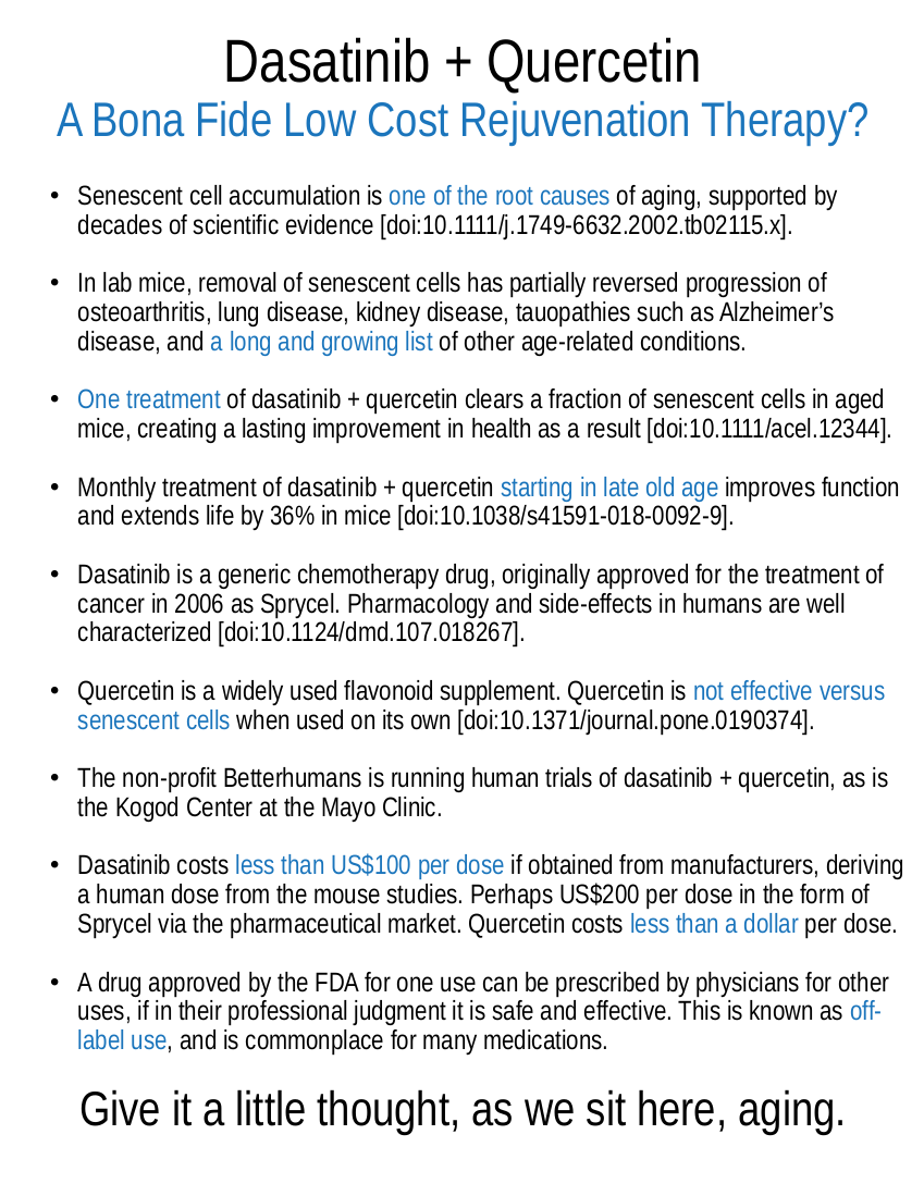
Sardinian Population Data Provides Evidence for a Reduced Burden of Infectious Disease to Slow the Pace of Aging

Aging is defined as an increase over time in the risk of death due to intrinsic causes. By this measure, aging has slowed over the past 150 years in most populations, particularly during the transition from an era of expensive calories and high rate of infection to an era of cheap calories and widespread use of effective antibiotics. A 60-year old today is far less impacted by aging and exhibits a far lower mortality risk than was the case for a 60-year old of two centuries past. To what degree is this late life outcome the result of improved nutrition versus a reduced burden of infection?

Given the importance of inflammation in aging, and the known impact of infectious disease on immune health over the long term, the consensus is that infection over a lifetime is more important than nutrition, even if the major contributing factor is persistent infection by just a few pathogens. Researchers here support that view by analyzing historical data from the Sardinian population that transitioned the era in which antibiotics were first introduced without also greatly changing their nutritional status. The results indicate that the use of antibiotics to control infectious disease produced a slowing of aging, and the data allows some insight into details of the period of a few decades over which that slowing took hold.

In biology, the term senescence is used to indicate the progressive accumulation of molecular damage that takes place in an organism as time goes by. This results in a gradual growth in the risk of death (demographic aging). By studying the pace at which mortality accelerates, it is possible to infer the general characteristics of the senescence process and to investigate which factors accelerate or decelerate its progression. So far, three major explanations for the determinants of senescence have been proposed: the constant senescence hypothesis; the inflammaging theory; and the calorie or energy restriction theory.

According to the constant senescence hypothesis, the pace of senescence is a biological constant among humans. As a result, senescence cannot be accelerated or decelerated by exogenous factors. Instead, the inflammation theory claims that the number and intensity of immune system responses to antigenic load in a lifetime is a fundamental factor in regulating the pace of senescence. Thus, individuals who have experienced a higher exposure to antigenic load will also experience a more rapid aging process. Finally, the calorie restriction theory, which is based on a plethora of experiments on a vast range of mammals and non-mammals, explores a reduced daily calorie intake and its positive effect on aging. In particular, a reduction in daily calorie intake is thought to slow senescence.

From a theoretical point of view, the three theories can be empirically tested. This would require a comparison of the aging process in cohorts who have experienced different nutritional regimes and different disease loads. However, the coincidence of the epidemiological transition and advent of antibiotics, from infection as majority cause of death to age-related disease as majority cause of death, with the onset of the nutrition transition, from a low to a high calorie regime, makes it difficult to isolate the effects of these two contrasting forces on mortality acceleration.

The epidemiological transition in Sardinia is unusual in that it started without any substantial modification in nutritional levels. This makes Sardinia a quasi-natural experiment where we can test the constant senescence hypothesis against the inflammation theory, without the confounding effect of changes in nutritional levels. To implement the analysis, the longitudinal life tables from 80 years onwards for Sardinian cohorts born in the period 1866-1908 were reconstituted and used to estimate the Gamma-Gompertz model: this model assumes that the individual hazard function follows the Gompertz model and that frailty is Gamma-distributed. The β parameter of the Gamma-Gompertz model, the so-called rate of aging, measures the relative derivative of the force of mortality, and in this sense, it may be used to measure how fast mortality progresses with age.

The results show that the Sardinian population experienced a dramatic reduction in the rate of aging that coincides with the onset of the epidemiological transition. The reduction in the rate of aging in an epoch characterized by a rapid reduction in infectious disease burden (probably due to quinine) appears to be consistent, at least at first sight, with the inflammation theory. The very low levels of nutrition observed in Sardinia, coupled with the dramatic fall off in the disease burden in the last years of the 19th century, might help to explain why the decline in the Sardinian rate of aging has been so dramatic compared with other European regions. The explanations advanced in the literature to justify the high prevalence of male centenarians in Sardinia have emphasized the role played by genetic factors. The possibility that genetic factors played a role in the evolution of the rate of aging in Sardinia cannot be entirely ruled out then. However, the analysis presented in this paper suggests that the very low Sardinian rate of aging at the beginning of the 20th century may depend on other factors such as nutrition and disease load.

Link: https://doi.org/10.1186/s41118-018-0028-8

Debating the Microbial Hypothesis for Alzheimer's Disease

Why do only some older people develop the elevated levels of amyloid-β that start the amyloid cascade of Alzheimer's disease, leading to tau aggregation and consequent death and dysfunction of brain cells? If amyloid-β is the result of persistent infection by pathogens such as herpesviruses and lyme spirochetes that are, collectively, only present in 20% or so of the population, then perhaps that is the answer. This is the core of the microbial hypothesis of Alzheimer's disease, that amyloid-β is a feature of the innate immune system, and thus persistent infection of brain tissue will result in higher levels of amyloid over time.

The microbial hypothesis can be balanced against other views on the rise of amyloid-β aggregation with age, such as the contribution of immune aging, in which the immune cells responsible for clearing out these aggregates falter in that work. Or consider the evidence for drainage of cerebrospinal fluid to decline due to age-related changes in fluid passages, and thus aggregates can no longer be effectively removed from the brain via these routes. It is plausible that all of these theories, each backed by a good amount of evidence, are to some degree correct. Alzheimer's will turn out to be a condition with multiple significant causes, and addressing all or most of those causes will be required to produce reliable benefits across the patient population.

The "germ theory" of Alzheimer's has been fermenting in the literature for decades. Even early 20th century Czech physician Oskar Fischer - who, along with his German contemporary Alois Alzheimer, was integral in first describing the condition - noted a possible connection between the newly identified dementia and tuberculosis. If the germ theory gets traction, even in some Alzheimer's patients, it could trigger a seismic shift in how doctors and understand and treat the disease. For instance, would we see a day when dementia is prevented with a vaccine, or treated with antibiotics and antiviral medications? Some researchers think it's worth looking into.

The hallmark pathology of Alzheimer's is accumulation of a protein called amyloid in the brain. Many researchers have assumed these aggregates, or plaques, are simply a byproduct of some other process at the core of the disease. Other scientists posit that the protein itself contributes to the condition in some way. Researchers have shown that amyloid is lethal to viruses and bacteria in the test tube, and also in mice. Evidence suggests that the protein is part of our ancient immune system that like antibodies, ramps up its activity to help fend off unwanted pathogens.

So does that mean that the microbe is the cause of Alzheimer's, and amyloid a harmless reaction to it? It's not that simple. In many cases of Alzheimer's, microbes may be the initial seed that sets off a toxic tumble of molecular dominos. Early in the disease amyloid protein builds up to fight infection, yet too much of the protein begins to impair function of neurons in the brain. The excess amyloid then causes another protein, called tau, to form tangles, which further harm brain cells. The ultimate neurological insult in Alzheimer's is the body's reaction to this neurotoxic mess. All the excess protein revs up the immune system, causing inflammation - and it's this inflammation that does the most damage to the Alzheimer's-afflicted brain.

So what does this say about the future of treatment? Possibly a lot. Researchers envision a day when people are screened at, say, 50 years old. "If their brains are riddled with too much amyloid, we knock it down a bit with antiviral medications. It's just like how you are prescribed preventative drugs if your cholesterol is too high." Any treatment that disrupts the cascade leading to amyloid, tau, and inflammation could theoretically benefit an at-risk brain. The vast majority of Alzheimer's treatment trials have failed, including many targeting amyloid. But it could be that the patients included were too far along in their disease to reap any therapeutic benefit.

Link: https://www.npr.org/sections/health-shots/2018/09/09/645629133/infectious-theory-of-alzheimers-disease-draws-fresh-interest

The Price of Progress or the Waste of Regulation?

The average cost of delivering a new therapy from laboratory to clinic is increasing at a fast pace, more than doubling since the turn of the century according to some studies, to stand at $2.5 billion or more. This is not driven by the work of research and development becoming more expensive: if anything, the price of the tools of biotechnology is in free fall, even as capacity increases by orders of magnitude. Biotechnology has gone through, and is still going through, its own echoed version of the computing revolution of recent decades. A mix of advances in computational power and materials science means that a graduate student of today requires six months of lab time and a few tens of thousands of dollars to accomplish what would have taken a full biotech company, five years, and tens of millions of dollars back in the 1990s.

So what is going on here? Why, in the midst of a transformative revolution in life science technological capabilities, is the price of building new therapies spiraling ever upwards? Government is the answer, bureaucrats and their incentives, regulation that demands an impossible degree of removal of risk from what is an inherently risky activity. The regulators of the FDA and other, similar organizations only suffer censure when patient issues occur that are related to approved medicines. No such censure happens when they reject medicines that would have helped greatly, or when they raise the cost of development high enough for beneficial programs to be abandoned as economically infeasible.

Given these incentives, and the point that no medicine is without risk, especially when used by people who are old and frail, the natural result is that regulators demand ever greater proof, ever greater cost, so as to able to claim that they did all they could. They reject perfectly feasible therapies because the treatments can never be made sufficiently risk free to remove the threat of bad press for regulators. Appearance of effectiveness is the driver, not actual benefits to society, as is the case in any well-established bureaucracy. The cost of billions of dollars presently required to bring a therapy to the clinic is not the price of progress. It is the waste produced by regulation and regulatory capture.

It is possible to run a useful program to evaluate the safety of a therapy for a small fraction of the cost and effort demanded by the FDA. The outcome would be little different in risk profile than the present excessive FDA process; people focus on the issues of the past as a justification for the vastly increased regulatory burden of the present, but issues are still occurring even today! We can make this comparison between levels of regulation by looking at what happens elsewhere in the world, and what happened in the past. There is no sense in the present regulatory burden; it is a monster run out of control, a cancer of perverse incentives.

Regulators prevent patients from choosing whether or not to take educated risks. There is every reason to have multiple layers of regulation and cost for medical development. Patients could choose the therapies they wished to use based on the history of safety testing. But we are not permitted that freedom. Everyone must conform to a program of regulation that dramatically slows the pace of progress. In an age in which rejuvenation biotechnologies are possible, plausible, and on the horizon, this suppression of technological progress is particularly unacceptable. It will kill all of us if allowed to continue, forcing us to join the countless lives already lost as a consequence of the regulatory slowdown in medical progress.

The open access paper here breaks down the costs and players in the development of new medical biotechnologies in much the same way as past studies, but with a focus on Alzheimer's disease. It is a useful primer to the environment in which development takes place, though one should always recall that this sort of work is inevitably affected by the relative detail and accessibility of sources of academic and governmental data versus the equivalent databases for private funding of research and development. That private funding is something like twice as large as public funding, very different in character and motivation, but far harder to break down and analyze.

The price of progress: Funding and financing Alzheimer's disease drug development

Prevention and treatment of Alzheimer's disease (AD) by 2025 has been articulated as a goal of the US government and has been endorsed by other countries. The failure rate of AD drug development is 99%; the failure rate of the development of disease-modifying therapies for AD is 100%. Despite these discouraging outcomes in drug development programs, the urgent need to address the socioeconomic crisis posed by AD requires that we continue to advance understanding of AD drug development.

To advance the research agenda in AD, financial resources are required including funding from government, industry, venture capital, foundations, and philanthropy. Federal research funding programs include the National Institutes of Health (NIH), National Science Foundation (NSF), Food and Drug Administration (FDA), Department of Defense, and Veterans Administration (VA). Private sector funding includes sources in the biopharma industry, venture capital, foundations, advocacy organizations, and support from philanthropists. Funding and financing resources form a complex financial ecosystem.

Total costs of an AD drug development program are estimated at $5.6 billion, and the process takes 13 years from preclinical studies to approval by the FDA. This compares to an estimated cost of cancer treatment development of $793.6 million per agent (assuming 9% cost of capital). Considering the pharmaceutical industry as a whole bringing a new agent to approval has an estimated cost of $2.8 billion. AD drug development costs substantially exceed most estimates for drugs in other therapeutic areas. Phase III trials are the most costly part of AD drug development, and pharmaceutical companies are among the few enterprises that can sustain such costs.

The principle public funder of research is the US NIH, investing more in health research than any other public enterprise in the world with an annual budget of approximately $34 billion U.S. dollars. Non-NIH federal agencies have smaller research budgets and grant portfolios related to AD. There is a mismatch between the cost of disease to society and the amount of research devoted to it. AD, for example, costs the US society more than $216 billion annually, and it has an NIH budget of $1.8 billion; for every $1 spent on AD, less than 1% of that amount is devoted to research.

Biotechnology companies can be defined as venture-backed drug development firms using technological applications centered on biological systems, living organisms, or their derivatives. Success in AD drug development will produce a very high return on investment. This possibility attracts venture capital to AD research, but the high rate of failure has kept this funding stream small. Venture capital funding in Central Nervous System disease declined 40% in the 2009-2013 period compared with the 2004-2008 period.

Angel investors or seed capital providers have high risk tolerance and supply small amounts of money to encourage novel ideas. If the concepts begin to mature and promise to lead to a successful program, venture capital may be attracted to allow more advanced drug development. Candidate therapies may pass from smaller to larger biotech companies as biotechs seek to strengthen their pipelines, progress toward vertically integrated Central Nervous System companies, or attract investors interested in a broader portfolio. This can be a healthy process allowing drugs to progress in testing before major pharmaceutical companies invest; however, the process also may lead to abuse by passing flawed agents from company to company and attracting capital from enthusiastic but under-informed investors.

The Alzheimer Association is the largest private noncorporate funder of AD research. In 2016, the association invested $90 million in research, including $25 million in new project investments and the rest in support of on-going multi-year commitments. Philanthropists make contributions to advocacy organizations or directly to universities and scientists to support research projects. Philanthropy plays a critically important role in the AD research ecosystem. Philanthropy often provides seed money for small projects that do not yet have preliminary data that would support a federal grant application. Philanthropy can fund high-risk/high-reward projects that might be too risky to receive funding from other sources such as the NIH.

The pharmaceutical industry is the largest funder of drug discovery and development research in the world, exceeding that of NIH or any other funding organization. Biopharma funds approximately 60% of all annual US research and development activities. The total annual research and development budget for biopharma (biotechnology and pharmaceutical industry) in 2016 was $75 billion. Over 70% of all AD clinical trials are sponsored or co-sponsored by the pharmaceutical industry. Payments from biopharma support much of the AD drug development ecosystem. New agents may be accessed through academic medical center collaborations, in-house discovery teams, acquisitions of biotechnology companies, mergers with other pharmaceutical companies, in-licensing of promising compounds, and partnering and co-development arrangements. Each of these has corresponding financial support by the pharmaceutical company.

The extreme expense of current drug development for AD is not sustainable, discourages companies from working in the AD research arena, dissuades venture capital from investing in AD drug development, and diminishes the opportunity to advance new therapies for patients with AD. Innovation is needed to improve the financial underpinnings of AD drug development and translational research.

Reprogramming Cells into Keratinocytes Turns Non-Healing Wounds into Skin

Researchers here report an interesting application of in situ cell programming. Knowing that keratinocytes do a lot of the heavy lifting in the coordination of skin healing, they reprogrammed cells at the surface of non-healing wounds, transforming them into keratinocytes capable of guiding the regeneration of skin. This is thought to be a way to aid healing in older individuals, or in other cases where chronic inflammation disrupts the normal processes of regeneration. Certainly this approach is notable for regenerating the full structure of skin, something that has only been achieved by one or two other methodologies to date.

Scientists have developed a technique to directly convert the cells in an open wound into new skin cells. The approach relies on reprogramming the cells to a stem-cell-like state and could be useful for healing skin damage, countering the effects of aging and helping us to better understand skin cancer. "Our observations constitute an initial proof of principle for in vivo regeneration of an entire three-dimensional tissue like the skin, not just individual cell types as previously shown."

The scientists knew that a critical step in wound recovery was the migration - or transplantation - of basal keratinocytes into wounds. These stem-cell-like cells act as precursors to the different types of skin cells. But large, severe wounds that have lost multiple layers of skin no longer have any basal keratinocytes. And even as these wounds heal, the cells multiplying in the area are mainly involved in wound closure and inflammation, rather than rebuilding healthy skin.

The researchers first compared the levels of different proteins of the two cell types (inflammatory versus keratinocytes) to get a sense of what they'd need to change to reprogram the cells' identities. They pinpointed 55 "reprogramming factors" (proteins and RNA molecules) that were potentially involved in defining the distinct identity of the basal keratinocytes. Then, through trial and error and further experiments on each potential reprogramming factor, they narrowed the list down to four factors that could mediate the conversion to basal keratinocytes.

When the team topically treated skin ulcers on mice with the four factors, the ulcers grew healthy skin (known as epithelia) within 18 days. Over time, the epithelia expanded and connected to the surrounding skin, even in large ulcers. At three and six months later, the generated cells behaved like healthy skin cells in a number of molecular, genetic, and cellular tests. The researchers are planning more studies to optimize the technique and begin testing it in additional ulcer models.

Link: https://www.salk.edu/news-release/the-alchemy-of-healing-researchers-turn-open-wounds-into-skin/

An Interview with David Gobel of the Methuselah Foundation

David Gobel, one of the pillars of our longevity science and advocacy community, cofounded the Methuselah Foundation with Aubrey de Grey way back when, and continues to run that organization today. Over the years he has supervised a diverse set of grants, projects, and successful investments in tissue engineering and aging research, including the first SENS rejuvenation research programs, prior to the launch of the SENS Research Foundation. With the recent influx of capital to new companies seeking to produce therapies that target mechanisms of aging, investment at the Methuselah Foundation has expanded to become the Methuselah Fund, a hybrid for-profit/non-profit vehicle that will continue the work of accelerating progress towards meaningful rejuvenation therapies.

How did your involvement in life extension begin; did you realize the problem of aging yourself, or were you introduced to it by someone else?

It started because of my awareness that the healthcare system was broken, like the growth of an unplanned city that has no rhyme or reason. Our healthcare system reacts to system failures rather than preventing them, because that is more lucrative. The incentives push science in poor directions, and then these become inferior technologies and treatments. I came to the conclusion that we need a system reset. After much research and reflection, it became my conviction that this reset should be to delay and reverse aging and rejuvenate robust health. I believe this will result in reduced suffering and the greatest opportunity for individual and civilizational growth.

Methuselah Foundation has given millions of dollars to regenerative medicine research, backing ventures such as Organovo, Oisin Biotechnologies, and SENS Research Foundation. Would you like to tell us about some of the results that these companies have obtained thanks to your charity?

Well, Organovo invented and is now selling high-fidelity 3D human liver and kidney tissues to the research market, is providing contract services, and is on track to deliver a 3D liver patch to the clinic in two years. Another portfolio alumnus, Silverstone Matchgrid, has saved the lives of over 1,000 people due to our investment in its paired kidney donation software. This software is now used in over 35 hospitals in the U.S., Europe, and soon, Saudi Arabia. I don't think I need to say anything about SENS Foundation - it is fantastic, and we at Methuselah Foundation couldn't be prouder of its success and contributions.

We have very high expectations for Oisin Bio and OncoSenX. We anticipate that it will be in Phase 1 safety trials by mid-2019. We hope to provide it to some patients much sooner than previously possible, as the FDA is liberalizing treatment availability via the recently passed "Right to Try" legislation. Leucadia Therapeutics is a startup focused on defeating Alzheimer's disease. This is progressing and promising. We hope to have major news later this year. Rather than go on, I'd like to say that we at Methuselah Foundation tend to be modest about proclaiming our successes. We prefer that the companies and scientists behind them get famous.

Can you tell us about the Methuselah Fund and how its mission differs from that of Methuselah Foundation?

The Methuselah Fund, or M Fund, is designed to give donors a chance to get a return on equity now that the longevity field is maturing. Many of our donors have been faithfully donating for years, and now that opportunities are emerging, we wanted to give them the first opportunity to invest. We are delighted to announce that we just successfully closed the M Fund's Founder's Round. We now have four companies in our portfolio and have been looking at helping form some promising new ventures. We are particularly proud to say that every single one of our members is a mission-driven individual who wants, more than anything, to see an end to the aging problem.

You were the first to put forward the concept of longevity escape velocity, or LEV. How far are we from LEV, assuming the current pace of research and no serious showstoppers?

I anticipate that within 3 years, some interventions will be available via safety trials and that people who are treated will receive benefits that put them on a path toward LEV. I believe things will accelerate from there, as vastly more attention is triggered by early advances. We are seeing the first glimmers of this already.

Link: https://www.leafscience.org/an-interview-with-david-gobel/

Mechanisms Involved in the Aging of Hematopoietic Stem Cells

Stem cell populations decline in activity with age, and the hematopoietic stem cells resident in bone marrow, responsible for generating blood and immune cells, are no exception. Their decline is one of the contributing factors leading to immunosenescence and inflammaging, the aging of the immune system. With age, the immune system becomes less effective in its tasks of destroying pathogens and errant cells, but also becomes chronically overactive at the same time. The result is inflammation, disruption of tissue regeneration, growing risk of cancer, increasing numbers of senescent cells, and vulnerability to infection.

Restoration of the hematopoietic stem cell population is one of the three necessary arms of immune rejuvenation. This will require advances in control over cell behavior, but is not too far beyond the present state of the art. The first generation of cell therapies resulted in cell transplants that near all die rather than engraft and participate in tissue maintenance. Today's stem cell therapies for the most part produce benefits due to temporary shifts in cell signaling brought about by the transplanted cells prior to their death. Reliable approaches by which large fractions of transplanted hematopoietic stem cells survive and take up residence will be needed. Transplanted cells will still suffer the consequences of a damaged surrounding tissue environment, but repairing that is a broader topic: it will need the other facets of the SENS damage repair approach to aging.

The other two arms of immune rejuvenation are regrowth of the thymus and clearance of malfunctioning immune cells. The thymus atrophies with age, but is required for the maturation of T cells of the adaptive immune system. These cells are initially created by hematopoietic stem cells in the bone marrow, migrate to the thymus, and there transformed into T cells of various types. As the thymus fades away this supply of T cells diminishes, ensuring that ever fewer naive T cells are available to tackle new threats. Lacking reinforcements, a growing fraction of the T cell population becomes senescent, exhausted, or uselessly specialized to persistent viruses such as cytomegalovirus. Other T cells malfunction and attack tissues, generating the varied and poorly mapped forms of autoimmunity that occur in late life.

Given a way to rapidly replace the entire immune cell population - restoration of the thymus and hematopoietic stem cell population may well be sufficient - it would make sense to destroy all of an older individual's immune cells. This would wipe the slate clean, removing all forms of damage and misconfiguration in immune cell populations. Vaccinations would have to be undertaken again, but that is a small price to pay for the opportunity to turn back immune system aging.

Updates on Old and Weary Haematopoiesis

Haematopoiesis is the process of the generation of all differentiated blood cells in the organism, including red blood cells, platelets, innate immune cells, and lymphocytes; all found to fade in functionality in aged individuals. Haematopoiesis is carried out by a rare population of haematopoietic stem cells (HSCs), which in adults, reside mainly in the bone marrow. There, they either remain dormant, i.e., in a quiescent state, or undergo proliferation and differentiation, depending on their cell-intrinsic transcriptional programs and the external cues from the surroundings.

Adult HSCs seem to be a heterogeneous subset of mainly multipotent and unipotent progenitors affiliated to specific lineages, and the ratio of their skewing shifts when homeostasis is perturbed. HSC maintenance relies on the support from the microenvironment or niche, necessary to preserve the self-renewing potential of HSCs. Extensive research on HSC niches composition shows that they are closely related to the vasculature in the bone marrow, with mainly endothelial, perivascular, and mesenchymal stromal cells secreting factors that support HSC maintenance. In this scenario, the effects of ageing on haematopoiesis may be the result of age-related alterations in all blood cell subsets, including HSCs and progenitors, as well as in the HSC niche.

In mice, the number of phenotypically defined HSCs can increase up to tenfold with ageing. In contrast, their functionality in terms of self-renewal and repopulating ability is remarkably reduced. Clonal HSC composition in old mice shows increased variability of clones derived from a single stem cell with smaller size per clone, when compared to young mice. Competitive transplantation of these HSCs proved that young HSCs perform better, with three-fold higher yield of mature granulocytes and lymphocytes. Furthermore, age-related defective HSCs seem to be able to differentiate into the myeloid lineage, but are incapable of the balanced generation of lymphocytes following transplantation. Thus, HSC defects are reflected in insufficiencies in their progeny of differentiated cells and contribute to poorer systemic performance of the haematopoietic system, i.e., immunosenescence.

At the molecular level, DNA damage and telomere shortening seem to be major mechanisms underlying the age-related decrease in the functionality and durability of HSCs. DNA damage accumulation is intimately related to increased reactive oxygen species (ROS) levels. In fact, HSCs reside in hypoxic bone marrow niches, which maintain their long-term self-renewal by mechanisms such as limiting their ROS production. Stressors, such as infections or chronic blood loss, shift HSCs from the quiescent to cycling state, which consequently leads to increased ROS levels and DNA damage.

Inflammageing is the characteristic process of chronic inflammation that has been described in aged individuals, with an increase of inflammatory cytokine levels that correlate with morbidity and age-related diseases. The HSC compartment is tightly connected to inflammatory processes, as a producer of innate immune cells. Furthermore, HSCs express pattern recognition receptors required for the identification of dangers, and a variety of cytokines and their receptors. Activation of these signalling pathways elicits HSC differentiation and myeloid skewing, aimed at mediating rapid myeloid cell recovery. However, when not finely regulated, they may cause HSC exhaustion.

HSC survival and function relies on the support from the microenvironment or niche in the bone marrow. Stem cell niches are complex and unique structures, yet they share many features that include cellular interactions, secreted factors, extracellular matrix, physical factors, metabolic conditions, and importantly, processes of scarring and inflammation. The changes in the bone marrow niche of aged mice include differences in gene expression and molecular structure in perivascular cells, arteries, and capillaries. In aged mice, enhancement of the Notch signalling pathway in endothelial cells can partially address some of these changes. Niche-forming vessel improvements are followed by increased HSC numbers, but no changes in their functionality. This suggests that niche-based rejuvenating strategies may have only partial efficiency to recover HSCs to a youthful state.

In conclusion, HSC ageing is characterised by reduced self-renewal, myeloid and platelet HSC skewing, and expanded clonal haematopoiesis that is considered a preleukaemic state. The underlying molecular mechanisms seem to be related to increased oxidative stress due to ROS accumulation and DNA damage, which are influenced by both cell- and cell non-autonomous mechanisms such as prolonged exposure to infections, inflammageing, immunosenescence, and age-related changes in the HSC niche. Thus, HSC ageing seems to be multifactorial and we are only beginning to connect all the dots.

Enhanced Lysosomal Activity Turns Back the Decline in Neural Stem Cell Function

Stem cell activity falters with age. This is a feature of all of the stem cell populations studied to date, though whether this is the result of declining cell count or increasing quiescence varies by tissue type. Stem cells are responsible for providing a supply of daughter somatic cells to replenish losses and maintain tissue function. Their progressive failure to do so is one of the important contributing causes of aging.

Why do stem cells undergo this decline? Intrinsic damage to the stem cells themselves is certainly a factor, but in many populations it isn't as important as changes in the signaling environment that take place in reaction to rising levels of molecular damage throughout a tissue. That said, in the research here, improved lysosomal activity is demonstrated to improve neural stem cell function. This implies that improved autophagy, increased removal of wastes and damaged components, is the cause of restored function. Autophagy declines with age, and there have been other examples in which enabling greater lysosomal function restores loss of organ function - such as in the liver, by adding more receptors essential to lyosomal activity.

Protein homeostasis, or proteostasis, is critical to maintain cellular integrity and function. Dysregulation of the proteome, including accumulation of damaged and aggregated proteins, is a major hallmark of aging. Accumulation of protein aggregates is also associated with pathological conditions, including neurodegenerative diseases. Though not much is known about the etiology of aggregates in many cases, their clearance can extend lifespan and alleviate the symptoms of neurodegeneration in some model systems.

There are three main mechanisms or branches of the protein homeostasis and clearance network: the lysosome-autophagy proteolytic system, molecular chaperones, and the proteasome. Macroautophagy, generally referred to as autophagy, is a tightly regulated process by which cellular organelles, proteins, and cytoplasm are engulfed into autophagosomes for degradation and recycling. The lysosomal-autophagy pathway is also important for the degradation of potentially toxic protein aggregates. Cellular quality control through this system may be particularly important in tissue-specific stem cells, which are used for lifelong tissue regeneration and repair.

Evidence suggests that the flux through the autophagy-lysosomal system is necessary for the maintenance and lineage progression of the adult neural stem cell (NSC) pool. These findings also raised a number of interesting questions regarding the precise role of autophagy in the NSC lineage in the adult and aging brain. For example, is autophagy critical for all stages of neurogenesis, or are specific transitions during lineage progression particularly dependent on this process?

Comparison of the activated (aNSC) and quiescent (qNSC) neural stem cells revealed striking differences in the expression of genes involved in protein homeostasis between the two cell types. Further analysis revealed that genes specifically associated with lysosomal function were selectively upregulated in the quiescent population. This is in contrast to aNSCs, which had higher expression of various molecular chaperones and displayed a signature associated with the proteasome and ubiquitin-mediated proteolysis. The use of a reporter system with manipulation of autophagic flux revealed that qNSCs degrade their lysosomal contents at a much slower rate than aNSCs.

The correlation between lysosome activation and NSC activation raises the question of whether activation of lysosomes is sufficient to drive NSCs out of the quiescent state. NSC activation involves cell cycle re-entry in response to intrinsic or extrinsic cues from the neurogenic microenvironment, although the molecular mechanisms are not fully understood. Could lysosome activation be a novel intrinsic stimulus to break quiescence? Recent work provides compelling evidence that this may be the case.

The authors observed that blocking lysosomal acidification induced aggregate accumulation in qNSCs and significantly reduced their ability to become activated. In contrast, induction of autophagic flux reduced the quantity of aggregates and enhanced the response of qNSCs to activation cues. This evidence suggests that clearance of protein aggregates is sufficient to induce activation of qNSCs in response to growth factor stimulation, although it cannot be ruled out that other unidentified cargo are critical for activation. Nevertheless, pathological lysosome dysfunction and aggregate buildup may have a causative role in the age-associated decrease in NSC activation and neurogenesis.

Link: https://doi.org/10.1177/1179069518795874

Piperlongumine Decreases Cognitive Decline in Aged Mice

Piperlongumine is a candidate senolytic agent, demonstrated to selectively destroy senescent cells in cell culture. Its ability to destroy senescent cells in vivo has not yet been confirmed, however, which would normally make it worthy of only academic interest. A sizable fraction of potential therapies fail to make the leap from cell culture to animal study. That said, unlike any of the senolytic candidates so far proven in animal studies, piperlongumine is a natural product, an extract of the long pepper. If it is usefully senolytic in mammals, then the regulatory path to widespread availability is much shorter and much less expensive than is the case for small molecule drugs.

Given this, there is considerable interest among patient advocates in the senolytic ability of piperlongumine in vivo. All it needs is an animal study with suitable accompanying measurements, and then it will be a matter of unleashing the supplement industry to work with regulators, mass manufacture, package, and distribute, giving them something worthwhile to do for a change. Unfortunately, while interesting, this study is not the study that we are still waiting for. The authors show that piperlongumine can achieve exactly the sort of results one would expect of a senolytic for cognitive decline in mice, mention senescent cells in passing, but do not assess whether or not the observed benefits resulted from clearance of senescent cells. This is frustrating, to say the least. The results here should be compared with the effects of the dasatinib and quercetin combination on neurodegeneration in a mouse model of Alzheimer's disease. It adds to the plausibility of piperlongumine as a useful senolytic, but plausibility is not proof.

In both normal aging and under pathological conditions, cognitive decline can diminish the quality of life. In the present study, we found that treatment with piperlongumine (PL), isolated from the long pepper, significantly improved cognitive function in novel object recognition and performance in nest building in 25-month-old female mice. These effects appear to be partly due to the modulation of neuronal activity and neurogenesis in the hippocampus.

PL is a primary constituent of Piper longum, which has been reported to kill multiple types of cancer cells through the targeting of the stress response to reactive oxygen species (ROS). Senescent cells can drive hyperplastic pathology and promote age-related neurodegeneration. Recently, PL has been reported to be a potential novel lead for the development of senolytic agents and the selective depletion of senescence cells as an anti-aging strategy may prevent cancer and aging-related degenerative diseases. Although in this study, we did not investigate the anti-tumour activities of PL in aged mice, PL treatment may be beneficial through the apoptosis of age-related senescence cells.

Cellular senescence is associated with oxidative stress and inflammation. An increase in the expression of GFAP has been the most common change to be observed in astrocytes with aging. The results of this study demonstrated that PL did not affect the size of area occupied by glia, such as microglia and astrocytes, in the hippocampus of the aged mice. We also observed that lipid peroxidation in the hippocampus was not altered in the aged mice. However, previously, we have demonstrated that PL effectively decreases astrogliosis and microglia activation in the parietal cortex in animal models of Alzheimer's disease. The results indicated that the inflammation and microglia activation that was triggered by pathological conditions were effectively suppressed by PL treatment.

In the present study, there were few DCX-positive neuroblasts in the dentate gyrus of 25-month-old female mice, but, the aged mice treated with PL exhibited significantly higher number of DCX-positive cells in the dentate gyrus than in controls. These results suggest that PL may have an effect on neurogenesis by preventing or reversing age-related decline. The precise mechanism of action through which PL improves cognitive function remains unclear. Further studies, therefore, are warranted to investigate the effects of PL on neurogenesis.

Link: https://doi.org/10.3892/ijmm.2018.3782

Rejuvenation Biotechnology as a Full Employment Program for Ethicists

Whenever I am told by ethicists that enabling people to live longer is a threat to society, a complex development that must be held back and studied so as to understand how best to allow it to progress, if at all, I have the feeling that I'm being held up for money. Ethics is, I feel, someone undermined in this day and age by the incentives that operate on the ethicist as a professional, with an office and a titled position in one or another institution. If he or she fails to find thorny problems that will require years of careful study, then he or she is out of a job. As a consequence I think a sizable proportion of the more modern incarnation of the field is essentially nonsense.

Acting to reduce the suffering and death of aging, by far the greatest cause of human pain and loss, isn't ethically complicated at all. It is the simplest thing in the world. Are we for or against suffering and death? Against? Good. Then we should bring an end to aging. That really is all there is to it, and all that has to be said on the matter. Medical science is close enough to the goal of rejuvenation therapies that no amount of effort deployed to other means of reducing suffering and death can be anywhere near as efficient a use of resources. Yet, strangely, those other approaches still receive far more attention. So we advocate for an adjustment of priorities: less war, less waste, more life science.

The author of New Methuselahs is one of those folk cheerfully carving out a portion of their living by making the ethics of rejuvenation appear much more complex than is actually the case. There is no problem that could possibly arise from ending aging that would be worse than what presently occurs as a result of aging; the hundred thousand lives lost daily, the hundreds of millions suffering pain, loss of capacity, loss of dignity as their bodies and minds corrode. The threat of overpopulation that is constantly brought up is a Malthusian dream, not a reality. Frequently predicted overpopulation and resource exhaustion has never come to pass, current trends head in the opposite direction, and the demographic models show that ending aging doesn't result in rapid population growth. If anything it is a madness of our era that we collectively have the capacity to do something about the death and suffering of aging, but would rather talk than act.

New Methuselahs: the Ethics of Life Extension

Life extension - slowing or halting human aging - is now being taken seriously by many scientists. Although no techniques to slow human aging yet exist, researchers have successfully slowed aging in yeast, mice, and fruit flies, and have determined that humans share aging-related genes with these species. In New Methuselahs, John Davis offers a philosophical discussion of the ethical issues raised by the possibility of human life extension. Why consider these issues now, before human life extension is a reality? Davis points out that, even today, we are making policy and funding decisions about human life extension research that have ethical implications. With New Methuselahs, he provides a comprehensive guide to these issues, offering policy recommendations and a qualified defense of life extension.

After an overview of the ethics and science of life extension, Davis considers such issues as the desirability of extended life; whether refusing extended life is a form of suicide; the Malthusian threat of overpopulation; equal access to life extension; and life extension and the right against harm. In the end, Davis sides neither with those who argue that there are no moral objections to life enhancement nor with those who argue that the moral objections are so strong that we should never develop it. Davis argues that life extension is, on balance, a good thing and that we should fund life extension research aggressively, and he proposes a feasible and just policy for preventing an overpopulation crisis.

Want to live longer? Consider the ethics

Life extension - using science to slow or halt human aging so that people live far longer than they do naturally - may one day be possible. Big business is taking this possibility seriously. From my perspective as a philosopher, this poses two ethical questions. First, is extended life good? Second, could extending life harm others?

Not everyone is convinced that extending life would be good. In a 2013 survey, some respondents worried that it might become boring, or that they would miss out on the benefits of growing old, such as gaining wisdom and learning to accept death. On the other hand, not everyone is persuaded that extended life would be a bad life. I'm not. But that's not the point. No one is proposing to force anyone to use life extension, and - out of respect for liberty - no one should be prevented from using it.

However, our liberty right is limited by the "harm principle." The harm principle says that the right to individual liberty is limited by a duty not to harm others. There are many possible harms: Dictators might live far too long, society might become too conservative and risk-averse, and pensions might have to be limited, to name a few. One that stands out to me is the injustice of unequal access.

It is unjust when some people live longer than the poor because they have better health care. It would be far more unjust if the rich could live several decades or centuries longer than anyone else. Some philosophers suggest that society should prevent inequality by banning life extension. This is equality by denial - if not everyone can get it, then no one gets it. However, "leveling-down" - achieving equality by making some people worse off without making anyone better off - is unjust. Indeed, most of us reject leveling-down in other situations. For example, there are not enough human organs for transplant, but no one thinks the answer is to ban organ transplants.

Another possible harm is that widespread life extension might make death worse for some people. All else being equal, it is better to die at 90 than nine. At 90 you're not missing out on many years, but at nine you lose most of your potential life. In a world where some people get life extension and some don't, what's the right measure for how many years death takes from you? If so, then the fact that some people can get life extension makes your death somewhat worse. This is a more subtle kind of harm than living in an overpopulated world, but it's a harm all the same.

However, not just any harm is enough to outweigh liberty. After all, expensive new medical treatments can extend a normal lifespan, but even if that makes death slightly worse for those who can't afford those treatments, no one thinks such treatments should be banned. I believe that life extension is a good thing, but it does pose threats to society that must be taken seriously.

An Overview of the Biochemistry of Muscle Aging

This popular science article covers some of the major research topics related to sarcopenia, the loss of muscle mass and strength that occurs with age. A great deal is known of the biochemistry of muscle aging, the signals and mechanisms involved in muscle stem cell activity and muscle growth, and how they change with age. A great deal more remains to be discovered, and fitting together what is already known into a coherent whole is a still a work in progress. Any proposed layering of cause and effect is speculative at best, and it is usually unclear as to where exactly any newly described signal or mechanism fits. It it is probably the case, here as elsewhere, that the fastest path to improved knowledge is to start in on manipulating the aging of muscle: adjust a mechanism in isolation of the others and analyze the results.

Up to a quarter of adults over the age of 60 and half of those over 80 have thinner arms and legs than they did in their youth. The good news is that exercise can stave off and even reverse muscle loss and weakness. Recent research has demonstrated that physical activity can promote mitochondrial health, increase protein turnover, and restore levels of signaling molecules involved in muscle function. But while scientists know a lot about what goes wrong in aging, and know that exercise can slow the inevitable, the details of this relationship are just starting to come into focus.

Mature muscle fibers are post-mitotic, meaning they do not divide anymore. As a result, in adulthood both muscle growth and repair are made possible only by the presence of muscle stem cells known as satellite cells. Elderly human satellite cells show dramatic changes in their epigenetic fingerprint. One gene, called sprouty 1, is known to be an important regulator of cell quiescence. Reduced sprouty 1 expression can limit satellite cell self-renewal and may partially explain the progressive decline in the number of satellite cells observed in human muscles during aging. Indeed, stimulation of sprouty 1 expression prevents age-related loss of satellite cells and counteracts age-related degeneration of neuromuscular junctions in mice.

Other likely culprits of muscle aging are the mitochondria, the powerhouses of muscle. To work efficiently, skeletal muscle needs a sufficient number of fully functional mitochondria. These organelles represent around 5 percent to 12 percent of the volume of human muscle fibers, depending on activity and muscle specialization (fast-twitch versus slow-twitch). And research suggests that abnormalities in mitochondrial morphology, number, and function are closely related to the loss of muscle mass observed in the elderly.

In 2005, researchers combined the circulation of young and old mice and found that factors in the blood of young mice were able to rejuvenate muscle repair in aged mice. It is now well known that the levels of circulating hormones and growth factors drastically decrease with age and that this has an effect on muscle aging. Indeed, hormone replacement therapy can efficiently reverse muscle aging, in part by activating pathways involved in protein synthesis. Moreover, the muscle itself is a secretory endocrine organ. Myokine proteins produced by the muscle when it contracts can act locally on muscle cells or other types of cells such as fibroblasts and inflammatory cells to coordinate muscle physiology and repair, or they can have effects in distant organs, such as the brain.

Although several of these myokines have been identified-in culture, human muscle fibers secrete up to 965 different proteins-researchers have only just begun to understand their role in muscle aging. The first myokine to be identified, interleukin-6 (IL-6), participates in muscle maintenance by decreasing levels of inflammatory cytokines in the muscle environment, while increasing insulin-stimulated glucose uptake and fatty-acid oxidation.

Researchers recently discovered a novel myokine, which they termed apelin. The researchers have demonstrated that this peptide can correct many of the pathways that are deregulated in aging muscle. When injected into old mice, apelin boosted the formation of new mitochondria, stimulated protein synthesis, autophagy, and other key metabolic pathways, and enhanced the regenerative capacity of aging muscle by increasing the number and function of satellite cells. Levels of circulating apelin declined during aging in humans, suggesting that restoring apelin levels to those measured in young adults may ameliorate sarcopenia.

Link: https://www.the-scientist.com/features/how-muscles-age--and-how-exercise-can-slow-it-64708

An Overview of the Present State of Development of Senotherapeutics

Senotherapeutics are treatments that in some way reduce the burden of senescent cell accumulation in old tissues. This is a broader category than senolytics, therapies that destroy senescent cells, and includes efforts to modulate the harmful signaling of senescent cells without destroying them. I'd say that latter strategy has little to recommend it at the present time; one would need evidence for significant vital populations of senescent cells in the brain to start to think about modulation rather than destruction. So far the approach of targeted destruction is doing very well in mouse studies, robustly producing rejuvenation and extension of healthy life span, even using therapeutics that are far from optimal in comparison to the improved versions now under development.

This paper is not open access, but in a world in which the copyright heretics of Sci-Hub continue to endure, journal paywalls now present little hindrance for the curious. I point it out because in addition to the initial overview of the biochemistry of cellular senescence in the context of aging, it also contains well presented tables of current senotherapeutics, their evidence, and their progress towards the clinic. This is a useful resource for those thinking seriously about self-experimentation or putting together pilot clinical trials.

Accumulating evidence suggests that, in contrast to the cell-autonomous tumor-suppressive mechanism of senescence, the paracrine effects of senescent cells themselves, particularly those mediated by the senescence-associated secretory phenotype (SASP), are responsible for aging-related pathologies, among which cancer has attracted increasing attention. Optimizing the beneficial impact while minimizing the deleterious effects of cellular senescence remains a serious challenge for multiple fields of scientific and clinical research.

Transient induction of cellular senescence, followed by tissue remodeling and senescent cell elimination by the immune system, is beneficial because it facilitates removal of damaged cells from the affected tissue. However, chronic senescence or inability to eliminate the senescent cells is frequently observed in aged individuals or in pathological contexts, leading to the accumulation of senescent cells which produce adverse effects.

Increasing evidence shows that both pro-senescence and anti-senescence therapies can be beneficial to tissue homeostasis. For instance, in the case of cancer, pro-senescence therapies can minimize the damage by limiting aberrant activities such as hyperactive proliferation, and more specifically by preventing or delaying events of carcinogenesis, while anti-senescence treatments may help to remove accumulated senescent cells and allow tissue regeneration. Of note, the term "anti-senescence" in this field of research does not mean that senescence is blocked or prevented, but means that when senescence is engaged it is subsequently pushed into apoptosis.

A two-step anticancer strategy was recently suggested in which senescence-inducing treatments are followed by senotherapy, thus providing a novel option to maximize therapeutic efficacy and improve clinical outcome. Although the incidence of senescence can improve long-term outcomes for cancer, the potentially harmful properties of senescent cells persisting in vivo make their quantitative elimination an outstanding therapeutic priority.

The most promising senolytics appear to be inhibitors of pro-survival BCL family proteins, probably because senescent cells physiologically need these factors to circumvent apoptosis for long-term survival. This class of agents has undergone extensive investigation in patients with chronic leukemia, with final FDA approval of a selective BCL-2 inhibitor, venetoclax. However, venetoclax is not a potent senolytic agent in vitro, whereas its homolog navitoclax has recently been disclosed to be one of the strongest senolytics. Navitoclax effectively inhibits BCL-2, BCL-xL, and BCL-W, suggesting that senolysis requires suppression of a wider range of anti-apoptotic effectors than of BCL-2 alone. It is rational to propose a broad spectrum of BCL protein inhibitors as a potential senolysis treatment in patients, but such molecules would need to exhibit acceptable toxicity through new or optimized formulation, delivery, or administration schedule.

Link: https://doi.org/10.1016/j.molmed.2018.08.002

Inducing Axons to Connect Through Scar Tissue in a Mouse Model of Spinal Injury

The two primary challenges in nerve regeneration are firstly to induce nerve tissue to regrow at all, and secondly to find a way to deal with the blockade of scarring that forms around injury sites. The existence of this scar tissue is why it is the case that some progress has been made in treatment for recent nerve injury, but very little can yet be done for patients with older injuries. In that context, the recent research results noted here are exciting, an advance that offers tangible hope to the many people who presently live with loss of function due to severed or damaged nerves. This is still very early stage work, however, and we all know that it takes long years to move from initial demonstrations in animal models to clinical trials to general availability.

Regeneration of the spinal cord has been a heavily advocated and well funded goal for as long as Fight Aging! has existed. Those us of a certain age no doubt recall the Christopher & Dana Reeve Foundation in the period in which its principals were more vocal and present in the media, in the early days of high hopes for stem cell research, and prior to Christopher Reeve's untimely death as a consequence of his spinal injury. That organization remains active, and is one amongst many supporting this line of research and development. It is a sad truth that when regulation of medicine forces a ten year or longer road from clinical readiness to clinical availability, added to the time needed to build working therapies, it is the case that the hopes of the present generation of patients only become a reality for the next generation of patients.

New therapy spurs nerve fibers to regrow through scar tissue, transmit signals after spinal cord injury in rodents

Researchers have identified a three-pronged treatment that triggers axons - the tiny fibers that link nerve cells and enable them to communicate - to regrow after spinal cord injury in rodents. Not only did the axons grow through scars, they could also transmit signals across the damaged tissue. "Previous studies had tested each of the three treatments separately, but never together. The combination proved to be the key."

Many decades of research have shown that a human's nerve fibers need three things to grow: genetic programming to switch on axon growth; a molecular pathway for the fibers to grab and grow along; and a trail of protein "bread crumbs" that spur the axons to grow in a particular direction. All three of these conditions are active when humans develop in the womb. After birth, these processes shut down, but the genes that control the growth programs are still sleeping in the body. The goal was to reawaken these genes and then launch the entire process anew with the three-pronged approach.

Not only had axons grown robustly through the scar tissue, but many fibers also had penetrated into the remaining spinal cord tissue on the other side of the lesion and made new connections with neurons there. When we stimulated the animal's spinal cord with a low electrical current above the injury site, the regrown axons conducted 20 percent of normal electrical activity below the lesion. In contrast, the untreated animals exhibited none. Despite the finding suggesting that the newly formed connections can conduct signals across the injury, the rodents' ability to move did not improve. "We expect that these regrown axons will behave like axons newly grown during development - they do not immediately support coordinated functions. Much like a newborn must learn to walk, axons that regrow after injury will require training and practice before they can recover function."

Required growth facilitators propel axon regeneration across complete spinal cord injury

Transected axons fail to regrow across anatomically complete spinal cord injuries (SCI) in adults. Diverse molecules can partially facilitate or attenuate axon growth during development or after injury, but efficient reversal of this regrowth failure remains elusive. Here we show that three factors that are essential for axon growth during development but are attenuated or lacking in adults - (i) neuron intrinsic growth capacity, (ii) growth-supportive substrate and (iii) chemoattraction - are all individually required and, in combination, are sufficient to stimulate robust axon regrowth across anatomically complete SCI lesions in adult rodents.

We reactivated the growth capacity of mature descending propriospinal neurons with osteopontin, insulin-like growth factor 1 and ciliary-derived neurotrophic factor before SCI; induced growth-supportive substrates with fibroblast growth factor 2 and epidermal growth factor; and chemoattracted propriospinal axons with glial-derived neurotrophic factor delivered via spatially and temporally controlled release from biomaterial depots, placed sequentially after SCI. We show in both mice and rats that providing these three mechanisms in combination, but not individually, stimulated robust propriospinal axon regrowth through astrocyte scar borders and across lesion cores of non-neural tissue that was over 100-fold greater than controls. Stimulated, supported and chemoattracted propriospinal axons regrew a full spinal segment beyond lesion centres, passed well into spared neural tissue, formed terminal-like contacts exhibiting synaptic markers and conveyed a significant return of electrophysiological conduction capacity across lesions.

An Inflammatory Feedback Loop in the Aging Brain Contributes to Neurodegeneration

Scientists here report on a mechanism that might explain some fraction of the rising levels of chronic inflammation observed in the aging brain - though as in most such research, it is a proximate cause, and it isn't very clear as to how it relates to the known root causes of aging. Whatever that relationship might be, it is clear enough that with advancing age the immune system falls into a state of continual, inappropriate activation and inflammation. This disrupts many important processes in the normal maintenance of tissue function, and particularly so in the brain, where immune cells undertake a greater range of important activities than is the case elsewhere in the body.

The activity of microglial cells plays an important role in brain aging. These cells are part of the brain's immune defense: For example, they detect and digest bacteria, but also eliminate diseased or defective nerve cells. They also use messenger substances to alert other defense cells and thus initiate a concerted campaign to protect the brain: an inflammation. This protective mechanism has undesirable side effects; it can also cause damage to healthy brain tissue. Inflammations are therefore usually strictly controlled.

Endocannabinoids play an important role in this control. These are messenger substances produced by the body that act as a kind of brake signal: They prevent the inflammatory activity of the glial cells. Endocannabinoids develop their effect by binding to special receptors. There are two different types, called CB1 and CB2. However, microglial cells have virtually no CB1 and very low level of CB2 receptors. Researchers have now found that the brake signals do not communicate directly with the glial cells, but via middlemen - a certain group of neurons, because this group has a large number of CB1 receptors.

This is how it might work in mice: As soon as microglial cells detect a bacterial attack or neuronal damage, they switch to inflammation mode. They produce endocannabinoids, which activate the CB1 receptor of the neurons in their vicinity. This way, they inform the nerve cells about their presence and activity. The neurons may then be able to limit the immune response. The scientists were able to show that neurons similarly regulator the other major glial cell type, the astroglial cells. During ageing the production of cannabinoids declines reaching a low level in old individuals. This could lead to a kind of vicious circle. Since the neuronal CB1 receptors are no longer sufficiently activated, the glial cells are almost constantly in inflammatory mode. More regulatory neurons die as a result, so the immune response is less regulated and may become free-running.

It may be possible to break this vicious circle with drugs in the future. Tetrahydrocannabinol (THC) is a powerful CB1 receptor activator, even in low doses. Last year, researchers were able to demonstrate that THC can reverse the aging processes in the brains of mice. This result now suggest that an anti-inflammatory effect of THC may play a role in its positive effect on the ageing brain.

Link: https://www.uni-bonn.de/news/218-2018

Incidence of Stroke is Associated with a Doubling of Dementia Risk

Aspects of aging, such as specific age-related conditions, arise from shared root causes. If an individual exhibits one given outcome of aging, then they are more likely to also exhibit others that arise from the same underlying processes. Thus we should not be surprised to see that incidence of stroke is correlated with incidence of dementia. We don't have to suggest that stroke-induced damage to the brain, and the inflammatory and other reactions to that damage, can accelerate the onset of dementia. We can instead argue that both stroke and dementia are consequences of the aging of the cardiovascular system, and there likely to occur in close proximity to one another. In fact both of these explanations are likely to be true.

A new study analysed data on stroke and dementia risk from 3.2 million people across the world, finding that people who have had a stroke are around twice as likely to develop dementia. The link between stroke and dementia persisted even after taking into account other dementia risk factors such as blood pressure, diabetes, and cardiovascular disease. Their findings give the strongest evidence to date that having a stroke significantly increases the risk of dementia.

The researchers analysed 36 studies where participants had a history of stroke, totalling data from 1.9 million people. In addition, they analysed a further 12 studies that looked at whether participants had a recent stroke over the study period, adding a further 1.3 million people. "We found that a history of stroke increases dementia risk by around 70%, and recent strokes more than doubled the risk. Given how common both stroke and dementia are, this strong link is an important finding. Improvements in stroke prevention and post-stroke care may therefore play a key role in dementia prevention."

Stroke characteristics such as the location and extent of brain damage may help to explain variation in dementia risk observed between studies, and there was some suggestion that dementia risk may be higher for men following stroke. "Around a third of dementia cases are thought to be potentially preventable, though this estimate does not take into account the risk associated with stroke. Our findings indicate that this figure could be even higher, and reinforce the importance of protecting the blood supply to the brain when attempting to reduce the global burden of dementia."

Link: http://www.exeter.ac.uk/news/featurednews/title_678124_en.html

Greater Life Expectancy Correlates with Greater Economic Productivity

The International Longevity Center in the UK turns out interesting white papers every so often. Note that the organization is funded by a number of pensions and insurance companies, sizable business concerns whose long term success depends on (a) correctly predicting the future of human aging, and, (b) preventing the short term incentives of politicians and executives from steering them over a cliff. The first point requires research, and the second point requires presenting that research publicly and loudly. This is a time of great uncertainty for the pensions and life insurance industry, an era in which accelerated technological innovation in the medical life sciences makes prediction difficult in comparison to the state of affairs a few decades past. It is clear that life spans will leap upwards at some point, but when?

The latest white paper from the International Longevity Center runs the numbers to show that increasing life expectancy correlates with increased productivity in developed countries. The authors suggest that this results from a greater return on investment in education, in that educated people have more time in which to be productive following their education, and this tends to encourage greater investment in education as a path to productivity. This is an effect that can be suppressed by laws that force retirement at a set age, an iniquity that is thankfully fading away but nonetheless still exists in some professions and parts of the world.

One important underlying point in all this is that increased longevity is not a matter of adding years of disability to the end of life; it can only be achieved robustly by extending the span of productive healthy life. Aging is caused by accumulated molecular damage, and to the degree that this damage accumulation can be slowed, both healthy and overall life span is increased. That slowing of damage has progressed steadily and incrementally over the past few generations, an incidental side-effect of improved medicine and increased wealth. The goal of present rejuvenation research and development programs is to step beyond mere slowing to be able to repair the damage, and thus reverse aging. That will lead to the expected great leap upwards in life expectancy for people already in later life. The first of these technologies are already in commercial development, but until they are tested no-one can say for sure how effective they will be. Nor is it possible to make firm predictions on the timing of the following therapies, still early in development.

Towards a longevity dividend: Life expectancy and productivity across developed countries

The positive relationship between income and life expectancy has been demonstrated across many different time periods but the causes of the relationship are much debated. In particular, there is a pertinent question about the direction of the relationship - does higher income lead to higher life expectancy or does higher life expectancy lead to higher income? This report is devoted to exploring the latter relationship. More specifically, based on previous theory and evidence, we develop a statistical method for assessing the extent to which differences in life expectancy explain cross country variation in productivity - measured in terms of GDP per hour worked, per worker, and per capita. We also explore two of the potential channels through which life expectancy might influence productivity - increased educational attainment and greater participation in the labour market.

While our previous research focussed on possible reasons why different age dynamics might affect productivity differently, this report is focussed more exclusively on the role of life expectancy. According to the wider economic literature, there are many reasons why increased life expectancy might boost economic output. Healthier workers are likely to be more productive, while longer lives may result in greater incentives to invest in schooling. The latter point is worth emphasising - if parents only expect their child to live to 40, the expected lifelong returns to investing in their education is likely to be far lower than if they are expected to live to 80.

We explore the relationship between life expectancy and various measures of productivity across OECD countries between the years 1970-2015. We use all three measures of GDP (per hour worked, per worker, and per capita) because population dynamics may impact the productivity of the workforce differently to the productivity of the population as a whole. For instance, an increased share of older retired people is likely to act as a drag on GDP per capita but its effects on the productivity of the workforce itself remain contentious.

We found life expectancy to be positively associated with productivity and that this relationship was robust to different productivity measures, the inclusion of a range of explanatory and control variables and different instruments. Moreover, we found life expectancy to be a more powerful determinant of productivity than either the young or old age dependency ratios. When investigating the channels through which life expectancy boosts productivity, we found education to be more important than employment. Regarding the latter, due to changes to public policy, such as the abolition of default retirement ages and raising pensionable age, the link between life expectancy and employment at older ages has been recoupled so there is increased capacity for life expectancy improvements to translate into higher employment rates at older ages.

Overall, this analysis suggests that there may well be a longevity dividend, whereby improvements to health result in wider economic and productivity improvements. Improving health and raising life expectancy must therefore remain a key goal not only for a nation's health and wellbeing but also for the wider economy. This is important, since in many debates about long run government spending, health spending is simply seen as a drain on fiscal resources, yet if by raising life expectancy it results in productivity improvements, this could support increased tax revenue for the exchequer. Public policy and economic forecasters should consider how best to take into account the potential fiscal benefit of better health and not neglect it in discussions of our long run sustainability.

A Clinical Trial of Induced Pluripotent Stem Cells for Heart Disease Begins Next Year

Induced pluripotent stem cells (iPSCs) were from the very first seen as a promising biotechnology. The approach to reprogramming cells from a patient sample into iPSCs costs little and is easy for any life science lab to work with. This offers the potential to generate patient matched cells of any type in a reliable manner, which in turn enables development of a range of potential regenerative therapies. As is always the case, moving from the lab to the clinic has progressed at a very slow pace, however. The regulatory system demands absolute certainty and enormous expense, and the primary result is that it takes a very long time to make any sort of progress towards commercial application of technologies proven in the laboratory. Further, and as is the case here, these incentives direct researchers away from using patient-matched cells in favor of standard donor sources even when that causes worse outcomes for patients. The gap between what is possible and what is permitted increases with every passing year. At some point, and in some way, this must end.

Early next year, a small clinical trial will begin in Japan, marking the first time reprogrammed stem cells will be deployed to help regenerate injured hearts. A team will implant sheets - each consisting of 100 million stem-cell derived cardiomyocytes - onto the hearts of three patients with advanced heart failure. The cardiac study is only the second-ever clinical application of induced pluripotent stem (iPS) cells, the first being an iPS-cell transplant to treat macular degeneration of the eye, which also took place in Japan. While it is a big deal to pioneer such a technology clinically, the trial also has its risks, unknowns, and critics.

Japan's health ministry conditionally approved the heart experiment in May, with the goal of assessing the safety of the procedure. If the first trial and a later one enrolling 10 patients prove successful, the treatment will be made commercially available soon under a new fast-track system in Japan designed to speed up the development of regenerative therapies. Since the trial was announced, several Japanese researchers have voiced their concerns. One of them notes that the trail participants will receive iPS-derived cells from a donor, instead of from their own tissue, and will have to be placed on immunosuppressants for three months to prevent rejection. The researchers running the trials say that creating cardiomyocytes derived from a patient's own cells is not always an option, because the reprogramming process takes a long time. Providing off-the-shelf treatments is a more feasible route to address heart failure. "Cell therapy using a patient's own cells seems to be not suitable for industrialization."

While preclinical work with iPS cells has proven effective in improving heart function in mice, pig, and monkey models, it's not quite clear by which mechanism the cells are promoting muscle regeneration. It's still unknown whether these cells actually integrate into the heart and become beating heart cells, or whether they just release factors and help existing heart cells. Research in pigs suggests that iPS cell-derived cardiomyocytes promote regeneration of the heart by secreting certain cytokines that stimulate the native heart muscle to grow.

Link: https://www.the-scientist.com/news-opinion/first-ips-cell-trial-for-heart-disease-raises-excitement--concern-64743

Inching Towards the Regulatory Classification of Aging as a Disease

Sizable factions within the research and advocacy communities are very interested in having aging officially classified as a disease, meaning its inclusion in the International Classification of Diseases maintained by the World Health Organization, as that is the basis for the definition of disease used by national regulatory bodies. The view is that this would open the door to greater large-scale institutional funding, more relevant clinical trials for therapies targeting the mechanisms of aging, and that this greater level of funding and activity will percolate back down the chain of research and development to accelerate progress. I think this a reasonable argument to make, though I would advocate for greater effort to be placed on finding a way to bypass the system rather than change it directly - the threat of competition tends to be more effective than petitions as a way to force change.

Lobbyists have made more progress towards classifying aging as a disease. The World Health Organization (WHO) has implemented the extension code "Ageing-related" (XT9T) in the latest version of the International Classification of Diseases (ICD). The previous version, the ICD-10, was released in 1983 and is now replaced by the new version, the ICD-11, which is expected to serve the medical community for many years, much as its predecessor has.

In the new ICD code, 'ageing-related' means "caused by pathological processes which persistently lead to the loss of organism's adaptation and progress in older ages". This is an important step forward for our field because ICD codes are a prerequisite for the registration of new drugs and therapies. It also marks the recognition that aging is a pathological process and represents a solid step forward in overcoming regulatory barriers to developing therapies that directly target the aging processes themselves.

WHO reports that by 2050, around 2 billion people (22% of the world's population) will be age 60 or over. This is commonly referred to as the "Silver Tsunami", and it is a real concern because society will become increasingly unable to cope with the rising numbers of elderly and potentially sick people, as aging is the greatest risk factor for multiple age-related diseases. The solution to this problem is to develop therapies that address the aging processes to keep older people healthy, active, and contributing to society rather than being burdens on healthcare systems, and that is without even considering the personal benefit of keeping people alive and healthy.

While the inclusion of the new code in the ICD-11 cannot be regarded as the WHO officially accepting aging as a disease, it does show that the WHO recognizes that aging is the primary risk factor for age-related diseases. There is also considerable debate as to if aging is a disease or not; we propose that it is a co-morbid syndrome. A syndrome is a set of medical signs and symptoms that are correlated with each other and, often, with a particular disease or disorder. This really does describe aging perfectly: it is a group of symptoms that consistently occur together and is a condition characterized by a set of associated symptoms. Ultimately, aging is an umbrella term describing a range of pathological changes; it may struggle to be accepted as a disease, but it already qualifies as a syndrome.

Link: https://www.leafscience.org/a-step-closer-to-aging-being-classified-as-a-disease/