Does Obesity Literally Accelerate Aging?

It is well known that carrying excess visceral fat tissue increases risk of age-related disease, shortens life expectancy, and raises lifetime medical expenditure. The more fat tissue, the worse the outcome, but even being modestly overweight rather than obese still produces a negative impact on long term health. This is the story told in a great many epidemiological studies with large patient populations. Does this mean that obesity accelerates aging, however? It might be surprising to find out that this isn't a question that has an easy or a straightforward answer.

In order to talk about whether aging is accelerated, one has to have a strong understanding of what causes aging. If we can list specific causative mechanisms of aging, and then measure their state, then we might be able to say whether or not aging is accelerated or slowed by a given circumstance. In the SENS view of aging, the root cause is accumulation of cell and tissue damage that arises as a side-effect of the normal operation of cellular metabolism. Things like the presence of lingering senescent cells or cross-links in the extracellular matrix. We can make the argument that a lifestyle choice that increases the pace at which senescent cells emerge in tissues is in fact an acceleration of aging. We can similarly argue that environmental circumstances such as smoking or chemotherapy that do the same have some component of accelerated aging in the harm that they cause.

Excess visceral fat tissue does in fact add to the presence of senescent cells. It also causes chronic inflammation via several other mechanisms, distinct from that of the inflammatory signaling produced by senescent cells. The chronic inflammation of aging is a downstream consequence of causes of aging, but it is a prominent feature of aging and causes further issues in and of itself, speeding up the progression of all of the common age-related conditions. Could upregulating inflammation directly, without going via one of the underlying causes of aging, be called an acceleration of aging? Perhaps not. Perhaps it should just be called harm and damage, and fall into the same category as breaking a bone and the long-term consequences that result from that sort of injury. So we might say that fat tissue accelerates aging in some senses, but in others it is not an acceleration of aging, just a harm.

This may be a matter of semantics and definitional games. The lesson at the end of the day is to avoid putting on excess weight, as even therapies targeting the causes of aging cannot prevent all of the long-term damage that being overweight will generate. Different perspectives are always interesting, however. Today's open access paper, noted below, looks at the question of whether or not obesity accelerates aging through the filter of the Hallmarks of Aging, a more recent catalog of potential causes and mechanisms of aging that overlaps to some degree with the causes of aging listed in the SENS proposals, but has significant differences. Some of the Hallmarks are clearly downstream consequences or markers of the progression of aging from the SENS perspective, for example.

Obesity May Accelerate the Aging Process

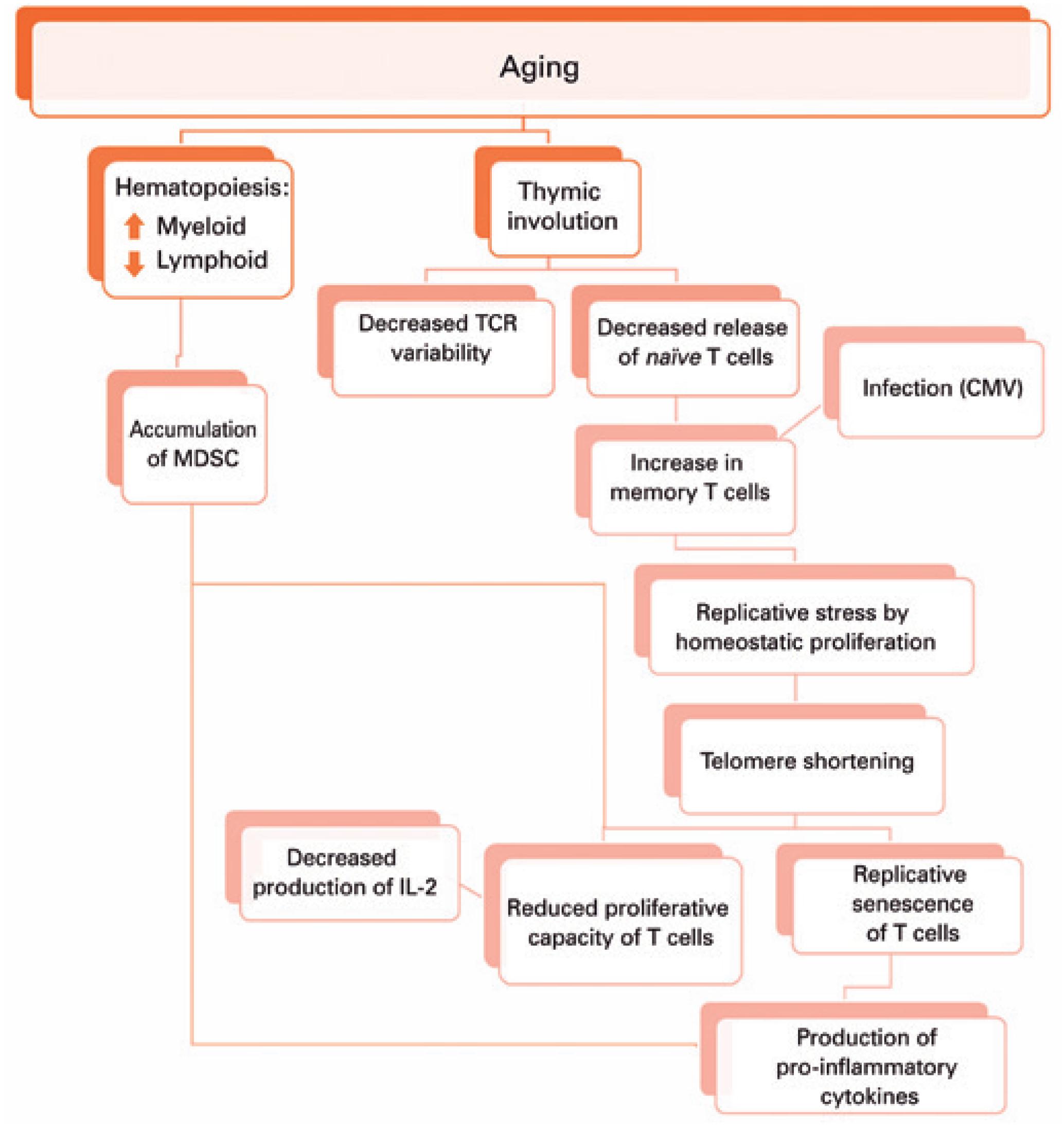
It has been suggested that obesity not only increases the onset of metabolic imbalances, but also decreases life span and impacts cellular processes in a manner similar to aging. A defining characteristic of aging is the gradual loss of physiological integrity, which results in increased vulnerability to disease and death. This loss of physiological integrity underlies multiple pathologies, including cancer, diabetes, cardiovascular disorders, and neurodegenerative disease. Recently, nine hallmarks which define the aging process have been described. We will briefly discuss each of the hallmarks of aging and the potential interactions between each hallmark and obesity.

Based on the evidence, two distinct hypotheses can be proposed. One is that the cellular responses provoked by an excess of nutrients cause obesity, and that obesity is responsible for accelerating the pace of aging. Supporting this hypothesis are the observations that knocking out the fat-specific insulin receptor, to produce extremely lean mice, and removal of visceral fat in rats increased life span; additionally, calorie restriction on lean strains of rats, had only a minor effects on lifespan. The alternative possibility is that the cellular responses provoked by an excess of nutrients are responsible for increasing the pace of aging. This common soil shared by both aging and obesity has been named "adipaging", and there is some evidence of commonalities: hyperglycaemia, for example, induces senescence and the SASP in endothelial cells and macrophages while glucose reduction prevents replicative senescence in human mesenchymal stem cells.

Telomere Attribution

Obesity causes oxidative stress and inflammation, which may increase the rate of telomere shortening. Although the association is weak or moderate, results show a trend toward a negative association between obesity, in particular central obesity, and telomere length. Human studies indicate that telomere shortening is directly correlated to adiposity, and telomere length is inversely associated with BMI. However, this association is not linear across the age and it is stronger in younger compared to older individuals. We feel that although the results cumulatively show a tendency toward an inverse correlation between obesity and telomere length; it is more prudent to conclude that the available studies are heterogeneous and show a weak statistical significance.

Epigenetic Alteration

Several studies demonstrated that obesity is associated with extensive changes in gene expression in multiple tissues and that increased BMI is associated with an altered methylation of specific genes. For instance, it was shown that obesity is associated with methylation changes in blood leukocyte DNA that could lead to immune dysfunction. Investigation of the association between BMI and epigenetic age in blood cells demonstrated that BMI is positively associated with epigenetic aging in middle-aged individuals. The impact of obesity on epigenetic aging is also described: obesity accelerates epigenetic changes associated with aging in the human liver resulting in an apparent age acceleration of 2.7 years for a 10-point increase in BMI, supporting the idea that obesity may accelerate the aging process.

Mitochondrial Dysfunction

Obesity has also been associated with mitochondrial dysfunction. Calorie restriction, conversely, which increases longevity, maintains mitochondrial function. Several studies showed that obesity induces a reduction in mitochondrial biogenesis and a decreased mitochondrial oxidative capacity in adipocytes of both rodents and humans. In obese individuals, reduced mitochondrial biogenesis is associated with metabolic alterations, low-grade inflammation, and insulin resistance. Several lines of evidence suggest that obesity induces a shift toward a fission process linked to mitochondrial dysfunction in liver and skeletal muscle. In skeletal muscle of obese mice, an increased mitochondrial fission was observed and the activity of protein involved in mitochondrial dynamic was altered. Aging and obesity appear superimposable in their impact on mitochondria and it is reasonable to hypothesize that they could exert additive effects.

Cellular Senescence

It has been demonstrated that SA β-gal+ cells are more abundant in pre-adipocyte and endothelial cells isolated from obese compared to lean rats and human, moreover there is a positive correlation between BMI and adipose tissue SA β-gal activity and p53. There is an accumulation of senescent T cells and an increased number of macrophages in the inflammatory foci of the visceral adipose tissue of obese mice, and obese mice accumulate senescent glial cells in the brain. There appears to be a strong relationship between obesity and senescence. Obesity may promote the aging process by inducing senescence. Conversely, senescence and the resulting pro-inflammatory secretory phenotype could contribute to the morbidity associated with obesity and plays a role in the development of insulin resistance and diabetes. There is a vast literature in support of this view.

Deregulated Nutrient Sensing

In biogerontology, the IIS and mTOR pathway are considered "accelerators" of the aging process. There is accumulating literature suggesting that in obesity, these pathways are over-activated. In contrast, there is also accumulating literature showing that pro longevity pathways, such as the AMPK and sirtuins pathways are dampened by obesity. In conclusion, there is solid evidence that obesity deregulates cellular mechanisms related to nutrient sensing.

Altered Intercellular Communication

It is accepted that aging impacts the organism at the cellular level, but also decreases the capacity of cells of an organism to interact. During aging, there is a decreased communication at the neuronal, neuroendocrine, and endocrine levels. Two of the most compelling examples of impaired communication are inflammaging and immunosenescence. The inflammaging phenotype results in elevated cytokines. These cytokines can accelerate and propagate the aging process. The literature persuasively suggests that the accumulation of pro-inflammatory cells, in the adipose tissue of obese patients, through cytokines and extracellular vesicles, accelerates the rate of aging both in the adipose tissue itself and the entire organism.

Genomic Instability

The impact of obesity on genomic instability has been analyzed. Results from animal studies and studies in humans, monitoring DNA damage in lymphocytes and sperm, were analyzed. However, heterogeneity in the study design, methodology, and confounding factors, preclude the conclusion that an association exists between obesity and DNA damage. Nevertheless, the causal relation between excess of body weight and genomic instability is supported by mechanistic studies. Oxidative damage seems as the one mechanism regarded as the most relevant.

Loss Of Proteostasis

With age, the ability of many cells and organs to preserve proteostasis under resting and stressful conditions is gradually compromised. Key pathways affected by the aging process alter components of the proteostasis machinery, e.g., by inducing reduction of chaperones or proteasomal degradation. Obesity can induce prolonged or chronic unfolded protein response possibly mediated by proteasome dysfunctions. In the livers of mouse models of obesity, proteasome activity is reduced and polyubiquinated proteins accumulate. In these mice, impaired proteasome function leads to hepatic steatosis, hepatic insulin resistance, and unfolded protein response activation. Treatment with chemical chaperones partially reverted this phenotype.

Hearing Loss and Tau Levels in Alzheimer's Disease

There is a correlation between hearing loss and progression of dementia via conditions such as Alzheimer's disease. It remains an open question as to the direction of causation in this relationship - or indeed whether there is little to no causation, and this is a case of two independent manifestations of the same underlying process of damage and dysfunction. Many aspects of aging are correlated simply because aging is, at root, caused by the accumulation of a small number of forms of cell and tissue damage. If a greater degree of any one type of damage is present, then all of the consequences of that damage will tend to be further advanced and more severe.

Age-related hearing loss (ARHL) has been considered as a promising modifiable risk factor for cognitive impairment and dementia. Nonetheless, the relationship between ARHL and Alzheimer's disease (AD) is still controversial. Besides the insufficient statistical power due to small sample size, their relationship might be further complicated by misclassification bias due to misdiagnosis, given that (1) AD was defined in previous observational studies mostly without pathological evidence, such as amyloid PET imaging or cerebrospinal fluid (CSF) biomarkers; (2) aged subjects with hearing loss (HL) might be more intellectually capable than the cognitive tests suggest. Therefore, investigating the association between ARHL and AD biomarkers might be less biased and more informative about the causal relationship.

Degeneration of the auditory system was reported in AD decades ago. In addition to confirming the prior findings that ARHL is associated with temporal lobe atrophy, we demonstrated for the first time, a strong link between ARHL and the amount of tau and phosphorylated tau (ptau) in CSF as well as reserve capability of entorhinal cortex. These influences seemed to be more obvious in the non-demented stage of the AD continuum. We did not find a significant relationship between ARHL and Aβ levels.

While AHRL can contribute to depression that may exacerbate the cognitive impairment and neurodegeneration biomarker profile, our results suggested that the neurodegenerative effects of ARHL might be driven by accelerating cerebrospinal fluid tau levels and atrophy of entorhinal cortex. Furthermore, our findings suggest that prevention or management of ARHL in preclinical and prodromal stage of AD might be effective in combating neurodegeneration.

Link: https://doi.org/10.18632/aging.101971

Bacterial Responses to Damage Provide Insight into the Ancient Origins of Aging

Aging is an accumulation of molecular damage and its consequences. Even single-celled life such as bacteria ages, quite differently from complex multicellular organisms, of course, but nonetheless in a way that is determined by strategies for coping with damage accumulation. Observing bacteria can provide insight into the ancient evolutionary origins of aging: why it exists, and how aging in single-celled life set down the foundations for aging in multicellular life.

Cellular aging, a progressive functional decline driven by damage accumulation, often culminates in the mortality of a cell lineage. Certain lineages, however, are able to sustain long-lasting immortality, as prominently exemplified by stem cells. Here, we show that Escherichia coli cell lineages exhibit comparable patterns of mortality and immortality. Through single-cell microscopy and microfluidic techniques, we find that these patterns are explained by the dynamics of damage accumulation and asymmetric partitioning between daughter cells.

Experimental data from long-term microscopy of bacterial lineages revealed that, in the presence of intracellular damage, each cellular division produces two physiologically asymmetric daughters. This asymmetry is generated because the damage harbored by the mother is biased toward the old cell pole, causing the daughter that inherits this pole - termed the old daughter - to age. Its sibling, on the other hand, rejuvenates through the inheritance of a lower damage load, being called the new daughter. Therefore, by partitioning damage with asymmetry, bacterial populations engage in a trade-off in which the fast growth of new daughters is sustained at the expense of the declining cellular function of old daughters.

At low damage accumulation rates, both aging and rejuvenating lineages retain immortality by reaching their respective states of physiological equilibrium. We show that both asymmetry and equilibrium are present in repair mutants lacking certain repair chaperones, suggesting that intact repair capacity is not essential for immortal proliferation. We show that this growth equilibrium, however, is displaced by extrinsic damage in a dosage-dependent response. Moreover, we demonstrate that aging lineages become mortal when damage accumulation rates surpass a threshold, whereas rejuvenating lineages within the same population remain immortal. Thus, the processes of damage accumulation and partitioning through asymmetric cell division are essential in the determination of proliferative mortality and immortality in bacterial populations. This study provides further evidence for the characterization of cellular aging as a general process, affecting prokaryotes and eukaryotes alike and according to similar evolutionary constraints.

Link: https://doi.org/10.1371/journal.pbio.3000266

Embryonic Stem Cell Exosomes Clear Senescent Cells and Promote Wound Healing

Skin ulcers and other forms of non-healing wound are a major problem for the elderly. Chronic inflammation, the presence of senescent cells, decline in stem cell function, and other features of aging conspire to degrade regenerative capacity. First generation stem cell therapies have shown some utility in promoting regeneration in older individuals, but it appears that benefits are near entirely mediated by the signals delivered by transplanted cells in the short period of time before they die. Thus, why not just deliver the signals, and skip the cells? This is an easier task from a logistical point of view.

As it turns out, a sizable fraction of signals carried between cells are transported within extracellular vesicles such as exosomes. These are small membrane-wrapped packages containing a highly varied mix of proteins that is yet to be catalogued in any extensive and reliable way. Harvesting exosomes from a cell culture and then delivering them to a patient is a very viable form of therapy, however, with far fewer attendant challenges than delivering cells. In the past few years, researchers have demonstrated benefits in numerous animal studies.

Today's open access paper is interesting for the effect on senescent cells noted when exosomes are delivered to ulcers in mice. These wounds exhibit significant numbers of senescent cells, and it might be presumed that these cells are disruptive to the healing process. Normally, in young animals, senescent cells are created during the healing process, but are quickly destroyed after delivering pro-growth signals that help to coordinate regeneration. When they linger, however, they instead generate chronic inflammation and interfere in other ways with regenerative processes. After delivery of exosomes, however, there are fewer senescent cells and improved regeneration. Is this reduction in senescent cells because the exosomes cause them to self-destruct, or because they help the immune system to destroy them? That is a question for further research, but it is most interesting to see that we might consider delivery of exosomes from embryonic stem cells to be a senolytic therapy to some degree.

Human embryonic stem cell-derived exosomes promote pressure ulcer healing in aged mice by rejuvenating senescent endothelial cells

Aging is an inevitable biological process. Senescent cells accumulating in various tissues during aging contribute to organismal aging and disrupt wound healing after injury. Pressure ulcer wounds, particularly for elderly populations, have been reported to heal poorly, because of aging-related changes in skin tissue. Stem cells, holding great therapeutic promise for various aging-related disorders, have been demonstrated to accelerate wound healing in aged mice, though the underlying mechanisms remain unclear. And, whether stem cell-derived exosomes could promote wound healing in aged individuals is barely reported. In this study, exosomes from human embryonic stem cells (ESC-Exos) were locally applied to treat pressure ulcer wounds in an aged mice model induced by D-gal treatment. We found that chronic ESC-Exos treatment effectively rejuvenate endothelial cell senescence and promote angiogenesis, enhancing wound healing.

Angiogenesis, the process by which new blood vessels are formed, plays vital roles in wound healing. We have previously reported that the underlying mechanisms of tissue recovery after exosome treatment partly involve exosome-mediated pro-angiogenesis effects, including cutaneous wound healing, ischemic hindlimb injury repair, and bone regeneration. Vascular endothelial cells are major effector cells in the angiogenic process of pressure ulcer healing; aging-related endothelial dysfunction and impaired angiogenesis likely contribute to delayed wound healing in the elderly. And applying anti-aging agents to wound beds could rejuvenate cutaneous cell viability, promote neo-vascularization, and enhance wound healing in aged skin. Thus, rejuvenating endothelial senescent cells and reversing aging-associated angiogenic dysfunction seem to comprise a promising therapeutic approach for wound healing in aged individuals.

In our study, we found that the number of senescent endothelial cells at wound beds was significantly reduced after chronic application of ESC-Exos. Also, D-gal-induced senescence in HUVECs was used to evaluate the rejuvenative effects of ESC-Exos in vitro; we found that endothelial senescence is correlated with a decrease in endothelial function (e.g., proliferative, migrative, and tube formation capacities), which is in accordance with the results of previous research. Moreover, chronic ESC-Exos treatment could reduce the aging hallmarks and recover the compromised function. Thus, the therapeutic effects of ESC-Exos on pressure ulcer healing in aged skin may be mainly attributed to their function in rejuvenating endothelial senescent cells and recovering angiogenic function.

Delivery of Mesenchymal Stem Cell Exosomes is Protective Against D-Galactose Accelerated Cardiac Aging in Mice

D-galactose is used in laboratory studies to accelerate aging in mice. As for any method of accelerating aging, it is really just a way of inducing cell and tissue damage in the hopes that the higher level manifestations of disease and system failure are roughly equivalent. This depends on the distribution and types of damage: natural aging is a given mix, and all of the methods of accelerating aging produce a different mix, sometimes very different. The damage induced by D-galactose isn't as distant from normal aging as, say, DNA repair deficiencies known as progeroid syndromes: it produces a greater burden of oxidative stress, senescent cells, chronic inflammation, and metabolic dysfunction via a variety of mechanisms. These are all important in normal aging.

Here, researchers show that delivery of exosomes derived from mesenchymal stem cells is protective against the cardiac aging induced in mice by D-galactose. This effect may translate to normal aging, but that must still be tested. The most widely available of present stem cell therapies produce benefits via the signals generated by the transplanted stem cells; these cells die quite quickly rather than integrate into tissues. Given this, why not just deliver the signals? Much of cell signaling is carried via extracellular vesicles such as exosomes, and harvesting exosomes for use in therapy is a somewhat simpler prospect than the transplantation of cells. Thus this is an area of energetic exploration, and we might expect that much of the range of present day stem cell therapies will be replaced in the years ahead with some form of extracellular vesicle therapy.

Aging is a risk factor for cardiovascular disease, and oxidative stress has been considered as a possible mechanism underlying aging-related pathologies. It was hypothesized that oxidative stress is associated with inflammation, which is an important contributor of aging. However, the signaling pathway connecting oxidative stress, inflammation, and aging remains undefined, and there is no effective therapeutic approach to alleviate aging-associated cardiovascular disease. Tumor necrosis factor-α (TNF-α), one of major inflammatory cytokines, is regulated by nuclear factor kappa B (NF-κB). It was reported that ischemic injury triggers the activation of NF-κB, which activates the transcription of inflammatory cytokines such as TNF-α. However, whether NF-κB regulates TNF-α in the aging process is not known.

It is known that mesenchymal stem cells (MSC) can improve heart function after infarction, and the beneficial effect of MSCs is mediated by paracrine factors which are transported by exosomes. Exosomes contain functional miRNAs and long noncoding RNAs (lncRNA) and serve as intercellular shuttles to deliver important messages to alter the gene expression and cellular functions of distant organs. We and others have reported that bone marrow MSC-derived exosomes improve heart function after infarction, and several miRNA-mediated exosomes' repair functions. However, it is unknown whether exosomes could prevent aging-induced cardiac dysfunction.

Because lncRNAs are more tissue-specific and developmental stage-specific compared to miRNA, we chose to investigate the role of lncRNA in exosomes. More recently, one report showed that lncRNA metastasis-associated lung adenocarcinoma transcript 1 (MALAT1) is associated with the aging process. However, it is unknown whether MSC exosomes contain lncRNA MALAT1 and whether lncRNA MALAT1 in exosomes could have a functional role in preventing aging-induced cardiac dysfunction. In this study, we explored whether umbilical mesenchymal stem cell (UMSC) derived exosomes could prevent aging-induced cardiac dysfunction and determined whether the potential mechanism was mediated by the exosome/lncRNA MALAT1/NF-κB/TNF-α pathway.

We discovered that human umbilical cord mesenchymal stem cell- (UMSC-) derived exosomes prevent aging-induced cardiac dysfunction. Silencer RNA against lncRNA MALAT1 blocked the beneficial effects of exosomes. In summary, we discovered that UMSC-derived exosomes prevent aging-induced cardiac dysfunction by releasing novel lncRNA MALAT1, which in turn inhibits the NF-κB/TNF-α signaling pathway. These findings will lead to the development of therapies that delay aging and progression of age-related diseases.

Link: https://doi.org/10.1155/2019/9739258

HSV-1 Accelerates the Formation of Amyloid Plaques in Mice

Why do only some people suffer Alzheimer's disease? The condition appears to be caused in its earliest stages by progressively increased levels of amyloid-β plaques in the brain, and different people have different degrees of this form of damage. Why does amyloid-β accumulate? It may be due to impaired drainage of cerebrospinal fluid, and thus a failure to clear out this and other forms of metabolic waste. In addition, amyloid-β may play a role in the innate immune response to infection. People with persistent infections such as herpesviruses will tend to generate more amyloid-β over time. As supporting evidence for this latter view of Alzheimer's disease as a consequence of lingering infection, researchers here demonstrate that the herpesvirus HSV-1 is capable of accelerating the formation of amyloid-β plaques in mice.

New research shows that viruses interact with proteins in the biological fluids of their host which results in a layer of proteins on the viral surface. This coat of proteins makes the virus more infectious and facilitates the formation of plaques characteristic of neurodegenerative diseases such as Alzheimer's disease. Before entering a host cell, viruses are just nanometer-sized particles, very similar to artificial nanoparticles used in medical applications for diagnosis and therapy. Scientists have found that viruses and nanoparticles share another important property; they both become covered by a layer of proteins when they encounter the biological fluids of their host before they find their target cell. This layer of proteins on the surface influence their biological activity significantly.

Researchers studied the protein corona of respiratory syncytial virus (RSV) in different biological fluids. The virus remains unchanged on the genetic level, but acquires different identities by accumulating different protein coronae on its surface depending on its environment. This makes it possible for the virus to use extracellular host factors for its benefit, and many of these different coronae make RSV more infectious.

Researchers also found that viruses such as RSV and herpes simplex virus type 1 (HSV-1) can bind a special class of proteins called amyloid proteins. Amyloid proteins aggregate into plaques that play a part in Alzheimer's disease where they lead to neuronal cell death. The mechanism behind the connection between viruses and amyloid plaques has been hard to find till now, but researchers found that HSV-1 is able to accelerate the transformation of soluble amyloid proteins into thread-like structures that constitute the amyloid plaques. In animal models of Alzheimer's disease, they saw that mice developed the disease within 48 hours of infection in the brain. In absence of an HSV-1 infection the process normally takes several months.

Link: https://www.eurekalert.org/pub_releases/2019-05/su-cop052319.php

Differential Access to Healthcare has Surprisingly Little Effect on Mortality

Today's open access review paper summarizes the results and methodologies of a number of epidemiological studies in which the authors found there to be surprisingly little variation in mortality resulting from unequal access to healthcare. The analysis of data attributes something like 5% to 15% of overall variation in mortality to differences in healthcare access. Lifestyle choices such as smoking, diet, exercise, and obesity are the largest contribution, accounting for perhaps as much as half or more of the total variation in mortality across populations.

What might we conclude from this sort of analysis? One possibility is that access to healthcare is in fact not all that unequal where it really matters, such as treatment of dangerous infectious disease. The truly vital services, those that are proven, low cost thanks to expiration of patents and economies of scale in production, and that have the most significant effects on mortality in specific cases, are available to near everyone in the study populations. That also implies that those paying for more expensive healthcare services are, on average, obtaining little benefit for the added expense, beyond the signaling effects that attend any conspicuous form of high end consumption.

Another possibility, quite familiar to this audience, is that when it comes to age-related diseases, the medical technologies of the past few decades are just not all that good. Treatments have failed to address the causes of aging, and instead took on the impossible task of trying patch over the consequences in a failing system. The result, with very few exceptions, such as treatments to control blood pressure and blood cholesterol, is therapies offering only marginal, unreliable benefits and little impact to mortality. It remains the case that in the matter of aging, maintaining fitness and slimness is more reliable or even more effective than most of what has been offered by medical science over recent decades. Only with the advent of true rejuvenation therapies, those targeting important mechanisms of aging, such as senolytic treatments that selectively clear senescent cells, will this state of affairs begin to change.

Contributions of Health Care to Longevity: A Review of Four Estimation Methods

It is often argued that improvements in population health, and life expectancy in particular, are best pursued via investments in medical services. Over the last few decades evidence has accumulated, showing that more powerful determinants of health and life expectancy lie elsewhere. Making high-yield investments to extend life expectancy requires an understanding of the relative contributions of health care and other determinants of health to health outcomes. It is estimated that a lack of access to medical care accounts for only about 10% of premature deaths. The methodology underlying these estimates, however, remains obscure. In this article we review four different estimates of the contributions of health care to premature mortality and other health outcomes.

The estimates converge around Schroeder's conclusion that health care accounts for between 5% and 15% of the variation in premature death. The various methods were consistent in showing that social and behavioral factors account for a much higher percentage of the variation in premature mortality than health care does. For example, the McGinnis/Schroeder method estimates that social circumstances account for about 15% of the variance in early mortality. The Wennberg method estimates that social circumstances account for 29% of variability, and the Park model estimates that social effects account for 46%. Similarly, the McGinnis/Schroeder method estimates that behavior patterns account for 40% of the variability in early mortality, the Wennberg method estimates 65%, and the Park method estimates 29%. In sum, these methods indicate that social and behavioral factors account for substantially more of the variability in premature mortality than health care does.

The suggestion that health care services account for only a small percentage of the variation in national life expectancy has important implications. Both personal and institutional health care expenditures are justified by confidence that health care spending enhances longevity and other indices of population health. Efforts to model the value of health care spending often assume that 100% of the variation in health outcomes is attributable to health care services. Even the most sophisticated models assume that 50% of the variation in population health is attributable to health care. Our analyses reaffirm the belief that health care is one component of a larger set of influences on health outcomes.

Epigenetic Changes May Act to Accelerate Progression of Alzheimer's Disease

Observing epigenetic changes in cells is to observe their reactions to circumstances, as epigenetic mechanisms determine the timing and amount of proteins produced from their genetic blueprints. Protein levels are the switches and dials of the machinery of the cell, determining behavior. These epigenetic changes have consequences, but it is important to remember that they are not root causes. They are a middle portion in a longer process, and thus most likely not the best place to intervene. The present state of technology makes it much easier to examine epigenetic changes than to trace back to root causes, unfortunately, which might tend to bias the medical development emerging from the research community towards less useful approaches.

The primary neuropathological signs of Alzheimer's disease (AD) are intraneuronal neurofibrillary tangles and extracellular β-amyloid (Aβ) plaques, along with accompanying synaptic and neuronal loss. In general, the distribution of neurofibrillary tangles in the AD brain follows a stereotypic pattern; beginning in the entorhinal/perirhinal cortex, progressing to limbic structures including the hippocampus, and then finally spreading neocortically across the frontal, temporal, and parietal cortex. Loss of neurons and severity of cognitive impairments in AD correspond closely with the burden of tangle pathology.

The neurodegenerative process is also mediated by excessive production and accumulation of Aβ peptides forming plaques. Generation of pathogenic Aβ peptides requires β-secretase (BACE1), which cleaves amyloid precursor protein (APP); the rate-limiting step in Aβ production. Synaptic dysfunction in AD, which is evident long before substantial neuronal loss, has been attributed to elevated BACE1 levels prompting the overproduction of toxic Aβ at synaptic terminals. Recently, it has been demonstrated that Aβ plaques create an environment that enhances the aggregation of tau, which in turn forms intracellular neurofibrillary tangles. Consequently, Aβ and neurofibrillary tangles jointly cooperate in the progression of AD. However, AD is not a normal part of aging and the biological mechanisms causing some individuals, but not others, to develop disease pathology remain unclear.

Epigenetic mechanisms could contribute to AD, as many manifestations of aging, including age-dependent diseases, have an epigenetic basis. Epigenetic marks like DNA methylation regulate gene transcription, are responsive to environmental changes, and show widespread remodeling during aging. Enhancers are genomic elements that modulate the complex spatial and temporal expression of genes, and are subject to epigenetic regulation. Prior genome-wide studies examining DNA methylation changes in the AD brain report a significant overlap between differential methylation and enhancer elements, suggesting that epigenetic disruption of enhancer function contributes to AD. Hence, in this study we perform a genome-wide analysis of DNA methylation at enhancers in neurons from AD brain.

We identify 1224 differentially methylated enhancer regions; most of which are hypomethylated in AD neurons. Methylation losses occur in normal aging neurons, but are accelerated in AD. Integration of epigenetic and transcriptomic data demonstrates a pro-apoptotic reactivation of the cell cycle in post-mitotic AD neurons. Furthermore, AD neurons have a large cluster of significantly hypomethylated enhancers in the DSCAML1 gene that targets BACE1. Hypomethylation of these enhancers in AD is associated with an upregulation of BACE1 transcripts and an increase in amyloid plaques, neurofibrillary tangles, and cognitive decline.

Link: https://doi.org/10.1038/s41467-019-10101-7

Thymic Epithelial Cell Exosomes As a Tool to Regrow the Aged Thymus

The thymus is where T cells of the adaptive immune system mature: thymocytes are generated in the bone marrow, migrate to the thymus, and become T cells there. Unfortunately, the thymus atrophies with age, and the resultant reduction in the supply of new T cells is most likely an important contributing cause of the age-related decline of the immune system. Over the years, the research community has investigated a broad range of methods by which the thymus might be regrown, most of which focus on providing signal proteins or regulatory proteins in order to spur greater replication and activity of the thymic epithelial cells that carry out the important work of T cell maturation. Researchers here demonstrate a novel approach in this category, using exosomes that home to the thymus and, based on results in cell studies, may then act to spur some degree of regrowth.

Transcription factor FoxN1 is the mastermind of thymus organogenesis and identity, and is also an acknowledged direct molecular target of the glycolipoprotein Wnt4. As a consequence, Wnt4 plays a key role during embryonic thymus development and the maintenance of its identity in adulthood. Thymic epithelial cells secrete less Wnt4, while their Frizzled receptors (Fz4 and Fz6) become up-regulated indicating a potential compensatory mechanism and possibly enhanced Wnt4-binding. This loss of Wnt4 expression weakens thymic epithelial identity and allows for thymic adipose involution to occur. This latter process leads to the expansion of thymic adipose tissue orchestrated by transcription factor PPARgamma. The Wnt/b-catenin pathway and PPARgamma have been reported to act as mutual inhibitors of one another in several tissue contexts, including the thymus. We have previously shown that the addition of exogenous Wnt4 reinforces thymic epithelial identity and confers resistance in a steroid-induced model of senescence through suppressing PPARgamma.

Recent publications of various tissue contexts have suggested that Wnt molecules (including Wnt4) travel in conjunction with extracellular vesicles (EVs), more specifically exosomes. It has also been reported that a significant portion of the Wnts - including Wnt4 - may actually be displayed on exosomal surfaces. EVs are released by most cell types of all phyla and mediate various biological effects. Biological functions attributed with exosomes encompass several physiological and pathological conditions, including cell and tissue regeneration. The thymus epithelium has also been reported to be a rich source of exosomes with key immunological relevance e.g., in thymocyte selection. Yet to date, TEC (thymic epithelial cell) exosomes have not been linked with thymus tissue regeneration.

Our goal was to evaluate the Wnt4 and miR27b levels of Wnt4-transgenic thymic epithelial cell (TEC)-derived exosomes, show their regenerative potential against age-related thymic degeneration, and visualize their binding and distribution both in vitro and in vivo. First, transgenic exosomes were harvested from Wnt4 over-expressing TECs and analyzed by transmission electron microscopy. For functional studies, steroid-induced TECs were used as cellular aging models in which steroid-triggered cellular aging was efficiently prevented by transgenic exosomes. Finally, DiI lipid-stained exosomes were applied on the mouse thymus sections and also iv-injected into mice, for in vitro binding and in vivo tracking, respectively. In vivo injected DiI lipid-stained transgenic exosomes showed detectable homing to the thymus.

In summary, our findings indicate that exosomal Wnt4 and miR27b can efficiently counteract thymic adipose involution. Although extrapolation of mouse results to the human setting needs caution, our results appoint transgenic TEC exosomes as promising tools of immune rejuvenation.

Link: https://doi.org/10.3389/fimmu.2019.00862

Decreased Cerebrospinal Fluid Flow is Associated with Cognitive Decline

Many neurodegenerative conditions are associated with the accumulation of forms of metabolic waste in the central nervous system, protein aggregates that form solid deposits between or within cells. Tauopathies such as frontotemporal dementia are associated with tau aggregates, synucleinopathies such as Parkinson's disease with α-synuclein, and amyloidoses with varying forms of amyloid, such as the amyloid-β found in elevated amounts in Alzheimer's disease patients. Alzheimer's itself is an amyloidosis that also becomes a tauopathy in its later stages. These protein aggregates and their surrounding halos of harmful biochemistry disrupt normal brain function and, in the worse cases, kill neurons. Eventually they kill the patient.

With the exception of certain inherited conditions, in which cellular biochemistry is broken due to an unfortunate and unlucky mutation, why is it that protein aggregates form in significant amounts only in older individuals? This seems an important question to keep in mind when working towards therapies for neurodegenerative conditions. In Alzheimer's disease, amyloid-β builds up for a decade or more prior to the point at which its consequences become noticeable. But why? In recent years researchers have found ever more supporting evidence for the hypothesis that impaired drainage of cerebrospinal fluid is an important factor. Metabolic wastes in the brain can be carried away for disposal via the various pathways for drainage of cerebrospinal fluid. These pathways falter or become occluded with age, however, and the degree to which that happens in any given individual may well be an important determinant of risk of dementia.

Several groups are working on approaches to the treatment and prevention of neurodegenerative conditions based on the impaired drainage hypothesis. Some of these lines of work have left the laboratories and entered commercial development. To pick two examples, Leucadia Therapeutics is quite far along towards means of restoring cerebrospinal fluid drainage through the cribriform plate, while EnClear Therapies is working on filtration of harmful metabolic waste from cerebrospinal fluid in a process akin to apheresis of blood. We can hope that these first efforts will be joined by others in the years ahead, and also hope that means of rejuvenation that target the underlying molecular damage of aging will prove to at least partially reverse loss of drainage of cerebrospinal fluid.

Decreased Cerebrospinal Fluid Flow Is Associated With Cognitive Deficit in Elderly Patients

The cerebrospinal fluid (CSF) is an important part of the central nervous system, as it allows exchange of water, small molecules, and proteins between the brain parenchyma and arterial and venous blood, by either passive diffusion or active transport. The CSF therefore plays an important role in regulating brain homeostasis, waste clearance, as well as intracranial pressure and blood supply. During aging, CSF turnover can be disrupted which could contribute to the etiology of age-related neurocognitive disorders. Several studies revealed that patients with Alzheimer's disease (AD) have disrupted CSF pressure, turnover, and oscillations. Moreover, biomarkers for AD are found in the CSF, and their abundance was shown to have predictive value for clinical progression.

The increase of intracranial pressure during the cardiac cycle causes a flow from the blood and brain interstitial fluid to the CSF, and a net CSF flow toward its extracerebral compartment and venous blood. Since this CSF flow is important for protein clearance from the brain, it is possible that impaired CSF flow could be associated with cognitive decline. Moreover, CSF flow is linked with brain perfusion, defects of which are known causes of neurocognitive disorders in the elderly. A number of studies suggested that the choroid plexus and the ventricular walls degenerate with the progression of AD, but none could determine whether disrupted CSF flow causes cognitive decline, or whether it is a by-product of AD or normal aging.

To the authors' knowledge, there are no published studies that investigated the relationship between CSF flow alterations and cognitive deficit in the elderly, adjusting for cardiovascular risk factors for the development of neurocognitive disorders. The purpose of this study was therefore to evaluate the association of CSF flow in the brain ventricles and cervical spine with cognitive deficit in a cohort of elderly patients admitted to our geriatric unit for non-acute reasons. The hypothesis was that reduced CSF flow would be associated with cognitive deficit.

The cohort comprised 71 women and 21 men, aged 73 to 96 years. Patients with lower CSF flow had significantly worse memory, visuo-constructive capacities, and verbal fluency. It is therefore possible that CSF flow alterations are responsible for at least a part of the cognitive deficit observed in our patients. Better diagnosis and treatment of CSF flow alterations in geriatric patients suffering from neurocognitive disorders is therefore recommended.

A Review of the State of Stem Cell Therapy for Stroke Patients

The author of this open access review asks whether or not we can consider stem cell therapy to aid recovery from stroke to be a solved problem. Given that clinical trials are underway, is it just a matter of time and we can all agree that viable treatments exist? Unfortunately matters might not be that cut and dried, and recent clinical trials have failed for reasons that can be hypothesized to center around differences in the production of cells for transplantation. Nothing is ever straightforward in biology and medicine. Further, in the long term, why would we ever want medical technologies that only work after the damage is done? The more desirable goal in regenerative medicine is to prevent the deterioration that causes stroke and other traumatic damage to the brain, and thus never wind up in the position of needing greatly enhanced regenerative capacities.

In the late 1980s, researchers ushered one of the pioneering laboratory investigations in cell therapy for stroke, demonstrating the survival of rat fetal neocortical grafts in ischemic adult rat cortex. Subsequent studies showed that these grafted fetal cells integrated with the ischemic brain received afferent fibers and vascularization from the host intact tissue and responded to contralateral sensory stimulation with increased metabolic activity. Equally promising are the observations that stroke animals transplanted with fetal striatal cells into the ischemic striatum displayed some improvements in a simple cognitive task of passive avoidance, as well as in a more complex water maze learning test.

Over the next four decades of preclinical research, additional evidence of graft survival, migration, differentiation, and functional integration in the ischemic brain, modest anatomical reconstruction, and remodeling of brain circuitry, neurochemical, physiological, and behavioral recovery have been documented. Several mechanisms have also been postulated to mediate the therapeutic effects of cell transplants in stroke; although initially designed as a cell replacement for dead or ischemic cells, the current view puts robust bystander effects of the grafted cells to secrete therapeutic substances.

The recognition that stroke not only affects neurons but also other neural cell types, especially vascular cells, prompted the search for alternative regenerative processes that rescue in tandem neural and vascular cells, under the theme of attenuating the impaired neurovascular unit. Toward stimulating these non-neuronal repair processes, the stem cells' by-stander effects have been proposed, including the grafted cells' ability to secrete substances that promote neurogenesis, angiogenesis, vasculogenesis, anti-inflammation, among other therapeutic substances. Over the last five years, additional novel stem cell component-based mechanisms have been demonstrated to accompany stem cell therapy, such as the transfer of stem cell-derived mitochondria, exosomes, microvesicles, and microRNAs into the ischemic area.

Although safety of the grafted cells has been overwhelmingly documented, efficacy has not been forthcoming. This cell-based regenerative medicine remains designated as "experimental" in the clinic. Equally disappointing, two recently concluded clinical trials indicated stem cells are safe but not effective in stroke patients. These failed clinical trials may be due to a loss in translation of optimal laboratory stem cell transplantation protocols to clinical trial designs. The Good Manufacturing Practice (GMP)-manufactured stem cells are likely different from the laboratory-grade stem cells, in that the phenotype and biological properties originally designed to treat a specific disease in the laboratory may now have a different disease indication in the clinic. This highlights the importance of strict adherence to the basic science findings of optimal transplant regimen of cell dose, timing, and route of delivery in enhancing the functional outcomes of cell therapy.

Link: https://doi.org/10.1002/sctm.19-0076

A Dysfunctional T Cell Population Associated with Impaired Vaccination Response

When surveying immunological research of the past decade or two, there are many cases in which specific subsets of adaptive immune system cell populations can be identified as problematic or actively harmful in older individuals. This goes beyond the obvious candidates such as senescent and exhausted T cells, and includes such things as the inflammatory T regulatory cells that emerge following heart injury. Researchers here describe another apparently harmful population of T cells associated with a failed influenza vaccine response. Would a targeted removal of these cells help? Since targeted removal of problem immune cells has helped in other circumstances and other studies, it sounds worth a try.

Decline in immune function has been well described in the setting of physiologic aging manifesting as impaired vaccine responses and diminution of antibody (Ab)-secreting cells with reduced numbers of lymph node germinal centers (GCs). CD4 T cells provide help to antigen-primed B cells to undergo proliferation, isotype switching, and somatic hypermutation resulting in the generation of long-lived plasma cells and memory B cells (MBCs).

This help is mediated by a specialized CD4 T-cell subset known as T follicular helper (Tfh) cells, characterized by the expression of CXCR5, which is required for the cells to migrate to the GC. We and others have described a circulating counterpart of CXCR5+ Tfh cells known as peripheral Tfh (pTfh) cells that are easily accessible from patient blood samples and are able to induce B cell differentiation. Studies in healthy adults have documented the importance of pTfh expansion at day 7 or day 28 post vaccination for their association with influenza vaccine response.

In order to understand the Ab response to influenza vaccine and the effect of aging with or without HIV infection, we conducted the present study in young and old HIV+ and HIV-uninfected healthy control [HC] participants who had already been classified as vaccine responders (VRs) and vaccine nonresponders (VNRs) based on their serologic responses to seasonal influenza vaccine. We focused on antigen-specific pTfh (Ag.pTfh). In this study, ex vivo quantitative and qualitative assessment of Ag.pTfh revealed key features of Ag.pTfh that favored vaccine responsiveness. In VRs, magnitude of response was impacted by both quality and quantity of Ag.pTfh cells, and these were compromised in old age in HCs and in young and old HIV+ individuals. In VNRs, in contrast, Ag.pTfh were heavily weighted towards an inflammatory phenotype irrespective of age or HIV status.

Our findings demonstrate that dysfunctional Ag.pTfh cells with an altered IL-21/IL-2 axis contribute to inadequate vaccine responses. Approaches for targeting inflammation or expanding functional Tfh may improve vaccine responses in aging and those aging with HIV infection.

Link: https://doi.org/10.1371/journal.pbio.3000257

Werner Syndrome versus Natural Aging

In science, a model is a system that is close enough to reality that one can learn something useful from it. It is almost always cost-effective to use models rather than the real thing as a test bed, even if the differences sometimes lead to misleading results. Medical and life science researchers put a great deal of effort into producing animal models of human diseases, a way to explore causes and treatments within available budgets. In some cases this is just a matter of standardization, as a given condition with very similar mechanisms exists in multiple species besides our own. In others, such as Alzheimer's disease, the models must be highly artificial, as none of the relevant mechanisms of the human condition exist naturally in the commonly used laboratory species. Artificial models tend to be far more prone to delivering misleading results, unfortunately.

Aging is an interesting case in modeling, in that the cost concerns are much greater here than in most other conditions. It is expensive in time and funding to generate old animals, and then watch what happens as aging progresses. So researchers have exploited the range of conditions known as DNA repair deficiencies, such as Hutchinson-Gilford progeria syndrome and Werner syndrome, in which cellular dysfunction leads to what appears, superficially, to be accelerated aging. But this is not accelerated aging. Aging is a specific balance of various forms of cellular damage and persistent metabolic waste. DNA repair deficiencies certainly have a surfeit of cellular damage, but it is of types either not seen in normal aging, or not present in any significant degree in normal aging. So some aspects of aging turn out to look somewhat similar, such as cardiovascular disease arising from general tissue dysfunction, but others are far from the same.

At the end of the day, we will need to treat natural aging as a medical condition. This will be accomplished by repairing its specific causative damage, preferably in some order of relative importance. Thus I feel that deeply examining DNA repair deficiencies cannot greatly help here: it is good for patients with these conditions, and thus should be accomplished, but it adds little to efforts to help everyone else. The damage is different in type and priority. The research and development communities will progress more rapidly in the matter of aging by studying aging and its causes, not by studying DNA repair deficiencies that have little in common with aging under the hood.

Today's popular science article on Werner syndrome is interesting for linking this topic with that of the epigenetic clock, the discovery that some epigenetic changes are characteristic of aging. They can be used to measure age, in fact, with a fair degree of accuracy. From the point of view of researchers who see aging as caused by an accumulation of molecular damage, these epigenetic changes are a measure of aging, a reaction to damage. Epigenetic change no doubt causes further downstream changes in tissue function, either good or bad depending on how well they compensate for the presence of damage, but they are not the cause of aging. Yet many in the research community do see epigenetic change as a suitable target for intervention in aging, and arguably they are doing a good job of persuading research groups and raising funds for this strategy. Yet I feel that this sort of approach to the treatment of aging is doomed to a far lesser degree of success than a strategy of targeting the deeper root causes. Force epigenetic changes back to a more youthful configuration, and the underlying damage is still there, still causing all of the other harms it is capable of.

The man who is ageing too fast

Nobuaki Nagashima was in his mid-20s when he began to feel like his body was breaking down. He was based in Hokkaido, the northernmost prefecture of Japan, where for 12 years he had been a member of the military, vigorously practising training drills out in the snow. It happened bit by bit - cataracts at the age of 25, pains in his hips at 28, skin problems on his leg at 30. At 33, he was diagnosed with Werner syndrome, a disease that causes the body to age too fast. Among other things, it shows as wrinkles, weight loss, greying hair, and balding. It's also known to cause hardening of the arteries, heart failure, diabetes, and cancer.

DNA, and the histones that package it up, can acquire chemical marks. These don't change the underlying genes, but they do have the power to silence or to amplify a gene's activity. Steve Horvath, professor of human genetics and biostatistics at the University of California, Los Angeles, has used one type of these, called methylation marks, to create an "epigenetic clock" that, he says, looks beyond the external signs of ageing like wrinkles or grey hair, to more accurately measure how biologically old you are. The marks can be read from blood, urine, organ or skin tissue samples. Horvath's team analysed blood cells from 18 people with Werner syndrome. It was as if the methylation marking was happening on fast-forward: the cells had an epigenetic age notably higher than those from a control group without Werner.

Scientists now understand that WRN is key to how the whole cell, how all our DNA works - in reading, copying, unfolding and repairing. Disruption to WRN leads to widespread instability throughout the genome. "The integrity of the DNA is altered, and you get more mutations... more deletions and aberrations. This is all over the cells. Big pieces are cut out and rearranged." The abnormalities are not just in the DNA but in the epigenetic marks around it too. The million-dollar question is whether these marks are imprints of diseases and ageing or whether the marks cause diseases and ageing - and ultimately death. And if the latter, could editing or removing epigenetic marks prevent or reverse any part of ageing or age-related disease?

Before we can even answer that, the fact is, we know relatively little about the processes through which epigenetic marks are actually added and why. Horvath sees methylation marks as like the face of a clock, not necessarily the underlying mechanism that makes it tick. The nuts and bolts may be indicated by clues like the WRN gene, and other researchers have been getting further glimpses beneath the surface. There's a feverishness around the idea of resetting or reprogramming the epigenetic clock, Horvath tells me. He sees huge potential in all of it, but says it has the feel of a gold rush. "Everybody has a shovel in their hand."

Evidence for Adult Neurogenesis in Humans Even in Very Late Life

The past year or so has seen an energetic debate over whether or not new neurons are generated in the adult human brain, a process known as neurogenesis. This process is well known and well studied in mice, and thought to be very important in the resilience and maintenance of brain tissue. The human data has always been limited, however, due to the challenges inherent in working with brain tissue in living people, and it was assumed was that the mouse data was representative of the state of neurogenesis in other mammals. In this environment, the publication of a careful study that seemed to rule out the existence of neurogenesis in adult humans produced some upheaval, and spurred many other teams to assess the human brain with greater rigor than was previously the case.

So far, all of the following studies published so far do in fact show evidence of adult neurogenesis in humans. This is the better of the two outcomes, as the regenerative medicine community has based a great deal of work on the prospect of being able to upregulate neurogenesis in order to better repair injuries to the central nervous system, or partially reverse the decline of cognitive function in the aging brain. The study here is particularly reassuring, as it shows that even in very late life there are signs that new neurons are being generated in the brain.

The idea that new neurons continue to form into middle age, let alone past adolescence, is controversial, as previous studies have shown conflicting results. A new study is the first to find evidence of significant numbers of neural stem cells and newly developing neurons present in the hippocampal tissue of older adults, including those with disorders that affect the hippocampus, which is involved in the formation of memories and in learning. Researchers also found that people who scored better on measures of cognitive function had more newly developing neurons in the hippocampus compared to those who scored lower on these tests, regardless of levels of brain pathology.

The researchers think that lower levels of neurogenesis in the hippocampus are associated with symptoms of cognitive decline and reduced synaptic plasticity rather than with the degree of pathology in the brain. For patients with Alzheimer's disease, pathological hallmarks include deposits of neurotoxic proteins in the brain. "In brains from people with no cognitive decline who scored well on tests of cognitive function, these people tended to have higher levels of new neural development at the time of their death, regardless of their level of pathology. The mix of the effects of pathology and neurogenesis is complex and we don't understand exactly how the two interconnect, but there is clearly a lot of variation from individual to individual. The fact that we found that neural stem cells and new neurons are present in the hippocampus of older adults means that if we can find a way to enhance neurogenesis, through a small molecule, for example, we may be able to slow or prevent cognitive decline in older adults, especially when it starts, which is when interventions can be most effective."

The researchers looked at post-mortem hippocampal tissue from 18 people with an average age of 90.6 years. They stained the tissue for neural stem cells and also for newly developing neurons. They found, on average, approximately 2,000 neural progenitor cells per brain. They also found an average of 150,000 developing neurons. Analysis of a subset of these developing neurons revealed that the number of proliferating developing neurons is significantly lower in people with cognitive impairment and Alzheimer's disease. The scientists are now interested in finding out whether the new neurons discovered in the brains of older adults are behaving the way new neurons do in younger brains.

Link: https://today.uic.edu/new-neurons-form-in-the-brain-into-the-tenth-decade-of-life-even-in-people-with-alzheimers

Reporting on Efforts to Design an XPRIZE for Longevity

The principals of the XPRIZE Foundation have been contemplating a longevity-focused research prize for many years now, but the process of design and set up never quite managed to make it all that far. By the look of things, that state of affairs might be changing. That the first working rejuvenation therapies are in clinical trials is something of a prompt for many organizations that needed either a little more supporting evidence or public approval to move forward with their plans relating to aging. Thus the XPRIZE Foundation held a gathering earlier this year in which members of the longevity science community came together to design a suitable research prize structure to encourage work on extending healthy longevity.

For those unfamiliar, the XPRIZE Foundation is famous for designing multi-million-dollar, global competitions to incentivize the development of technological breakthroughs. On April 29th and 30th, the XPRIZE Foundation hosted an event at its headquarters in Culver City, California that could have a profound effect on the evolving landscape of biorejuvenation research: the Future of Longevity Impact Roadmap Lab. With this event, the purpose of which was to gather subject matter experts to brainstorm a potential longevity-research prize, XPRIZE has turned its focus towards solving the critical problem of age-related diseases on society and extending healthy human lifespan for all.

The attendees were a diverse crowd, a veritable who's who of the broader pro-longevity movement: researchers such as Steve Horvath and Greg Fahy, investors such as Sergey Young (board member of XPRIZE and creator of the $100 million Longevity Vision Fund), long-time advocates such as myself, Aubrey de Grey, and Jim Strole, global policy makers, journalists, cryonicists such as Max More, transhumanists such as Zoltan Istvan and Natasha Vita-More, and of course XPRIZE founder Peter Diamandis.

To facilitate this, the attendees, numbering approximately 70, were divided into tables of four or five - each person tasked with generating a preliminary idea for a longevity-focused XPRIZE and further charged with convincing the rest of their table that their proposed idea should be the one put forth by their table to the rest of the group for consideration. My table happened to include Aubrey de Grey, and thus I knew that a lively discussion was all but assured.

The idea I personally put forth was a conceptually simple one: meaningful physiological remediation of dementia (not just proxy diagnostics or biomarkers) by 2030. I thought this was well suited to the the XPRIZE qualities of "bold, but feasible" and "define the problem, not the solution", and it has several other factors in its favor, namely that dementia is by far the most damaging aspect of aging in terms of protracted emotional suffering and large-scale socioeconomic effects, it is the one aspect of aging that everyone already unequivocally believes is horrific and needs solving, the existing system has failed to solve it for decades, many promising therapy angles have no traditional profit motive and thus will not come to market without additional incentive, success would be clear to validate, and curing it would create an amazing and hopeful narrative with which to enlist the entire world in overcoming all of the diseases of aging.

Aubrey apparently agreed, and with his vote of confidence, this idea became one of the prize concepts pitched to the entire group for consideration. Ideas arising from the other tables' groups covered a wide range of topics as well, included growing fully functional organs from stem cells, demonstrating the arrest of epigenetic markers of aging, successful brain transplantation, creation of an ageless mouse, and restoration of homeostatic and damage repair mechanisms in the elderly.

In terms of an ideal XPRIZE contest, the sought-after configuration was maximal impact and audacity, a proof-of-concept expected date achievable within 10 or 15 years, and with the shortest possible time period between proof-of-concept and widespread adoption. When all was said and done, two concepts stood out. These were the aforementioned proposals put forth by Aubrey and myself: limited but specifically measured human rejuvenation by 2032 and meaningful physiological remediation of dementia by 2030. Of course, with the current exercise completed and the attendees now back to their respective homes and workplaces, it remains to be seen just how the outcome will inform the immediate plans of the XPRIZE Foundation.

Link: https://www.leafscience.org/success-at-the-xprize-foundation/

Reviewing the Reserve Supply of Immature Neurons in the Adult Brain

To what degree can the adult brain restructure and regenerate itself? In one sense the components of the central nervous system, brain included, are clearly among the least regenerative of tissues in mammalian species. In another sense the brain is capable of significant compensatory change following damage. Further, the normal operation of the brain over time depends upon the plasticity of neural circuits in response to changing circumstances: learning, memory, and so forth.

The authors of today's open access research propose that these capacities for regeneration and change may arise not just from a supply of daughter cells created by neural stem cell populations, but also from a reserve population of immature neurons that are generated during early development and then retained throughout life. This hypothesis lacks solid evidence, but it is this sort of speculation - what is this apparently inactive cell population actually doing? - that drives further investigations.

Looking at the broader picture, it is a question of great interest to researchers in the field as to whether or not it is possible to upregulate the existing mechanisms of repair and plasticity in the central nervous system. Are there comparatively simple signal or regulatory proteins that can be targeted to change cell behavior in ways that provoke greater regeneration and maintenance in the aging brain? This is an open question for human medicine, though it is certainly the case that many studies in mice have provided promising data over the years. It remains to be seen as to where that work will lead.

Newly Generated and Non-Newly Generated "Immature" Neurons in the Mammalian Brain: A Possible Reservoir of Young Cells to Prevent Brain Aging and Disease?

The aging of the brain, especially in the light of a progressive increase of life expectancy, will impact the majority of people during their lifetime, putting at stake their later life and that of their relatives. This cannot be seen only as a health problem for patients but as a more general, worrisome, social, and economic burden. In spite of fast and substantial advancements in neuroscience/neurology research, resolutive therapeutic solutions are lacking.

For a long time, some hopes have been recognized in structural plasticity: The possibility for a "generally static" brain to undergo structural changes throughout life that may go beyond the modifications of synaptic contacts between pre-existing neuronal elements. During the last five decades, the discovery that the genesis of new neurons (adult neurogenesis) can still occur in some regions of the central nervous system (CNS) supported such hopes, suggesting that young, fresh neurons might replace the lost/damaged ones.

The real roles and functions of adult neurogenesis are far from being elucidated, and it appears clear that the new neurons can mainly serve physiological functions within the neural circuits, rather than being useful for repair. Interestingly, and adding further complexity, non-newly generated, immature neurons sharing the same molecular markers of the newly born cells are also present in the mature brain.

Independently from any specific physiological function (at present unknown), the novel population of "immature" neurons (nng-INs) raise interest in the general context of mammalian structural plasticity, potentially representing an endogenous reserve of "young", plastic cells present in cortical and subcortical brain regions. Finding more about such cells, especially regarding their topographical and phylogenetic distribution, their fate with increasing age, and the external/internal stimuli that might modulate them, would open new roads for preventive and/or therapeutic approaches against age-related brain damage and cognitive decline.

A current hypothesis is that in the large-brained, long-living humans the neurons generated at young ages might mature slowly, maintaining plasticity and immaturity for very long periods. Hence, immature neurons, intended as both newly generated (in neurogenic sites) and non-newly generated (nng-INs in cortex and subcortical regions), might represent a form of "reserve" of young neurons in the absence of continuous cell division. In this context, solid evidence suggests that "adult" neurogenesis in mammals should not be considered as a constitutive, continuous process taking place at the same rate throughout life, but rather as an extension of embryonic neurogenesis, which can persist for different postnatal periods by decreasing (even ceasing) at different ages and in different brain regions.

There is no sharp boundary between developmental processes and subsequent tissue maintenance and aging processes and some events, such as adult neurogenesis, have all the hallmarks of late developmental processes. In that sense, adult neurogenesis is not at all similar to the cell renewal/regenerative processes known to occur in other stem cell systems, such as the skin, blood, or bone; rather, it is characterized by progressive neural stem cell/progenitor depletion, the cell addition being directed at the completion of organ or tissue formation, not at the replacement of lost cells. This aspect is more prominent and precocious in large-brained mammals, especially humans.

Mitochondrial DNA Damage in Age-Related Macular Degeneration

One of the early features of age-related macular degeneration, in which the retina degenerates, causing progressive blindness, is a rising level of oxidative stress in the retinal pigment epithelium. Researchers here consider a role for mitochondrial DNA damage in the generation of this oxidative damage. Mitochondria are the power plants of the cell, descendants of ancient symbiotic bacteria that still retain a little of their original DNA. They carry out energetic chemical operations that result in a flow of oxidative molecules as a by-product. Damage to mitochondrial DNA that causes loss of proteins essential to the molecular machinery inside a mitochondrion can lead to a sizable leap in production of oxidative molecules, not just by mitochondria, but exported by the cell into the surrounding tissue.

Age-related macular degeneration (AMD) is a complex eye disease that affects millions of people worldwide and is the main reason for legal blindness and vision loss in the elderly in developed countries. Although the cause of AMD pathogenesis is not known, oxidative stress-related damage to retinal pigment epithelium (RPE) is considered an early event in AMD induction. However, the precise cause of such damage and of the induction of oxidative stress, including related oxidative effects occurring in RPE and the onset and progression of AMD, are not well understood.

Many results point to mitochondria as a source of elevated levels of reactive oxygen species (ROS) in AMD. This ROS increase can be associated with aging and effects induced by other AMD risk factors and is correlated with damage to mitochondrial DNA. Therefore, mitochondrial DNA (mtDNA) damage can be an essential element of AMD pathogenesis. This is supported by many studies that show a greater susceptibility of mtDNA than nuclear DNA to DNA-damaging agents in AMD. Therefore, the mitochondrial DNA damage reaction (mtDDR) is important in AMD prevention and in slowing down its progression as is ROS-targeting AMD therapy. However, we know far less about mtDNA than its nuclear counterparts. Further research should measure DNA damage in order to compare it in mitochondria and the nucleus, as current methods have serious disadvantages.

Link: https://doi.org/10.3390/ijms20102374

How Might Nutrient Rich Diets Turn Our Gut Bacteria Against Us?

Nutrient rich diets are harmful, even if only considering the accumulation of visceral fat tissue that results from eating more calories than are strictly necessary for sustained periods of time. Visceral fat tissue produces chronic inflammation, and that in turn accelerates progression of all of the common age-related conditions. High nutrient diets also have an effect on gut bacteria, however, and it is becoming apparent that the state of these bacterial populations has a noteworthy influence on the course of long-term health. This may be as large an effect as that of exercise, but this remains to be determined in certainty.

Together with our microbes, we form a synergist relation, which is termed holobiont or metaorganism. Disturbance of this host-microbe homeostasis can lead to dysbiosis (microbial imbalance on or inside the host) and/or disease development. It is well documented that inflammatory diseases are accompanied by changes in microbial density or microbial community composition. However, comprehensive sequencing approaches have not yet led to the identification of a key pathogen, nor to the discovery of a specific pathobiome that is responsible for the disease. On the contrary, it is becoming more and more apparent that our associated microbiota is not as specific as we thought and that, even within the same individual, microbial community composition underlies strong temporal variability.

Inflammatory diseases, such as inflammatory bowel diseases, are dramatically increasing worldwide, but an understanding of the underlying factors is lacking. We here present an ecoevolutionary perspective on the emergence of inflammatory diseases. We propose that adaptation has led to fine-tuned host-microbe interactions, which are maintained by secreted host metabolites nourishing the associated microbes.

A constant elevation of nutrients in the gut environment leads to an increased activity and changed functionality of the microbiota, thus severely disturbing host-microbe interactions and leading to dysbiosis and disease development. In the past, starvation and pathogen infections, causing diarrhea, were common incidences that reset the gut bacterial community to its "human-specific-baseline." However, these natural clearing mechanisms have been virtually eradicated in developed countries, allowing a constant uncontrolled growth of bacteria. This leads to an increase of bacterial products that stimulate the immune system and ultimately might initiate inflammatory reactions.

Link: https://doi.org/10.1128/mBio.00355-19

Cofilin May Link Amyloid-β Aggregation and Tau Aggregation in Alzheimer's Disease

The early stage of Alzheimer's disease is characterized by the slowly increasing aggregation of amyloid-β into solid deposits, something that may occur due to failing clearance of metabolic waste from the brain via drainage paths for cerebrospinal fluid. The complex biochemistry surrounding amyloid-β is damaging to the operation of brain cells, but not damaging enough to cause more than mild cognitive impairment in and of itself. Unfortunately, the presence of amyloid-β also in some way creates the foundation for the second stage of the condition, in which a modified form of tau protein forms aggregates known as neurofibrillary tangles. These aggregates and their surrounding biochemistry are far more harmful, causing major neural dysfunction and cell death in the ultimately fatal end stages of Alzheimer's disease.

How does amyloid-β aggregation cause tau aggregation? The answer is unlikely to be simple, and unlikely to involve only one mechanism, as little in biochemistry is anything other than complicated. There is a good deal of evidence to suggest that chronic inflammation and associated dysfunction of immune cells such as microglia in the central nervous system are important bridging mechanisms between amyloid-β and tau. For example, clearing out senescent microglia and thus reducing neuroinflammation turns back tau aggregation in mouse models. Given present progress in senolytic therapies, it won't be too long now before the research community finds out how well this approach does in humans.

What about other mechanisms, however? Today's research suggests that amyloid-β disrupts the normal activity of tau in a previously unsuspected way. Tau is a normally a part of the cellular cytoskeleton, the microtubules that support cell structure. The presence of amyloid-β encourages another protein, cofilin, to disrupt the microtubules and thus free up tau from its usual location and behavior. The evidence from mice in this study supports the view that this process is important in the generation of the altered forms of tau that eventually form neurofibrillary tangles. How does this process interact with neuroinflammation and bad behavior on the part of microglia? That remains to be determined.

Cofilin may be early culprit in tauopathy process leading to brain cell death

The two primary hallmarks of Alzheimer's disease are clumps of sticky amyloid-beta (Aβ) protein fragments known as amyloid plaques and neurofibrillary tangles of a protein called tau. Abnormal accumulations of both proteins are needed to drive the death of brain cells, or neurons. But scientists still have a lot to learn about how amyloid impacts tau to promote widespread neurotoxicity, which destroys cognitive abilities like thinking, remembering and reasoning in patients with Alzheimer's. While investigating the molecular relationship between amyloid and tau, neuroscientists have now discovered that the Aβ-activated enzyme cofilin plays an essential intermediary role in worsening tau pathology.

The research introduces a new twist on the traditional view that phosphorylation of tau is the most important early event in tau's detachment from brain cell-supporting microtubules and its subsequent build-up into neurofibrillary tangles. These toxic tau tangles disrupt brain cells' ability to communicate, eventually killing them. Without microtubules, axons and dendrites could not assemble and maintain the elaborate, rapidly changing shapes needed for neural network communication, or signaling. Tau molecules are like the railroad track ties that stabilize and hold train rails (microtubules) in place.

Using a mouse model for early-stage tauopathy, researchers showed that Aβ-activated cofilin promotes tauopathy by displacing the tau molecules directly binding to microtubules, destabilizes microtubule dynamics, and disrupts synaptic function - all key factors in Alzheimer's disease progression. Unactivated cofilin did not. The researchers also demonstrated that genetically reducing cofilin helped prevent the tau aggregation leading to Alzheimer's-like brain damage in mice. "Our data suggests that cofilin kicks tau off the microtubules, a process that possibly begins even before tau phosphorylation. That's a bit of a reconfiguration of the canonical model of how the pathway leading to tauopathy works."

Activated cofilin exacerbates tau pathology by impairing tau-mediated microtubule dynamics

Alzheimer's disease (AD) is a progressive neurodegenerative disorder and the most common form of dementia. While the accumulation of Aβ is pivotal to the etiology of AD, both the microtubule-associated protein tau (MAPT) and the F-actin severing protein cofilin are necessary for the deleterious effects of Aβ. However, the molecular link between tau and cofilin remains unclear. In this study, we found that cofilin competes with tau for direct microtubule binding in vitro, in cells, and in vivo, which inhibits tau-induced microtubule assembly. Genetic reduction of cofilin mitigates tauopathy and synaptic defects in Tau-P301S mice and movement deficits in tau transgenic C. elegans. The pathogenic effects of cofilin are selectively mediated by activated cofilin, as active but not inactive cofilin selectively interacts with tubulin, destabilizes microtubules, and promotes tauopathy. These results therefore indicate that activated cofilin plays an essential intermediary role in neurotoxic signaling that promotes tauopathy.

Walking Pace Correlates with Life Expectancy

Walking pace, like grip strength, is one of the simple measures used by physicians to assess the progression of frailty in old age. Researchers here report on epidemiological data that shows an association between life expectancy and walking pace, in that older individuals who walk more slowly tend to have a shorter life expectancy. This is only to be expected: a slower pace tends to arise due to the presence of chronic age-related disease, lack of fitness, and in general a higher burden of cell and tissue damage, all of which are known to lead to a greater mortality risk.

People who report that they have a slower walking pace have a lower life expectancy than fast walkers. The research, using data from the UK Biobank of 474,919 people recruited within the UK, found those with a habitually fast walking pace have a long life expectancy across all levels of weight status - from underweight to morbidly obese. Underweight individuals with a slow walking pace had the lowest life expectancy (an average of 64.8 years for men, 72.4 years for women). The same pattern of results was found for waist circumference measurements. This is the first time research has associated fast walking pace with a longer life expectancy regardless of a person's body weight or obesity status.

"Our findings could help clarify the relative importance of physical fitness compared to body weight on life expectancy of individuals. In other words, the findings suggest that perhaps physical fitness is a better indicator of life expectancy than body mass index (BMI), and that encouraging the population to engage in brisk walking may add years to their lives. Studies published so far have mainly shown the impact of body weight and physical fitness on mortality in terms of relative risk, for example a 20 per cent relative increase of risk of death for every 5 unit BMI increase, compared to a reference value of a BMI of 25 (the threshold between normal weight and overweight). However, it is not always easy to interpret a "relative risk". Reporting in terms of life expectancy, conversely, is easier to interpret and gives a better idea of the separate and joint importance of body mass index and physical fitness."

Link: https://www.leicestershospitals.nhs.uk/aboutus/our-news/press-release-centre/?entryid8=67262

The Pension Industry Will Change Radically, Willingly or Otherwise

Promises to pay at a future date are a dangerous tool in the hands of politicians and state employees, those who suffer little to no personal consequences when past promises are revealed to be based on faulty assumptions and thin air. Either someone ends up paying, usually the taxpayers, or the promises are broken. Pensions are, of course, just such a promise. The pensions industry in the US is a good example of the way in which entitlement schemes run awry even without any sort of external shock to the system, such as large numbers of pension recipients suddenly living 5-10 years longer than the models predict. This seems likely to happen in the relatively near future, given progress towards the effective targeting of mechanisms of aging by the research and medical development communities. There must and will be radical change in pensions, either willingly or otherwise.

If you work in social security, it's possible that your nightmares are full of undying elderly people who keep knocking on your door for pensions that you have no way of paying out. Tossing and turning in your bed, you beg for mercy, explaining that there's just too many old people who need pensions and not enough young people who could cover for it with their contributions; the money's just not there to sustain a social security system that, when it was conceived in the mid-1930s, didn't expect that many people would ever make it into their 80s and 90s.

When you wake up, you're relieved to realize that there can't be any such thing as people who have ever-worsening degenerative diseases yet never die from them, but that doesn't make your problem all that better; you still have quite a few old people, living longer than the pension system had anticipated, to pay pensions to, and the bad news is that in as little as about 30 years, the number of 65+ people worldwide will skyrocket to around 2.1 billion, growing faster than all younger groups put together. Where in the world is your institution going to find the budget?

Suppose for a moment that human aging never existed and that, barring accidents and communicable diseases, people went on living for centuries - their health, independence, and most importantly, ability to work, remaining pretty much constant over time. In a scenario like this, it's difficult to imagine why any government would go through the trouble of setting up a pension system that works the way the current one does. Paying out money to perfectly able-bodied people to do nothing for the rest of their lives just because they're over 65 would make no sense at all.

Thus retirement exists out of necessity more than desire. The health of average retirees doesn't interfere just with their ability to work but also to enjoy life in general. Most people over the age of 65 suffer two or more chronic illnesses; the risk of developing diabetes, cancer, cardiovascular diseases, dementia, and so on skyrockets with age. Many people imagine a longer, drawn-out old age in which ill health and the consequent medical expenses and pensions are extended accordingly, just as in the nightmares of social security planners. This is most definitely not what life extension is about, and it's obvious that extending old age as it is right now would not be a solution to the problem of pensions.

However, lifespan and healthspan - that is, the length of your life and the portion of life you spend in good health - are causally connected; you don't just drop dead because you're 80 or 90 irrespective of how healthy you are. The reason we tend to die at around those ages is that our bodies accumulate different kinds of damage in a stochastic fashion; as time goes by, the odds of developing diseases or conditions that eventually become fatal go higher and higher, even though which specific condition will kill you depends a lot on your genetics, lifestyle, and personal history. The idea behind life extension isn't to just "stretch" lifespan; rather, the idea is to extend healthspan, that is preserving young-adult-like good health well into your 80s or 90s, and the logical consequence of being perfectly healthy for longer is that you will also live for longer.

Again, the fundamental reason that pensions exist is to economically support people who are no longer able to do it themselves. We need to have such a system in place if we don't want to abandon older people to their fate. If life extension treatments take ill health and age-related disabilities out of the equation entirely, pensions as we know them today will no longer be needed, because you will be able to support yourself through your own work regardless of your age.

Link: https://www.leafscience.org/the-future-of-pensions/

Considering the Experience of Being One of the Last Mortals

With the development of rejuvenation therapies underway, and accelerating, somewhere ahead lies a dividing line. Some people will be the last to age to death, too comprehensively damaged for the technologies of the time to recover. Everyone else will live indefinitely in youth and health, protected from aging by periodic repair of the underlying cell and tissue damage that causes dysfunction and disease. Where is that dividing line? No one can say in certainty. I look at the children of today, with long lives ahead of them, and find it hard to believe that in a hundred years the problem won't be solved well in time for them to live for as long as they choose. Equally, people in middle age today will certainly benefit greatly from the advent of first generation rejuvenation technologies, such as senolytics, each narrowly focused on one mechanism of aging. Yet I'm skeptical that matters will progress rapidly enough to rescue them. So somewhere between those two points are the people on the very edge; the last mortals.

In a sense this isn't terribly profound at all. It is the same story for every as yet uncontrolled medical condition, where the medical research community is working towards effective treatments that will arrive at some uncertain future date. There will be those who are the last to die, just as the therapies that save everyone else are rolling out. It is only the magnitude that is greater in the case of aging - a hundred or a thousand times greater. Does the fact that it affects everyone mean that there will be public disorder, disputes between the first immortals and the last mortals, where only private, personal existential crises exist today? I think claims of societal unrest as a result of the realization that your children will live indefinitely, while you yourself will not, are likely overwrought.

The Last Mortals

Ever-growing lifespans are the result of regular advances in medical science. In 1900 the three leading causes of death in the United States were pneumonia/influenza, tuberculosis, and diarrhoea. Only a century and a bit on, many of the major acute illnesses are tractable. Every month brings striking new medical advances. Increasingly, medical research is shifting from acute conditions such as influenza towards chronic conditions including diabetes and Alzheimer's. Ageing is the ultimate chronic condition, and there seems to be no reason, in principle at least, that would prevent us from discovering a means of halting or reversing ageing itself.

What if that all happens sooner rather than later? But what if it's not soon enough? Imagine that, after a few more breakthroughs, a scientific consensus emerges that we will have conquered illness and ageing by the year 2119; anyone alive in 2119 is likely to live for centuries, even millennia. You and I are very unlikely to make it to 2119. But we are likely to make it relatively close to that date - in fact, relative to the span of human history, we've already made it very close right now. Think that through, carefully. What would it mean to realize that you very nearly got to live forever, but didn't? What would it mean if, in our looming senescence, we were increasingly forced to share social space with young people whose anticipated allotment of time massively dwarfs our own? We would then be the last mortals.

To be precise, the kind of immortality I have in mind can be called biological immortality. A biologically immortal organism does not die from illness or ageing - though they may still die in a plane crash. If humans acquired biological immortality, our expected lifespans would jump to enormous lengths. Almost everyone would still eventually die; statistics dictate that if you fly on planes every few weeks for eternity, eventually one will crash. This point allows us to sidestep one of the perennial questions about immortality: is endless life something we'd really want? What is distinctive for biological immortals is that death becomes only a possibility, an option, not an inevitability on a fixed timetable. This sort of immortality, I would think, is definitely not a curse. To have the option of living healthily a very long time, possibly for as long as one could want (but no longer), seems like an unmitigated blessing.

Until now, the wish for immortality was mere fantasy. No one has ever lived beyond 122 years, and no one has reasonably expected to do so. But what happens once the scientists tell us that we're drawing near, that biological immortality will be ready in a generation or two - then what? Seneca told us to meet death cheerfully, because death is "demanded of us by circumstances" and cannot be controlled. Death's inevitability is what makes it unreasonable to trouble oneself. Yet, as I've been arguing, soon death may cease to be inevitable. It may become an option rather than a giver of orders. And, as the fantasy of immortality becomes a reasonable desire, this will generate not only new sorts of failed desires, but also new ways to become profoundly envious.

A Conservative View of the Present State of Senolytic Development for Rejuvenation

Here, one of the leading researchers working on the biochemistry of senescent cells - and their relevance to aging - considers the state of development of senolytic therapies. These are treatments, largely small molecule drugs at this stage, but also including suicide gene therapies, immunotherapies, and more, that are capable of selectively destroying some fraction of the senescent cells present in old tissues. There is tremendous enthusiasm in the scientific and development communities for the potential to create significant degrees of rejuvenation via this approach. The results in mice are far and away more impressive and reliable than anything else that has yet been tried in the matter of aging and age-related disease. Simple one time treatments with senolytics lead to significant extension of life span and reversal of aspects of age-related disease. Leading researchers, of course, have to be far more muted when writing for scientific journals, so the tone here is more cautious than enthused.

Healthy aging is limited by a lack of natural selection, which favors genetic programs that confer fitness early in life to maximize reproductive output. There is no selection for whether these alterations have detrimental effects later in life. One such program is cellular senescence, whereby cells become unable to divide. Cellular senescence enhances reproductive success by blocking cancer cell proliferation, but it decreases the health of the old by littering tissues with dysfunctional senescent cells (SNCs). In mice, the selective elimination of SNCs (senolysis) extends median life span and prevents or attenuates age-associated diseases. This has inspired the development of targeted senolytic drugs to eliminate the SNCs that drive age-associated disease in humans.

SNCs produce a bioactive "secretome," referred to as the senescence-associated secretory phenotype (SASP). This can disrupt normal tissue architecture and function through diverse mechanisms, including recruitment of inflammatory immune cells, remodeling of the extracellular matrix, induction of fibrosis, and inhibition of stem cell function. Paradoxically, although cellular senescence has evolved as a tumor protective program, the SASP can include factors that stimulate neoplastic cell growth, tumor angiogenesis, and metastasis, thereby promoting the development of late-life cancers. Indeed, elimination of SNCs with aging attenuates tumor formation in mice, raising the possibility that senolysis might be an effective strategy to treat cancer.

Given that our knowledge of SNCs in vivo is limited, how should researchers identify senolytic drug targets? One strategy is to identify vulnerabilities shared by cancer cells and SNCs and then use tailored variants of anticancer agents to target such vulnerabilities to selectively eliminate SNCs. Although cancer therapeutics that interfere with cell division are unsuitable as senolytic drugs, agents that block the pathways that cancer cells rely on for survival might be worth pursuing as senolytics. For example, resistance to apoptosis (a form of programmed cell death) is a feature shared by cancer cells and SNCs. Proof-of-principle evidence for the effectiveness of this strategy comes from targeting the BCL-2 protein family members: BCL-2, BCL-XL, and BCL-W. These antiapoptotic proteins are frequently overexpressed in both cancer cells and SNCs. Two targeted cancer therapeutic agents, ABT-263 and ABT-737, have been shown to selectively eliminate SNCs in mice by blocking the interactions of BCL-2, BCL-XL, and BCL-W.

Senolytic drugs that inhibit targets originally discovered in oncology could yield promising first-generation drugs to treat humans. However, this strategy may not accomplish the long-term goal of developing ideal senolytics that selectively, safely, and effectively eliminate SNCs upon systemic administration. Efforts to identify such "next-generation" senolytics could nonetheless benefit from general principles that have been used in anticancer drug discovery. For instance, it will be important to focus drug development on age-associated degenerative diseases in which SNCs are clear drivers of pathophysiology and in which senolysis could be disease modifying (e.g., osteoarthritis and atherosclerosis).

As knowledge of the fundamental biology and vulnerabilities of SNCs expands, the rational design of targeted senolytics is expected to yield therapies to eliminate SNCs that drive degeneration and disease. This positive outlook is based on successes in oncology and because the main limitation of cancer therapies - the clonal expansion of drug-resistant cells - does not apply to SNCs. Additional confidence comes from the recent progress in bringing senolytic agents into clinical trials. The first clinical trial is testing UBX0101 for the treatment of osteoarthritis of the knee. Success in these first clinical studies is the next critical milestone on the road to the development of treatments that can extend healthy longevity in people.

Link: https://doi.org/10.1126/science.aaw1299

Repeated Cycles of Incomplete Healing as a Cause of Aging

The authors of the open access paper here have an intriguing view of the way in which regenerative processes run awry with age, and thus contribute to the aging process. As is the case for many single mechanism proposals regarding aging, I think that the viewpoint is useful, but the mechanism in question is probably not as important to aging as proposed here - it is one of many issues. Nonetheless, this is an interesting example of the way in which it is hard to pin down the ordering of specific mechanisms in aging; it is quite possible to argue for A to cause B or B to cause A and present a good case for either. Here, for example, dysregulated regeneration is thought to be a cause of senescent cell accumulation, whereas it is equally possible to argue that the chronic inflammatory signaling produced by senescent cells disrupts the usual short-lived cycle of inflammation that is necessary to coordinate various cell populations necessary to the regenerative process.

The rate of biological aging varies cyclically and episodically in response to changing environmental conditions and the developmentally-controlled biological systems that sense and respond to those changes. Mitochondria and metabolism are fundamental regulators, and the cell is the fundamental unit of aging. However, aging occurs at all anatomical levels. At levels above the cell, aging in different tissues is qualitatively, quantitatively, and chronologically distinct. For example, the heart can age faster and differently than the kidney and vice versa. Two multicellular features of aging that are universal are: (1) a decrease in physiologic reserve capacity, and (2) a decline in the functional communication between cells and organ systems, leading to death.

Decreases in reserve capacity and communication impose kinetic limits on the rate of healing after new injuries, resulting in dyssynchronous and incomplete healing. Exercise mitigates against these losses, but recovery times continue to increase with age. Reinjury before complete healing results in the stacking of incomplete cycles of healing. Developmentally delayed and arrested cells accumulate in the three stages of the cell danger response (CDR1, 2, and 3) that make up the healing cycle. Cells stuck in the CDR create physical and metabolic separation - buffer zones of reduced communication - between previously adjoining, synergistic, and metabolically interdependent cells. Mis-repairs and senescent cells accumulate, and repeated iterations of incomplete cycles of healing lead to progressively dysfunctional cellular mosaics in aging tissues.

Metabolic cross-talk between mitochondria and the nucleus, and between neighboring and distant cells via signaling molecules called metabokines regulates the completeness of healing. Purinergic signaling and sphingolipids play key roles in this process. When viewed against the backdrop of the molecular features of the healing cycle, the incomplete healing model provides a new framework for understanding the hallmarks of aging and generates a number of testable hypotheses for new treatments.

Link: https://doi.org/10.3390/biology8020027

Journalists Have Very Fragmentary, Incomplete Views of the Longevity Industry

The lengthy and somewhat overwrought article I'll point out today is a good example of the way in which journalists fail when writing on the topic of the growing biotechnology industry that is making the first steps towards the medical control of aging. They talk to just a few people, and thus have a very narrow (generously) or absolutely incorrect (more accurately) view of what might be happening, the prospects for the future, and the shape of the field as a whole. In this case the few people are the folk at AgeLab at MIT, and George Church, with a focus on the veterinary deployment of gene therapies by Rejuvenate Bio, and a fairly traditional Alzheimer's researcher.

To speak directly, and without meaning to be cruel about it, AgeLab should not exist. It is an entity focused on coping with the realities of aging, making recommendations on small ways that older people might do a little better under the burden of aging. This is a waste of funding in a world in which there is even the slightest possibility of treating aging as a medical condition, and the present state of senolytics, among many other signs, shows that there is far more than a slight possibility of that outcome. Unfortunately AgeLab is far from the only organization set up on the premise that aging cannot be changed, and that the only thing to be done is cope. Holding it up in any discussion of where things might be going in the future is just silly. As rejuvenation works, the AgeLabs of the world will vanish, and rightfully so.

The genetic approach to aging, of using gene therapies of various sorts to adjust the operation of metabolism in late life is espoused by George Church and others. This seems to me just an incremental advance over small molecule calorie restriction mimetic or other stress response upregulation efforts. Gene therapy can be more precise, with fewer off-target effects, and a more flexible, direct development program. But at the end of the day this is still largely a case of altering metabolism to better resist aging rather than addressing the underlying causes of aging. This tweaking of metabolic processes simply cannot produce sizable benefits, as the underlying damage still exists, and the gene therapy can only tweak one set of mechanisms related to that damage, leaving all the others to fester. It will certainly look at lot better than the medicines of yesterday, which failed to even achieve this much, but why aim low? This type of approach to aging is the majority of the field still, but it is not the future of therapies for aging. The effect sizes won't be large enough and reliable enough in comparison to those of clearing senescent cells or other forms of damage repair.

The traditional Alzheimer's researchers, those associated with a few decades of failure to make progress towards therapies, can be pessimistic. If one talks to them, but not to the researchers running new ventures and new programs that offer real signs of progress in different approaches to treating the condition, then one comes away with the idea that everything is intractable and the field is making only slow progress, if it progresses at all. Similarly, in the bigger picture, one cannot look at the longevity industry, ignore the approach of rejuvenation through repair of damage, and come away with anything other than an incomplete view of what is taking place, an incorrect view of what is important for the future, and an incorrect view of what the plausible pace of progress might be in the years ahead.

Can We Live Longer but Stay Younger?

Where fifty years ago it was taken for granted that the problem of age was a problem of the inevitable running down of everything, entropy working its worst, now many researchers are inclined to think that the problem is "epigenetic": it's a problem in reading the information - the genetic code - in the cells. To use a metaphor of the Harvard geneticist David Sinclair, the information in each cell is digital and perfectly stored; it's the "readout," the active expression of the information, that's effectively analogue, and subject to occlusion by the equivalent of dirt and scratches on the plastic surface of a CD. Clear those off, he says, and the younger you, still intact in the information layer, jumps out - just as the younger Beatles jump out from a restored and remastered CD.

We don't have to micromanage the repair, the Harvard molecular biologist George Church observes: "If we think epigenetically, we can see that we can make the cells industriously do the repair themselves." He is among a group of engineer-entrepreneurs who are trying not to make better products for aging people but to make fewer aging people to sell products to. Perhaps aging is not a condition to be managed but a mistake to be fixed. Sinclair, for one, has successfully extended the life of yeast, and says that he is moving on to human trials. He is an evangelist for the advantages of what he calls "hormesis" - the practice of inducing metabolic stress by short intense exercise or intermittent fasting. "Every day, try to be hungry and out of breath" is his neatly epigenetic epigram.

Anti-aging research, in its "translational," or applied, form, seems to be proceeding along two main fronts: through "small molecules," meaning mostly dietary supplements that are intended to rev up the right proteins; and, perhaps more dramatically, through genetic engineering. Typically, genetic engineering involves adding or otherwise manipulating genes in a population of animals, often mice, perhaps by rejiggering a mouse's genome in embryo and then using it to breed a genetically altered strain. In mice studies, genetic modifications that cause the rodents to make greater amounts of a single protein, sirtuin 6, have resulted in longer life spans (although some scientists think that the intervention merely helped male mice to live as long as female mice).

Church and Noah Davidsohn, a former postdoc in his lab, have engaged in a secretive but much talked-about venture to make old dogs new. They have conducted gene therapy on beagles with the Tufts veterinary school, and are currently advertising for Cavalier King Charles spaniels, which are highly prone to an incurable age-related heart condition, mitral-valve disease; almost all of them develop it by the age of ten. Using a genetically modified virus, Church and Davidsohn's team will insert a piece of DNA into a dog's liver cells and get them to produce a protein meant to stop the heart disease from progressing. But the team has larger ambitions. It has been identifying other targets for gene-based interventions, studying a database of aging-related genes: genes that are overexpressed or underexpressed - that make too much or too little of a particular protein - as we grow old. In the CD replay of life, these are the notes that get muffled or amplified, and Davidsohn and Church want to restore them to their proper volume.

Many problems cling to this work, not least that there are surprisingly few "biomarkers" of increased longevity. One researcher makes a comparison with cancer research: we know a patient's cancer has been successfully treated when the cancer cells go away, but how do you know if you've made people live longer except by waiting decades and seeing when they die? Ideally, we'd find something that could be measured in a blood test, say, and was reliably correlated with someone's life span.

Church is optimistic about the genetic-engineering approach. "We know it can work because we've already had success reprogramming embryonic stem cells: you can take a really old cell and turn that back into a young cell. We're doing it now. Most of the work was done in mice, where we've extended the life of mice by a factor of two. It isn't seen as impressive, because it's mice, but now we're working on dogs. There are about nine different pathways that we've identified for cell rejuvenation, one of which eliminates senescent cells" - moldering cells that have stopped dividing and tend to spark inflammation, serving as a perpetual irritant to their neighbors.

Cellular Senescence in the Development of Cataracts

The ability to selectively destroy senescent cells through the use of senolytic therapies doesn't make greater understanding of the biochemistry of senescent cells irrelevant, but it does mean that we don't have to wait around for that greater understanding to arrive in order for the development of therapies to get started. Destroy the bad cells now, benefit the patients now, and let the ongoing research proceed at its own pace. The open access paper here is an example of that ongoing research, an exploration of the proteins that might be important in cellular senescence in cataracts, a prominent cause of age-related blindness. Regardless of the outcome here, senolytic therapies should be under development to treat cataracts now, not later.

Senescence is a leading cause of age-related cataract (ARC). The current study indicated that the senescence-associated protein, p53, total laminin (LM), LMα4, and transforming growth factor-beta1 (TGF-β1) in the cataractous anterior lens capsules (ALCs) increase with the grades of ARC. In cataractous ALCs, patient age, total LM, LMα4, TGF-β1, were all positively correlated with p53.

In lens epithelial cell senescence models, matrix metalloproteinase-9 (MMP-9) alleviated senescence by decreasing the expression of total LM and LMα4; TGF-β1 induced senescence by increasing the expression of total LM and LMα4. Furthermore, MMP-9 silencing increased p-p38 and LMα4 expression; anti-LMα4 globular domain antibody alleviated senescence by decreasing the expression of p-p38 and LMα4; pharmacological inhibition of p38 MAPK signaling alleviated senescence by decreasing the expression of LMα4. Finally, in cataractous ALCs, positive correlations were found between LMα4 and total LM, as well as between LMα4 and TGF-β1.

Taken together, our results implied that the elevated LMα4, which was possibly caused by the decreased MMP-9, increased TGF-β1 and activated p38 MAPK signaling during senescence, leading to the development of ARC. LMα4 and its regulatory factors show potential as targets for drug development for prevention and treatment of ARC.

Link: https://doi.org/10.18632/aging.101943

Another Cholesterol-Lowering Variant that Reduces Heart Disease Risk, but This One Has Unfortunate Side Effects

In recent years, researchers have discovered a number of human gene variants or mutations that significantly lower blood cholesterol, and this also the risk of heart disease, such as DSCAML1, ANGPTL4, and ASGR1. Why does this work? Oxidized cholesterol contributes to the development of atherosclerosis with advancing age, by causing macrophages to falter in their work of removing cholesterol from blood vessel walls, become inflammatory, transform into foam cells, and die, leaving debris that grows the lesions the cells are trying to repair. Reducing overall cholesterol works because it reduces oxidized cholesterol as well.

Yet this business of reducing blood cholesterol is unfortunately far from the most efficient way to tackle atherosclerosis. It can only slow it down, and not produce significant reversal of existing fatty lesions in blood vessel walls. Nonetheless, when lowered cholesterol levels are in place for the entire lifespan rather than just as a result of statin drugs in later life, and there is a considerable prevention effect, then effect sizes can be quite large. Sadly, the mutation in APOB noted here has unpleasant side-effects that make this gene and its protein a less desirable target for therapy than the others mentioned above.

A new study finds that protein-truncating variants in the apolipoprotein B (APOB) gene are linked to lower triglyceride and LDL cholesterol levels, and lower the risk of coronary heart disease by 72 percent. Protein-truncating variants in the APOB gene are among the causes of a disorder called familial hypobetalipoproteinemia (FHBL), which causes a person's body to produce less low-density lipoproteins (LDL) and triglyceride-rich lipoproteins. People with FHBL generally have very low LDL cholesterol, but are at high risk of fatty liver disease.

"An approved drug, Mipomersen, mimics the effects of having one of these variants in APOB, but due to the risk of fatty liver disease, clinical trials for cardiovascular outcomes won't be done. Using genetics, we provided evidence that targeting this gene could reduce the risk of coronary heart disease."

The researchers sequenced the APOB gene in members of 29 Japanese families with FHBL. Eight of the Japanese families had protein-truncating variants in APOB, and individuals with one of those variants had LDL cholesterol levels 55 mg/DL lower and triglyceride levels 53 percent lower than individuals who did not have an APOB variant. The researchers also sequenced the APOB gene in 57,973 participants of a dozen coronary heart disease case-control studies of people with African, European, and South Asian ancestries, 18,442 of whom had early-onset coronary heart disease. Again, they found that people with these APOB gene variants had lower LDL cholesterol and triglyceride levels. Only 0.038 percent of the people with coronary heart disease carried an APOB variant, while 0.092 percent of those without coronary heart disease did, indicating that carrying gene variants in APOB reduces the risk of coronary heart disease.

Link: https://www.eurekalert.org/pub_releases/2019-05/buso-bfr051319.php

Changes in T Cell Populations that Characterize the Progression of Immunosenescence

Immunosenescence is the name give to the age-related decline in effectiveness of the immune system. Some authors consider this to be distinct from inflammaging, the growth in chronic inflammation due to overactivation of the immune system in response to molecular damage and the presence of senescent cells, while others consider that chronic inflammation to be an aspect of immunosenescence. In today's open access paper, researchers review immunosenescence from the perspective of the adaptive immune system, here meaning detrimental changes in T cell populations. The contributing causes of these changes are given as (a) the atrophy of the thymus, (b) a growing bias towards production of myeloid rather than lymphoid cells in the bone marrow, and (c) the burden of persistent infection, particularly cytomegalovirus.

The progressive age-related atrophy of the thymus, known as thymic involution, may be the most important of these issues. Thymocytes created in the bone marrow migrate to the thymus, where they mature into T cells. As active thymic tissue is replaced with fat, the supply of new T cells diminishes. While the overall number of T cells remains much the same throughout life, these cells become increasingly dysfunctional due to a lack of replacements. Growing numbers of T cells in the old are senescent or exhausted, or uselessly specialized in large numbers to fight persistent pathogens such as cytomegalovirus. Ever fewer naive T cells capable of tackling new threats remain, and immune capability declines.

The importance of the thymus to immune aging is why, over the years, many research projects have sought to regrow and restore the thymus. Unfortunately none of these have yet resulted in reliable approaches in humans. Delivery of recombinant KGF was perhaps the most promising, given that it works very well to regrow the thymus in aged mice and non-human primates. The only human trial failed miserably, however, and no-one seems much interested in looking further into why this was the case. At the present time the closest approach to clinical application may that of Lygenesis: grow thymic tissue organoids and implant them into lymph nodes. I'm of the belief that upregulating FOXN1, a master regulator of thymic growth and function, is probably the best option, however. There is a long history of successfully achieving thymic regrowth via this method in mice, and the regulatory biochemistry appears to be the same in other mammalian species.

Immunosenescence: participation of T lymphocytes and myeloid-derived suppressor cells in aging-related immune response changes

Immunosenescence was initially defined as a group of changes that occur in the immune response during the aging process. The reason for that is the immune system was believed to collapse with the aging process, considering the increased susceptibility of these individuals to infectious diseases and developing cancer, reduced production of antibodies against specific antigens, increase in autoantibodies, decrease in T-lymphocyte proliferation, in addition to thymic involution. However, immunosenescence is currently defined by some researchers as remodeling of the immune system, suggesting plasticity of the immune system in the aging process. According to these researchers, the aging process does not necessarily bring an inevitable decline of immune functions; what happens is a rearrangement or an adaptation of the immune system to adjust the body that has been exposed to different pathogens throughout life. Depending on how successful that rearrangement or adaptation is, senior individuals can reach longevity with quality of life or, conversely, develop chronic diseases (comorbidities) and/or be often hospitalized due to severe infections.

This adaptation of the immune system brought by aging seems to result in reduced number and repertoire of T cells due to thymic involution, accumulation of memory T cells from chronic infections, homeostatic proliferation compensating for the number of naïve T cells, decreased proliferation capacity of T cells against stimuli, T cell replicative senescence and inflammaging, besides accumulation of myeloid-derived suppressor cells (MDSC).

As we get older, during hematopoiesis in the bone marrow, the myeloid lineage tends to increase, which can favor the accumulation of MDSC. These cells are able to suppress T cells proliferation and function, and produce pro-inflammatory cytokines. Moreover, there is thymus involution and replacement of thymic tissue by adipose tissue. Hence, there is reduced T cell receptor (TCR) variability and release of naïve T cells. The decreased thymic release of naïve cells, together with the immune response against infections throughout life, lead to the accumulation of memory T cells. In elderly individuals, both naïve and memory T cells can be maintained thanks to homeostatic proliferation, which shortens the telomeres of these cells, resulting in replicative senescence of T cells that produce pro-inflammatory cytokines, and promote inflammaging. The shortening of telomeres also decreases the proliferation capacity of T cells, which will produce less interleukin-2, further decreasing the proliferation of these cells.

Considering T cells are essential for the adequate response against pathogens and neoplasms, and for protection after vaccination, it seems reasonable that changes in T cells quantity, phenotype, and function play an important role in immunosenescence. By understanding each of the mechanisms originated by remodeling of the immune system brought by aging, we could use the cells addressed in the present study (T cells and MDSC) as early and minimally invasive biomarkers for aging-related diseases. The aim is to minimize the limitations of immunosenescence and ensure better treatment for the vulnerable elderly population.

Impaired Insulin Signaling and Chronic Inflammation in the Alzheimer's Brain

In past years, there has been considerable discussion of Alzheimer's disease as a type 3 diabetes. This is by no means a formal designation, but enough papers have put forward the concept that when a new version of diabetes was in fact discovered not so long ago, it had to be designated type 4. Why call Alzheimer's a form of diabetes? Because dysregulation of insulin metabolism appears to be a feature of the condition. In the paper here, these issues with insulin signaling are linked to the generation of chronic inflammation. This makes a great deal of sense in the broader context of what is known of Alzheimer's disease, as dysregulation of immune cells in the brain, and rising levels of inflammatory signaling, are thought to arise from the presence of amyloid-β and in turn generate tau aggregates and severe pathology in the brain. In effect, inflammation bridges the early, mild stages of the condition and the later severe stages and their very different biochemistries.

Recently, type 2 diabetes mellitus (T2DM) has been identified as a risk factor for Alzheimer's disease (AD). Epidemiological studies of patient data sets have found a clear correlation between T2DM and the risk of developing AD or other neurodegenerative disorders. In one study, 85% of AD patients had diabetes or showed increased fasting glucose levels, compared to 42% in age-matched controls. In longitudinal studies of cohorts of people, it was found that glucose intolerance was a good predictor for the development of dementia later in life.

When analyzing the brain tissue of AD patients, it was observed that insulin signaling was much desensitized, even in AD patients that did not have T2DM. One study found that the levels of insulin, IGF-1, and IGF-II were much reduced in brain tissue. In addition, levels of the insulin receptor, the insulin-receptor associated PI3-kinase, and activated Akt/PKB kinase were much reduced. A second study found increased levels of IGF-1 receptors and the localization of insulin receptors within cells rather than on the cell surface where they could function.

Insulin is an important growth factor that regulates cell growth, energy utilization, mitochondrial function and replacement, autophagy, oxidative stress management, synaptic plasticity, and cognitive function. Insulin desensitization, therefore, can enhance the risk of developing neurological disorders in later life. Other risk factors, such as high blood pressure or brain injury, also enhance the likelihood of developing AD. All these risk factors have one thing in common - they induce a chronic inflammation response in the brain. Insulin reduces the chronic inflammation response by inhibiting secondary cell signaling induced by pro-inflammatory cytokines. A desensitization of insulin signaling enhances the inflammation response and the desensitization observed in T2DM, therefore, not only compromises growth factor signaling, and energy utilization in the brain, but also facilitates the chronic inflammation response.

Link: https://doi.org/10.3389/fnagi.2019.00088

VCAM1 Levels Correlate with Parkinson's Disease Severity

Levels of VCAM1 in the bloodstream increase with age, and it appears to be an important signal molecule in at least the brain. Its expression is upregulated by inflammatory cytokines, and so is a marker of inflammatory disease. Chronic inflammation of course increases with age. Researchers have shown that blocking VCAM1 can prevent suppression of neurogenesis due to delivery of old blood plasma into young mice, which is an interesting result, as one might not expect detrimental reactions to inflammatory signaling to have such a narrow bottleneck of regulation. Would a method of interfering with VCAM1 assist in tissue maintenance and cognitive function in older individuals? That remains to be determined with any certainty. The work here showing a correlation between VCAM1 and severity of Parkinson's disease, a neurodegenerative condition, reinforces the point that high levels of VCAM1 are undesirable.

There is increasing evidence that Parkinson's disease (PD) pathology is accompanied by ongoing inflammatory processess. This neuroinflammatory component is particularly relevant for better understanding disease progression accordingly developing disease-modifying therapies. Therefore, the present study explored dysregulated inflammatory profiles in the peripheral blood cells and plasma of PD patients within the context of established clinical indicators. We performed a screening of selected cell-surface chemokine receptors and adhesion molecules in peripheral blood mononuclear cells (PBMCs) from PD patients and age-matched healthy controls in a flow cytometry-based assay. ELISA was used to quantify VCAM1 levels in the plasma of PD patients.

The present data illustrate the role soluble VCAM1 (sVCAM1) levels may play in PD pathology. The levels of sVCAM1 observed here were even higher than those reported for patients with rheumatoid arthritis, multiple sclerosis, and neuromyelitis optica. Although substantial evidence exists for the association between increased sVCAM1 and age and cognitive impairment, the use of age-matched healthy donors in this study has illustrated that the increase observed in PD is independent of physiological aging. Furthermore, sVCAM1 correlated with both disease stage and the motor aspects of daily living.

Whether elevated sVCAM1 levels actively drive disease progression in PD or are a consequence of it remains to be fully understood. Of note, VCAM1 has already been implicated to be a potential mediator of PD pathogenesis. Thus, whether targeting the VCAM1-VLA4-axis is a viable therapeutic avenue remains to be established. Indeed, promising evidence for the therapeutic potential of the VCAM1-VLA4 axis in age-related pathologies of the central nervous system already exists; it has been shown that blocking VCAM1 slows down normal brain aging, induces neurogenesis, and ameliorates neuroinflammation. Our chemotaxis assay revealed diminished lymphocytic migration in PD patients which may be indicative of compromised cellular adherence and infiltration of endothelial barriers. Therefore, additional investigations and in vivo studies addressing both the expression and functional state of VCAM1 on brain endothelial cells are necessary.

Link: https://doi.org/10.1186/s12974-019-1482-8

Is α-synuclein, Like Tau, Driven to Aggregate by the Activities of Inflammatory Microglia?

What are the important steps in the progression of neurodegenerative diseases characterized by the presence of protein aggregates? These aggregates are misfolded or otherwise altered proteins that precipitate to form solid deposits. This means α-synuclein in the case of Parkinson's disease, or amyloid-β and tau in the cause of Alzheimer's disease, to pick the best known examples. A growing body of evidence is pointing to dysfunction and inflammation in the immune cells known as microglia, a type of macrophage resident in the central nervous system. Like macrophages elsewhere in the body, microglia are responsible for chasing down pathogens and clearing up debris. They also participate in a range of other supporting activities that assist the function of neurons.

In Alzheimer's disease, there is compelling evidence for microglia to be driven into an inflammatory state by the presence of amyloid-β. They act as the bridge between the mild earlier stage of the condition, in which amyloid-β accumulates, and the later stage in which tau aggregates form and neurons die. It is the chronic inflammation and dysfunction of microglia in brain tissue that drives this more severe tau pathology. Inflammatory behavior in microglia appears to involve significant numbers of senescent microglia, and researchers have shown that removing these senescent cells can turn back tau pathology in mouse models and reduce levels of neuroinflammation. Lingering senescent cells of any cell type cause harm through secreting inflammatory and other signals, the senescence-associated secretory phenotype (SASP). This actively maintains a disordered tissue environment, and we'd all benefit from its removal in old age.

Given that microglia have this role in Alzheimer's disease, are they also causing similar issues in other neurodegenerative disease processes? Most likely yes. The article here examines the role of microglia in α-synuclein aggregation, an important part of the progression of Parkinson's disease. This continues to add support for the idea that senolytic therapies, capable of removing senescent cells and dampening the inflammation that they cause, will prove to be a useful treatment for neurodegenerative conditions. Indeed, they should be a useful preventative treatment prior to the advent of neurodegenerative disease. Chronic inflammation drives many of the common diseases of aging, and to the extent that the causes of that inflammation can be prevented, then age-related disease - and aging itself - will be pushed back.

Do Immune Cells Promote the Spread of α-Synuclein Pathology?

How does α-synuclein pathology spread? Researchers say immune cells bear some of the blame. Certain types of inflammation in the intestine modulate α-synuclein accumulation there. In mice, experimental colitis at a young age accelerated α-synuclein pathology in the brain 18 months later, consistent with the idea that misfolded protein can travel from gut to brain. Other research implicates brain immune cells in propagation. Mutant α-synuclein oligomers that were incapable of forming fibrils still stimulated aggregation in brain. They appeared to work their mischief by firing up inflammation, suggesting that microglia somehow mediate α-synuclein spread.

First, peripheral immunity. Scientists know that intestinal infections or inflammation can pump up α-synuclein production in the gut, perhaps as part of an antimicrobial defense. This strengthened the idea that Parkinson's disease might start in the intestine and travel from there to the brain. People who suffer from inflammatory bowel disorders are at elevated risk of Parkinson's disease, and genetic studies have found shared risk between the two. While the links are suggestive, no one had yet shown directly that gut inflammation triggered brain pathology.

Researchers provoked colitis in 3-month-old transgenic α-synuclein mice by adding dextran sulfate sodium (DSS) to their water. This irritant caused macrophages to invade the lining of the gut wall. In response, enteric neurons lying just below the mucosa, in the submucosal plexus, began to accumulate α-synuclein. The researchers aged the mice to 12 or 21 months. At 12 months, they saw no difference between the brains of control transgenics and those that had colitis as youngsters. By 21 months, however, the colitis group had six times more α-synuclein aggregates in brainstem regions than controls did. These mice had but half as many dopaminergic neurons as controls, suggestive of neurodegeneration.

Researchers are also interested in how α-synuclein aggregates propagate within the brain. When researchers injected aggregated material into mouse brain, it was quickly cleared to undetectable levels. Then, after an incubation period, aggregates appeared and spread through brain. The leading theory holds that this occurs through templated seeding of endogenous α-synuclein by the injected aggregates. To test this idea, researchers used a mutant form of α-synuclein, V40G, that forms unstructured oligomers but is incapable of forming fibrils. In a test tube, V40G blocks fibrillization of wild-type α-synuclein as well. Thus, this form should prevent templated seeding in vivo.

The researchers injected either V40G or wild-type α-synuclein into the striata of wild-type mice. To their surprise, V40G seeded aggregates even better than wild-type α-synuclein did. Four weeks after injection, mice that had received V40G had far more α-synuclein pathology in the rhinal cortex than did mice treated with wild-type protein. Why might this be? The researchers analyzed gene expression in injected brains to glean clues. They found heightened inflammatory and innate immune responses in V40G-treated animals relative to those treated with wild-type α-synuclein. Supporting this, levels of the inflammatory cytokine IL-1β shot up in numerous brain regions after V40G administration, and this spike preceded the spread of α-synuclein aggregates to these regions. Treating mice with the anti-inflammatory drug lenalidomide along with V40G prevented this spike in IL-1β.

Based on these findings, researchers proposed a new model of α-synuclein propagation. Perhaps α-synuclein oligomers kick off microglial activation and cytokine release, and this inflammatory microenvironment then aggravates nearby neurons, causing α-synuclein to clump up in their cell bodies. By this logic, rather than α-synuclein aggregates passing directly from neuron to neuron, microglia would be essentially the conveyor belt for α-synuclein pathology.

Nematodes are Probably Not Useful Models of Mitochondrial Aging

Mitochondria, the power plants of the cell, carry their own DNA, encoding a few proteins essential to mitochondrial operation. Mutational damage to these genes can result in broken mitochondria that take over cells and cause the export of oxidizing molecules, contributing to the progression of aging. Not all mitochondrial DNA damage is the same, however: point mutations versus deletion mutations, for example. Researchers have struggled to produce consistent data in mice and nematodes with increased levels of mitochondrial DNA damage of various sorts. Some mice engineered to have greater mutation rates in mitochondrial DNA exhibit accelerated aging, while others do not, with little sign of a coherent explanation as to why beyond the sentiment that short-lived species are not useful models in this case.

The work here in nematodes, using radiation to produce mitochondrial DNA damage, should probably taken as more in the same vein. The researchers find no correlation between damage levels and life span, and this may well be because they are not introducing the right sort of mutational damage that occurs over the course of aging in longer-lived species. It is thought that deletion mutations, or other equally drastic damage, is necessary, for example. But nematodes do not accumulate such damage over the course of their very short lives. They may just be a very poor model for any consideration of the mitochondrial contribution to the aging process.

The mitochondrial free radical theory of aging (mFRTA) proposes that accumulation of oxidative damage to macromolecules in mitochondria is a causative mechanism for aging. Accumulation of mitochondrial DNA (mtDNA) damage may be of particular interest in this context. While there is evidence for age-dependent accumulation of mtDNA damage, there have been only a limited number of investigations into mtDNA damage as a determinant of longevity. This lack of quantitative data regarding mtDNA damage is predominantly due to a lack of reliable assays to measure mtDNA damage.

Here, we report adaptation of a quantitative real-time polymerase chain reaction (qRT-PCR) assay for the detection of sequence-specific mtDNA damage in C. elegans and apply this method to investigate the role of mtDNA damage in the aging of nematodes. We compare damage levels in old and young animals and also between wild-type animals and long-lived mutant strains or strains with modifications in reactive oxygen species detoxification or production rates. We confirm an age-dependent increase in mtDNA damage levels in C. elegans but found that there is no simple relationship between mtDNA damage and lifespan.

In order to more directly test the relevance of mtDNA damage in the context of lifespan determination, we introduce damage to mtDNA directly by exposing young C. elegans to UV- or γ-radiation. Sufficiently high levels of UV-radiation cause extensive mtDNA damage and this indeed shortened C. elegans lifespan. However, we found that lower levels of this stressor still significantly increase mtDNA damage but without causing significant detriments and that some levels even resulted in lifespan extension and healthspan improvements.

This is consistent with the concept of hormesis; that exposure to mild stress, through evoking adaptive responses and strengthening stress defense mechanisms can lead to lifespan extension. However, it is worth noting that in our experiments, even under conditions where UV damage results in hormetic benefits, damage remained detectably elevated, even on the day following exposure. The lack of evidence for a tight relationship between mtDNA damage burden and lifespan in C. elegans is consistent with our recent finding that, most likely due to the short lifespan of nematodes, mtDNA deletion do not accumulate with age in C. elegans.

Link: https://doi.org/10.3389/fgene.2019.00311

Mitochondrial Function and the Association Between Health and Intelligence

Intelligent people tend to have a longer life expectancy. Is this because they also tend to have more education, be wealthier, and make better lifestyle choices? This web of correlations is hard to untangle. Might there also be underlying physical mechanisms that contribute to this well known association between intelligence and long-term health, however? Are more intelligent people a little more physically robust, on average? There is some evidence for this sort of effect to be present in other species, and some genetic studies suggest that common variants affect both traits, while twin studies also add evidence in favor of physical mechanisms that influence both intelligence and longevity.

Here, researchers argue that variations in mitochondrial function is the mechanism of greatest interest in this matter, as this can affect the energy-hungry tissues of both brain and heart muscle. Mitochondria are the power plants of the cell, packaging chemical energy store molecules to power cellular processes. It is well known that mitochondrial function is important in aging, and declines with age. If an individual has a slightly more efficient mitochondrial population, or mitochondria that are just a little more resilient to the molecular damage of aging, perhaps that will be enough for both improved brain function throughout development and adult life, and a slower decline into age-related disease and mortality.

For over 100 years, scientists have sought to understand what links a person's general intelligence, health and aging. In a new study, scientists suggest a model where mitochondria, or small energy producing parts of cells, could form the basis of this link. This insight could provide valuable information to researchers studying various genetic and environmental influences and alternative therapies for age-related diseases, such as Alzheimer's disease. "There are a lot of hypotheses on what this link is, but no model to link them all together. Mitochondria produce cellular energy in the human body, and energy availability is the lowest common denominator needed for the functioning of all biological systems. My model shows mitochondrial function might help explain the link between general intelligence, health, and aging."

The insight came while working on a way to better understand gender-specific vulnerabilities related to language and spatial abilities with certain prenatal and other stressors, which may also involve mitochondrial functioning. Mitochondria produce ATP, or cellular energy. They also respond to their environment, so habits such as regular exercise and a diet with fruits and vegetables can promote healthy mitochondria. "These systems are being used over and over again, and eventually their heavy use results in gradual decline. Knowing this, we can help explain the parallel changes in cognition and health associated with aging. Also with good mitochondrial function, the aging processes will occur much more slowly. Mitochondria have been relatively overlooked in the past, but are now considered to relate to psychiatric health and neurological diseases. Chronic stress can also damage mitochondria and that can affect the whole body - such as the brain and the heart - simultaneously."

Link: https://munews.missouri.edu/news-releases/2019/0508-intelligence-can-link-to-health-and-aging-mu-study-finds/

Poor Sense of Smell Correlates with Increased Mortality in Older Individuals

It is quite easy to find correlations between the many varied aspects of aging. People age at different rates, largely due to differences in lifestyle choices: exercise, calorie intake, smoking, and so forth. Genetics are less of an influence. While there is tremendous interest in the genetics of aging, I have to think that this is something of a case of a hammer in search of a nail. This is an era of genetic technologies and genetic data, in which the cost of the tools has fallen so low and the scope of the capabilities has expanded so greatly that everyone is tempted to use it in every possible circumstance. Yet outside of the unlucky minority who suffer severe inherited mutations, genetic variations only become important in later life, and even then the contribution of genetics to life expectancy is much smaller than that of lifestyle choices.

Nonetheless, the point is that different people age at different rates. For any given person, however, the many aspects of aging are fairly consistent with one another - nothing races ahead in isolation. Aging is a body-wide phenomenon of multiple processes of damage accumulation that proceed in an entangled fashion, feeding one another and all contributing to systemic downstream consequences, such as chronic inflammation or vascular dysfunction. In this sort of a system, if any one organ or biological system is more aged and damaged in a given individual, then it is very likely that all of the others are as well. This works for correlations with mortality as well as specific age-related diseases or metrics.

In the research results noted below, a poor sense of smell in older individuals correlates with a significantly raised risk of mortality over a ten year horizon. For the reasons given above, this shouldn't be terrible surprising. Loss of sense of smell is a reflection of levels of neurodegeneration, loss of function in the brain. That in turn tends to match up with loss of function elsewhere in the body, particularly in the cardiovascualar system. Failing sense of smell is further specifically associated with Alzheimer's disease, as the olfactory system in the brain is where the condition starts. You can look at the work of Leucadia Therapeutics for evidence that Alzheimer's disease begins in this way because clearance of cerebrospinal fluid in that part of the brain is impaired with age, leading to increased molecular waste and cellular dysfunction.

Poor Sense of Smell and Risk for Death in Older Adults

Many older adults have a poor sense of smell, which can affect their appetite, safety, and quality of life. It is also associated with increased risk for death and may be an early sign of some diseases, like Alzheimer's disease and Parkinson's disease. Most previous studies have studied people with a poor sense of smell for relatively short periods of time, and they did not examine whether there are differences by race or sex. We also need a better understanding of the factors that might explain the relationship between poor sense of smell and increased risk for death.

Researchers analyzed data on the members of an ongoing study that was done in 2 communities in the United States (Memphis, Tennessee, and Pittsburgh, Pennsylvania). There were 2289 adults, aged 71 to 82 years, at baseline. The participants completed a Brief Smell Identification Test (BSIT). As part of the test, they smelled 12 common odors and were asked to identify each odor from 1 of 4 options. Each correct response was given a point. Using the BSIT scores, the researchers classified the participants as having good, moderate, or poor sense of smell. Participants attended several clinical study visits, where they were examined and had cognitive tests. In these visits, patients were identified as having dementia or Parkinson's disease, and staff measured participants' body weights. The main end points for the study were death from any cause; death from dementia or Parkinson's disease; and death from cardiovascular disease, cancer, or respiratory causes.

A poor sense of smell was associated with older age, male sex, black race, alcohol drinking, and smoking. It was also associated with dementia, Parkinson's disease, and chronic kidney disease. Participants with a poor sense of smell had a nearly 50% higher risk for death at 10 years. A poor sense of smell was also associated with increased risk for death from dementia or Parkinson's disease and death from cardiovascular disease. The investigators did some exploratory statistical analyses and found that weight loss and a history of dementia or Parkinson's disease could explain only part of the relationship between poor sense of smell and death.

Relationship Between Poor Olfaction and Mortality Among Community-Dwelling Older Adults: A Cohort Study

To assess poor olfaction in relation to mortality in older adults and to investigate potential explanations, 2289 adults aged 71 to 82 years at baseline underwent the Brief Smell Identification Test in 1999 or 2000 (baseline). All-cause and cause-specific mortality was assessed at 3, 5, 10, and 13 years after baseline. During follow-up, 1211 participants died by year 13. Compared with participants with good olfaction, those with poor olfaction had a 46% higher cumulative risk for death at year 10 and a 30% higher risk at year 13.

However, the association was evident among participants who reported excellent to good health at baseline but not among those who reported fair to poor health. In analyses of cause-specific mortality, poor olfaction was associated with higher mortality from neurodegenerative and cardiovascular diseases. Mediation analyses showed that neurodegenerative diseases explained 22% and weight loss explained 6% of the higher 10-year mortality among participants with poor olfaction.

The DNA Damage Response Falters in Old Stem Cells

Efficient DNA repair is necessary to prevent cells from becoming dysfunctional or senescent in response to stochastic nuclear DNA damage. This is particularly important in stem cell populations, as there is no outside source to replace their losses, or repair persistent dysfunction. Researchers here note that the DNA damage response fails to trigger sufficiently in old intestinal stem cell populations, and this may be an underlying contributing cause of higher levels of cellular senescence in these cells.

Aging is related to disruption of tissue homeostasis, which increases the risks of developing inflammatory bowel diseases (IBDs), and colon cancer. However, the molecular mechanisms underlying this process are largely unknown. Various age-related dysfunctions of adult tissue-resident stem/progenitor cells (TSCs, also known as somatic stem cells) are associated with perturbation of tissue homeostasis. Restoration of stem cell functions has attracted much attention as a promising therapeutic strategy for geriatric diseases.

The intestinal epithelium is one of the most rapidly renewing tissues in the body. Lgr5-expressing intestinal stem cells (ISCs) in crypts differentiate into epithelial cells and thereby maintain intestinal homeostasis. Therefore, dysfunction of ISCs may be important for the disruption of intestinal homeostasis and subsequent induction of functional disorders. However, the influence of aging on the functions of ISCs and induction of diseases is largely unknown.

Recent studies demonstrated that accumulation of senescent cells promotes organismal aging. Cells become senescent in response to various aging stresses, such as oxidative stress, telomere shortening, inflammation, irradiation, exposure to chemicals, and the mitotic stress, all of which induce DNA damage. Numerous types of DNA damage occur naturally and are removed by the DNA damage response (DDR). This response induces DNA repair and apoptosis; therefore, its dysregulation leads to accumulation of damaged DNA and consequently cellular dysfunctions, including tumorigenesis. The mutation rate is highest in the small and large intestines. However, the influence of aging on the DDR in ISCs has not been studied.

Here, we compared induction of the DDR, inflammation, and mitochondrial biogenesis upon irradiation between young and old mouse ISCs in vivo. Induction of the DDR and expression of associated proteins were decreased in old ISCs. The DDR was sustained in old differentiated cells, suggesting that only the responsiveness to DNA damage was perturbed and DDR capacity was potentially sustained in old ISCs. Our results suggest that the competence of the DDR in ISCs declines with age in vivo.

Link: https://doi.org/10.1186/s41232-019-0096-y

Aging, Metabolic Rate, and the Differences Between Birds and Mammals

There is a strong association in mammalian species between metabolic rate, size, and life span. When pulling in bird species to compare, however, it is observed that they tend to have higher metabolic rates and longer life spans at a given size. So the question here is what exactly is going on in bird metabolism that allows for this more heated operation of cellular metabolism, necessary to meet the demands of flight, without the consequences to life span observed in mammalian species. The open access paper here is illustrative of research in this part of the comparative biology of aging field. Is there anything in this ongoing work on metabolism and aging that might one day lead to methods of extending mammalian life? Perhaps, perhaps not. Altering the operation of metabolism is a poor second best to repairing the damage that causes aging, but one never knows what might emerge from fundamental research at the end of the day.

Mitonuclear communication is at the heart of metabolic regulation, especially in fundamental processes such as cellular respiration. All endothermic organisms have evolved high metabolic rates for increased heat production. However, birds and mammals evolved endothermy independently of each other, and demonstrate some stark differences. Birds live significantly longer lives compared with mammals of similar body size, despite having higher metabolic rates, body temperatures, and blood glucose concentration.

The underlying physiological mechanisms that explain differences between mammals and birds are varied, and include differences at tissue- and cell-levels. For both of these groups, mass-specific basal metabolic rate (BMR) decreases with body size and body size accounts for much of the variation in BMR, however, much variation among species still remains to be explained. Because BMR is defined fundamentally as the sum of tissue metabolic rates, it follows that variation in BMR may relate to the relative size of central organs.

Alternatively, cellular machinery of the tissues of birds and mammals may differ. Metabolic intensity of tissues is thought to vary because of differences in numbers of mitochondria within cells, concentrations of metabolic enzymes, activity or quantity of the membrane sodium-potassium ATPase pump, and the number of double bonds in fatty acids of cell membranes. Because of differences in whole-organism metabolic rate, we may also expect differences within the rates of cellular processes, including oxidative stress.

Oxidative stress is a balance, inherent to all aerobic organisms, between the potential damage that could be accrued by reactive oxygen species (ROS) and the resources cells have to thwart that damage through the antioxidant system. This process has gained momentum in the ecological physiology literature because it has been implicated in determining rates of aging. Here, we sought to quantify parts of the oxidative stress system in a diverse group of birds and mammals. Our question was two-fold: does oxidative stress (a product of aerobic respiration and thus BMR) scale with body mass in these two groups? And are there differences in oxidative stress between birds and mammals?

Our first finding is that cellular metabolism and every parameter that we measured to quantify oxidative stress in birds and mammals does not scale with body mass. This implies that differences at the cellular level might make small contributions to scaling at the organ level, pointing to the fact that scaling of metabolism may reside in higher levels of organization. An obvious explanation may be that organ sizes between similarly-sized birds and mammals may be disproportionally larger in birds compared with mammals, leading to higher BMR.

Secondly, birds showed significantly lower basal cellular oxygen consumption, lipid oxidative damage, and lower activities of catalase. These results together imply several possible physiological mechanisms, none of which are mutually exclusive: (i) birds may have cells with significantly fewer mitochondria or with mitochondria that are more uncoupled; (ii) birds may be less burdened by ROS production compared with mammals; or (iii) birds may have membranes with lower membrane polyunsaturation compared with mammals.

Link: https://doi.org/10.1093/icb/icz017

Towards Restoration of Neural Stem Cell Function in the Old

Every tissue in the body supported by its own specialized small stem cell populations. The vast majority of cells in the body, known as somatic cells, are limited in the number of times they can divide. Their telomeres shorten with each cell division, and they become senescent or self-destruct when reaching the Hayflick limit on replication, triggered by short telomeres. Stem cells have no such limitation, and use telomerase to maintain telomere length regardless of the number of divisions they undergo. They divide asymmetrically to generate daughter somatic cells with long telomeres that can replace lost somatic cells in order to maintain tissue function. This split between a small privileged cell population and a large, limited cell population most likely evolved because it greatly reduces the risk of cancer.

Unfortunately, stem cell activity declines with age, producing a slow decline of tissue function, ultimately causing disease and death. This may also be an adaptation that exists to reduce cancer risk. From a mechanistic point of view, it appears to be a reaction to rising levels of molecular damage, and the consequences of that damage, such as chronic inflammation and other forms of altered signaling between cells. While some stem cell populations are damaged and diminished in and of themselves in older individuals, such as hematopoietic stem cells, others, such as muscle stem cells, have been shown to be just as capable in old age as in youth, but much less active. The stem cells lapse into extended quiescence and cease to create daughter somatic cells.

Neural stem cells appear more akin to muscle stem cells than hematopoietic stem cells in the matter of whether or not they still exist in old individuals and are capable of activity, given the right instructions. The activity of neural stem cells is an important portion of neuroplasticity, the ability of the brain to generate new neurons that integrate into existing neural circuits or form new neural circuits. This is the basis of cognitive function and also of repair in the brain. To the degree that the supply of new neurons declines, this is a slow road to neurodegeneration. Many other issues need to be fixed in the aging brain, such as chronic inflammation, slowed drainage of cerebrospinal fluid, and the aggregation of proteins associated with neurodegenerative conditions. Nonetheless, stem cell function must be restored in some way.

Prince Charming's kiss unlocking brain's regenerative potential?

As we age, our brains' stem cells 'fall asleep' and become harder to wake up when repairs are needed. Despite efforts to harness these cells to treat neurological damage, scientists have until recently been unsuccessful in decoding the underlying 'sleep' mechanism. Now, researchers studying brain chemistry in mice have revealed the ebb and flow of gene expression that may wake neural stem cells from their slumber.

The team focused their attention on protein Hes1, which is strongly expressed in the adult cells. This normally suppresses the production of other proteins such as Ascl1, small amounts of which are periodically produced by active stem cells. Monitoring the production of the two proteins over time, the team pinpointed a wave-like pattern that leads to stem cells waking up and turning into neurons in the brain. When they knocked out the genetic code needed to make Hes1, the cells started to make more Ascl1, which then activated almost all the neural stem cells.

"It is key that the same genes are responsible for both the active and quiescent states of these stem cells. Only the expression dynamics differ between the two. A better understanding of the regulatory mechanisms of these different expression dynamics could allow us to switch the dormant cells on as part of a treatment for a range of neurological disorders."

High Hes1 expression and resultant Ascl1 suppression regulate quiescent vs. active neural stem cells in the adult mouse brain

Somatic stem/progenitor cells are active in embryonic tissues but quiescent in many adult tissues. The detailed mechanisms that regulate active versus quiescent stem cell states are largely unknown. In active neural stem cells, Hes1 expression oscillates and drives cyclic expression of the proneural gene Ascl1, which activates cell proliferation. Here, we found that in quiescent neural stem cells in the adult mouse brain, Hes1 levels are oscillatory, although the peaks and troughs are higher than those in active neural stem cells, causing Ascl1 expression to be continuously suppressed.

Inactivation of Hes1 and its related genes up-regulates Ascl1 expression and increases neurogenesis. This causes rapid depletion of neural stem cells and premature termination of neurogenesis. Conversely, sustained Hes1 expression represses Ascl1, inhibits neurogenesis, and maintains quiescent neural stem cells. In contrast, induction of Ascl1 oscillations activates neural stem cells and increases neurogenesis in the adult mouse brain. Thus, Ascl1 oscillations, which normally depend on Hes1 oscillations, regulate the active state, while high Hes1 expression and resultant Ascl1 suppression promote quiescence in neural stem cells.

Rejuvenate Bio to Launch a Gene Therapy Trial for Heart Failure in Dogs

One of the many possible paths towards developing a new medical technology is to first focus on veterinary use. It is considerably less costly in time and resources to develop a therapy for dogs, say, than it is to develop a therapy for humans. Later, given robust success in veterinary medicine, the therapy can be brought into the sphere of human medicine. This is the approach taken by Rejuvenate Bio for their class of regenerative gene therapies. As noted here, the company is moving forward to trials in companion animals, starting later this year.

Back in 2015, the Church lab at Harvard began testing a variety of therapies focused on age reversal using CRISPR, a gene editing system that was much easier and faster to use than older techniques. Since then, Professor Church and his lab have conducted a myriad of experiments and gathered lots of data with which to plan future strategies for tackling aging. Last year, we learned that Rejuvenate Bio had already conducted some initial studies with beagles and were planning to reverse aging using CRISPR gene therapy. The goal was to move these studies forward to a larger scale as a step towards bringing similar therapies to humans to prevent age-related diseases.

Choosing to develop therapies for dogs helps pave the way for therapies that address the aging processes in humans and could support their approval, which would otherwise be much more challenging. If Rejuvenate Bio can produce robust data in dogs showing that some processes of aging have been reversed, it lends considerable justification for human trials. The company is also taking a different tack; instead of focusing on increasing lifespan, it is instead targeting an age-related disease. Rejuvenate Bio will be launching a gene therapy trial in dogs during the fall this year to combat mitral valve disease (MVD), a condition commonly encountered in the Cavalier King Charles Spaniel breed and directly caused by the aging processes. The study will initially focus on this particular breed and expand to include other dogs with MVD as time passes.

This gene therapy is focused on adding a new piece of DNA into the cells of the dogs in order to halt the buildup of fibrotic scar tissue in the heart, which is linked to the progression of MVD and other forms of heart failure. Fibrotic tissue is the result of imperfect repair, which occurs when a more complete repair is not possible due to a lack of replacement cells or high levels of inflammation. The therapy may also be useful for other heart conditions, such as dilated cardiomyopathy (DCM). If the initial results are successful, we could see more dog breeds included as well as other conditions, including DCM, added to the program.

Link: https://www.leafscience.org/anti-aging-gene-therapy-for-dogs-coming-this-fall/

Mitochondrial Dysfunction as a Contributing Cause of Osteoporosis

Bone is constantly remodeled throughout life through the actions of osteoblasts, cells that build bone, and osteoclasts, cells that break down bone. The proximate cause of osteoporosis, the age-related loss of bone mass and strength, is a growing imbalance between these cell types that favors osteoclasts. Why does this happen? Chronic inflammation generated by the presence of senescent cells appears to be one cause, as cells react to inflammation in ways that favor osteoclast ativity over osteoblast activity. Researchers here provide evidence for the age-related decline in mitochondrial function to be important as well, another mechanism that ensures more osteoclasts than osteoblasts are introduced into bone tissue.

Some risk factors for osteoporosis such as being older and female or having a family history of the condition cannot be avoided. But others can, like smoking cigarettes, consuming alcohol, taking certain medications, or being exposed to environmental pollutants. But until now researchers haven't gained a firm picture of how these exposures link up with bone loss. A new study reveals a mechanism by which these factors and osteoporosis may be linked. Damage to mitochondria - key cellular organelles and energy generators - leads to a surge in the creation of cells called osteoclasts, which are responsible for breaking down bone.

The scientists took a close look at how problems with mitochondria affected a type of immune cell known as macrophages. Macrophages are a front line for the immune system, engulfing and digesting foreign invaders to the body. But macrophages can also diversify, transforming into osteoclasts when the circumstances are right. To understand how mitochondrial damage could be linked to osteoporosis through the work of macrophages, the researchers induced damage to a key enzyme responsible for energy production in mitochondria, cytochrome oxidase C, in lab-grown mouse macrophages. Doing so led the macrophages to release a variety of signaling molecules associated with an inflammatory reaction and also seemed to encourage them to go down the path toward becoming osteoclasts.

Looking closely at what was going on, they observed an anomaly with a key molecule, RANK-L, that helps regulate the bone-rebuilding process and is released by bone-building cells as a means of inducing bone break-down. When mitochondria were damaged, they underwent stress signaling and transformed into osteoclasts at a much faster rate, even when RANK-L levels were low. These osteoclasts led to greater rates of bone resorption, or break down. The researchers confirmed their findings in a mouse model, showing that animals with a mutation that leads to dysfunctional mitochondria had increased production of osteoclasts. Because some of the same environmental risk factors that seem to promote osteoporosis, like smoking and some pharmaceuticals, can also impact mitochondrial function, the team posits that this stress signaling might be the pathway by which they are acting to affect bone health.

Link: https://penntoday.upenn.edu/news/link-between-mitochondrial-damage-and-osteoporosis

Anethole Trithione is a Mitochondrial ROS Blocker

Mitochondria, the power plants of the cell, generate reactive oxygen species (ROS) as a side effect of the energetic operations needed to package fuel supplies used by cellular processes. While ROS are necessary signals in many physiological circumstances, such as the beneficial reaction to exercise, excessive ROS generation can be harmful. Excessive ROS generation is also observed in aging. Suppressing that excessive ROS flux at its source, without affecting the beneficial signaling roles, has been demonstrated to be beneficial in disease states characterized by inflammation and high degrees of oxidative stress. It may also very modestly slow the progression of aging.

A number of mitochondrially targeted antioxidant compounds have been developed over the past fifteen years, and shown to produce at least some these benefits: MitoQ, SkQ1, SS31, and so forth. An alternative approach to delivering antioxidants to the mitochondria, to soak up ROS as they are generated, is to suppress the production of ROS. The challenge here is doing this without disrupting the normal function of mitochondria, which would of course be far more damaging than any potential realized benefit.

Regardless, a small class of mitochondrial ROS blocker compounds does exist, and here researchers show that the approved drug anethole trithione, also known as sulfarlem, and in this paper, confusingly, by the designation OP2113, is also a mitochondrial ROS blocker. It can achieve this goal without greatly altering mitochondrial function. It remains to be seen as to whether this compound can do as well as mitochondrially targeted antioxidants, should it or an improved version be further developed for this clinical use. It is worth remembering that even it it does, and can improve health to some degree, as suggested by human clinical trials, the effects on longevity will be vanishingly small in long-lived species such as our own. They are not large in flies or mice, species with a far greater plasticity of longevity.

An old medicine as a new drug to prevent mitochondrial complex I from producing oxygen radicals

The free radical theory of aging suggests that free radical-induced damage to cellular structures is a crucial event in aging; however, clinical trials on antioxidant supplementation in various populations have not successfully demonstrated an anti-aging effect. Current explanations include the lack of selectivity of available antioxidants for the various sources of oxygen radicals and the poor distribution of antioxidants to mitochondria, which are now believed to be both the primary sources of reactive oxygen species (ROS) and primary targets of ROS-induced damage. Indeed, mitochondrial dysfunction that occurs due to accumulation of oxidative damage is implicated in the pathogenesis of virtually all human age-related diseases.

Given the key role of age-dependent mitochondrial deterioration in aging, there is currently a great interest in approaches to protect mitochondria from ROS-mediated damage. Mitochondria are not only a major source of ROS but also particularly susceptible to oxidative damage. Consequently, mitochondria accumulate oxidative damage with age that contribute to mitochondrial dysfunction. Cells and even organelles possess several protection pathways against this ROS-mediated damage given that local protection is fundamental to circumvent the high reactivity of ROS. Therefore, mitochondria appear as the main victims of their own ROS production, and evidence suggests that the best mitochondrial protection will be obtained from inside mitochondria.

his conclusion has driven several potential therapeutic strategies to improve mitochondrial function in aging and pathologies. Antioxidants designed for accumulation by mitochondria in vivo have been developed and are currently being thoroughly tested for mitochondrial protection. The growing interest in ROS production associated with diseases has elicited numerous clinical trials that have also demonstrated that uncontained ROS reduction in cells is deleterious, and it appears that an adequate balance of ROS production is necessary for correct cell function. As a consequence, there is also a growing interest in the selective inhibition of ROS production of mitochondrial origin that would not affect cellular signalization involving either mitochondrial or cytosolic ROS production.

The molecule OP2113 (Anetholtrithion, or 5-(4-methoxyphenyl)dithiole-3-thione, CAS number 532-11-6) has been marketed in many countries and used in human therapy in certain countries including France, Germany, and China for its choleretic and sialogogic properties. Anetholtrithion also exhibits chemoprotective effects against cancer and various kinds of toxicity caused by some drugs and xenobiotics. These chemoprotective effects appear to be mainly due to its antioxidant properties. The most typical indications for which anetholtrithion is currently used include increasing salivary secretion in patients experiencing dry mouth.

However, until now, no precise mechanism of action has been described for this molecule. Considering the high lipophilicity of OP2113, which represents a promising characteristic for mitochondrial targeting, we investigated the effect of OP2113 on mitochondrial ROS/H2O2 production. Here we show that OP2113 decreases ROS/H2O2 production by isolated rat heart mitochondria. Interestingly, it does not act as an unspecific antioxidant molecule, but as a direct specific inhibitor of ROS production at complex I of the mitochondrial respiratory chain, without impairing electron transfer. This work represents the first demonstration of a drug authorized for use in humans that can prevent mitochondria from producing ROS/H2O2.

An Analysis of Six Decades of Change in the Variability of Human Life Span

Inequality is something of a fixation these days; all too many people think that addressing inequality via forced reallocation of the wealth that exists is more important than generating more wealth for all through technological progress. That way lies only ruins. The growth of capabilities and wealth provided by technological progress must be the most important goal, above all others, particularly if we are to develop and benefit from rejuvenation biotechnologies.

Still, all too many people focus on inequality to the exclusion of progress, and inequality, not progress, is the hot button topic of the moment. Thus this paper on variability of human life span over time is presented as a discussion on inequality. Nonetheless, after skipping the rhetoric, the data is quite interesting. The years since 1950 have seen staggering advances in the state of medical technology, unevenly distributed between regions of the world, but the long term direction near everywhere is onward and upward. Despite this uneven distribution of wealth and technology, it seems that most of the variation in human life span is not found between wealthy and less wealthy regions, which may be a surprise to some observers.

Living a long and healthy life is among the most highly valued and universal human goals, so the unparalleled longevity gains recorded all over the world during the last few decades are cause for celebration. While a huge body of scholarship has shed considerable light on the 'efficiency part' of the process (i.e., the global, regional and national trajectories in life expectancy over time are very well documented), much less is known about the 'equality part'. Since mortality can arguably be considered the ultimate measure of health, lifespan inequalities should be seen as the most fundamental manifestations of health disparities.

Studies on lifespan disparities usually focus their attention on differences occurring either between or within countries. The former approach typically compares the average health performance among a cross-section of countries (most often by comparing the corresponding life expectancies) and aims at understanding why population health is better in some countries than in others. In contrast, the latter approach explores the lifespan differences that might exist among the individuals within a given country. Surprisingly, the study of global lifespan inequality - that is, the study of variations in individuals' lifespan both within and between all world countries - is largely underdeveloped.

Our findings indicate that (i) there has been a sustained decline in overall lifespan inequality, (ii) adult lifespan variability has also declined, but some plateaus and trend reversals have been identified, and (iii) lifespan inequality among the elderly has increased virtually everywhere. All these changes have occurred against a backdrop of generalized mortality reductions. While such an increase in elderly lifespan inequality should be expected in the context of increasing longevity, it is nonetheless important to document which countries or regions are spearheading the process and which ones are lagging behind.

The increase in lifespan variability among the elderly was previously investigated in a selected group of highly industrialized countries. According to the authors of that study, the systematic increases in longevity alter the health profile of survivors in fundamental ways: advances in medicine, socioeconomic conditions, and public health planning have facilitated frailer individuals reaching more advanced ages, thus increasing the heterogeneity in health profiles among the elderly. As shown in this paper, it turns out that such mechanisms might have been operating not only in high-income settings but also across all world countries and regions, irrespective of their stage in the demographic or epidemiological transitions.

Decomposing global lifespan inequality levels into within- and between-country components, we observe that most of the world variability in ages at death can be explained by differences occurring within countries. Depending on the inequality measure and the period we choose, the within-country component explains approximately 85% and 95% of the total variation. This suggests that traditional narratives in global health disparities focusing on international variations in life expectancy neglect the major source of lifespan inequality: the source that takes place within countries. This is precisely the component that has experienced the most dramatic changes during the last six decades. Indeed, our counterfactual analyses suggest that the observed changes in global lifespan inequality can be largely attributable to the changes in within-country lifespan distributions, while the contributions of increasing longevity and differential population growth have played a relatively minor role.

Since most lifespan variability takes place within countries, focusing on the trends of central longevity indicators alone disregards the major source of variability, thus potentially arriving at overly simplistic conclusions. During recent decades, much progress has been made in increasing longevity while reducing age-at-death variability across the full lifespan and, to a lesser extent, across adult ages. However, we now appear to face a new challenge: the emergence of diverging trends in lifespan inequality among the elderly around the globe. While lifespan inequality is increasing among the elderly across virtually all world countries, longevity and heterogeneity in mortality among the old has increased faster in the richer regions of the globe.

Link: https://doi.org/10.1371/journal.pone.0215742

Evidence for the Mutation Accumulation Hypothesis of the Origin of Aging

Researchers here examine the growing vaults of genomic data for evidence to support the theory that aging evolves because evolutionary selection is inefficient when it comes to genes variants that have harmful effects in later life. Selection acts most readily on variants that aid reproductive success in early life. Thus variants that are damaging in late life accumulate, reinforcing an age-related decline of health and robustness. This is closely related to the concept of antagonistic pleiotropy, which refers to genes and biological systems that are beneficial in youth but become harmful in later life. These will tend to be selected for, with all of the attendant unpleasant consequences for individual members of the species.

Medawar's mutation accumulation hypothesis explains aging by the declining force of natural selection with age: Slightly deleterious germline mutations expressed in old age can drift to fixation and thereby lead to aging-related phenotypes. Although widely cited, empirical evidence for this hypothesis has remained limited. Here, we test one of its predictions that genes relatively highly expressed in old adults should be under weaker purifying selection than genes relatively highly expressed in young adults.

Combining 66 transcriptome datasets (including 16 tissues from five mammalian species) with sequence conservation estimates across mammals, here we report that the overall conservation level of expressed genes is lower at old age compared to young adulthood. This age-related decrease in transcriptome conservation (ADICT) is systematically observed in diverse mammalian tissues, including the brain, liver, lung, and artery, but not in others, most notably in the muscle and heart. Where observed, ADICT is driven partly by poorly conserved genes being up-regulated during aging. In general, the more often a gene is found up-regulated with age among tissues and species, the lower its evolutionary conservation. Poorly conserved and up-regulated genes have overlapping functional properties that include responses to age-associated tissue damage, such as apoptosis and inflammation. Meanwhile, these genes do not appear to be under positive selection.

Hence, genes contributing to old age phenotypes are found to harbor an excess of slightly deleterious alleles, at least in certain tissues. This supports the notion that genetic drift shapes aging in multicellular organisms, consistent with Medawar's mutation accumulation hypothesis.

Link: https://doi.org/10.1111/acel.12965

The Goal of Fixing the Power Plants of the Cell

The power plants of the cell are, of course, the mitochondria. Every cell has a herd of hundreds of mitochondria roaming its cytoplasm, working to generate ever more copies of the chemical energy store molecule adenosine triphosphate that is used power cellular processes. Mitochondria are the distant descendants of ancient symbiotic bacteria. Like bacteria they replicate by division, but also tend to fuse together and promiscuously pass around component parts. Since the original symbiosis, mitochondria have evolved into component parts of the cell. They have their own remnant DNA, but much of the original genome has migrated into the cell nucleus over evolutionary time. Further, mitochondria are monitored and recycled when worn or damaged by the cell's autophagic mechanisms, a constant process of quality control.

Mitochondrial function declines with age. In a minority of cells, mitochondrial DNA becomes damaged in ways that allow mutant mitochondria to outcompete their functional counterparts in the herd. The cell becomes pathological, exporting harmful oxidative molecules into the surrounding tissue. This contributes to conditions such as atherosclerosis via the creation of oxidized lipids that cause macrophages to become harmful, inflammatory foam cells. In the majority of cells, mitochondria undergo a form of general malaise, becoming structurally altered and less effective in their primary role of providing energy for the cell. This may be due to a failure of quality control mechanisms, which in turn may be due to declining mitochondrial fission, but the deeper roots of these issues are unclear.

It is generally acknowledged in the research community that at least slowing and preferably turning back the course of mitochondrial dysfunction in aging is a good idea. Mitochondrial dysfunction is quite clearly implicated in many age-related diseases, particularly neurodegenerative conditions. It may underlie more subtle and pervasive manifestations of aging such as declining stem cell function that leads to reduced tissue maintenance throughout the body, as well as the many downstream issues resulting from that. I have to say that, despite this consensus, all too little of the research community is working on means of addressing mitochondrial aging that have the potential for true rejuvenation of function.

Outside of the SENS rejuvenation research programs, the mainstream of the scientific community looks toward calorie restriction mimetics and other means of tinkering with mitochondrial function without addressing the root causes of decline. Increasing the amount of NAD+ in circulation in cells, for example, is presently popular. This will produce benefits in older individuals, and the initial trials seem promising in that respect, but it doesn't solve the underlying problems. Thus this approach cannot achieve more than modest improvements in health and longevity, as those underlying problems remain, to gnaw away at the function of cells and tissues in myriad ways. The open access paper here is an example of this sort of focus, in that it does not look beyond ways to alter mitochondrial metabolism, perhaps making mitochondria a little more active or a little more resilient in the face of underlying damage. We can and must do better than this.

Negative Conditioning of Mitochondrial Dysfunction in Age-related Neurodegenerative Diseases

In a bid to unravel how and why aging occurs, a plethora of different theories of aging have surfaced over the decades. The free radical theory, which was first proposed in 1957 is one of the most well-known and longstanding theories of aging. The free radical theory suggests that mitochondria play a crucial role in aging, as they are the main source of reactive oxygen species (ROS), leading to increased mitochondrial DNA (mtDNA) mutations. Such aging-associated mtDNA mutations thus perturb mitochondrial function resulting in pathological conditions. Mitochondria, the molecular batteries of the cell, play a crucial role in regulating the energy of the cells by producing adenosine triphosphate (ATP) through oxidative phosphorylation. Their prominent role in cell homeostasis in almost all tissues thus explains its postulated widespread effects on aging.

In light of the wide-ranging effects of aging and the associated neurodegenerative diseases on mitochondrial dysfunction, negative conditioning thus surfaces as a solution to tackle the problem. Despite the fact that the mechanism of action of neurodegenerative conditions on mitochondrial dysfunction remains to be elucidated, the possible mechanisms and the potential key molecules involved have been narrowed, and could lead to new avenues for therapeutic intervention to improve mitochondrial quality and function.

Molecular evidence of mitochondrial dysfunctions opens up possibilities for targeting specific molecules or complexes for biochemical or pharmacological therapeutic interventions. Given the neuroprotective function conferred by Parkin and PINK1, their deficiencies could be targeted to restore mitochondrial function in Parkinson's disease patients. For instance, nilotinib, a c-Abl tyrosine kinase inhibitor that is able to cross the blood brain barrier, can be used to increase Parkin levels. Parkin recruitment could also be increased by upregulating mutant PINK1 activity via kinetin triphosphate, an ATP analogue. Rapamycin is well-known to specifically inhibit the mammalian target of rapamycin (mTOR), which is a master regulator of growth and metabolism. Experimental evidence has shown that rapamycin reduced mitochondrial dysfunction after cerebral ischemia and this reduction was linked to significantly upregulated mitophagy.

Recently, researchers have looked at phytochemicals, natural compounds of vegetal origin, as a potential means of therapy. This approach is perceived to be closer to 'natural' treatment since the compounds are consumed in the diet, occur at physiological concentrations, or are known as traditional medicine. Notably, resveratrol, curcumin, quercetin, and sulforaphane are phytochemicals with the ability to contribute to negative conditioning of mitochondrial dysfunction. They do so by altering mitochondrial function and processes.

Dietary energy restriction (DR) by daily calorie restriction (CR) or intermittent fasting (IF) has been shown to extend lifespan and health span in various animal models. In addition, both CR and IF protect against age-related cardiovascular diseases and neurodegenerative diseases. Under CR, reactive oxygen species (ROS) generation has been observed to decrease especially at the liver and heart mitochondrial complex I in several studies. Such finding sheds light on how decreasing ROS can reduce disease occurrence. In an attempt to elucidate the molecular mechanism involved, numerous hypotheses have been put forth to explain how CR reduces ROS. One such hypothesis is that lowering the inner mitochondrial membrane potential along with the associated proton leak, may lead to a reduction in ROS generation. Due to reduced plasma concentration of hormones like thyroxin (T4) and insulin by CR, loss of double bonds in the membrane phospholipids is induced, resulting in a decline in the unsaturation/saturation index in several animal models tested. Such reduction increases membrane resistance to peroxidation injury thus lowering oxidative damage.

While numerous unresolved questions persist about the mechanistic link between neurodegenerative diseases and mitochondrial dysfunction, the fact that mitochondrial dysfunction plays a crucial role in the pathogenesis is clear. Mitochondrial dysfunction is a wide-ranging phenomenon that is triggered by a cohort of molecules, often incurring damage via multiple pathways. Despite decades of research on neurodegenerative diseases, treatment options remain purely symptomatic due to the unknown etiology. Given the common role played by mitochondrial dysfunction in neurodegenerative conditions, it provides a potential avenue for effective therapeutic intervention, and hopefully a platform for early intervention.

MicroRNA-199 Produces Significant Heart Regeneration in Pigs

This is one of the more promising animal studies of heart regeneration that I recall seeing in recent years, particularly given that it is accomplished in pigs, which are a good match in size for human tissues. The heart is one of the least regenerative organs in the mammalian body, and damage, such as that resulting from a heart attack, results in scar tissue and loss of function rather than healing. Here, researchers used a microRNA in order to provoke native cells into regenerative activities that would not normally take place. One of the major goals of the regenerative medicine community over the past two decades has to been to find ways to either deliver cells capable of regrowth or to deliver instructions to native cells that will cause them to heal the damaged tissues.

Myocardial infarction, more commonly known as a heart attack, caused by the sudden blocking of one of the cardiac coronary arteries, is the main cause of heart failure. At present, when a patient survives a heart attack, they are left with permanent structural damage to their heart through the formation of a scar, which can lead to heart failure in the future. This is in contrast to zebrafish and salamanders, which can regenerate the heart throughout life. In a new study, the team of investigators delivered a small piece of genetic material, called microRNA-199, to the heart of pigs, after a myocardial infarction which resulted in the almost complete recovery of cardiac function at one month later.

This is the first demonstration that cardiac regeneration can be achieved by administering an effective genetic drug that stimulates cardiac regeneration in a large animal, with heart anatomy and physiology like that of humans. "It is a very exciting moment for the field. After so many unsuccessful attempts at regenerating the heart using stem cells, which all have failed so far, for the first time we see real cardiac repair in a large animal. It will take some time before we can proceed to clinical trials. We still need to learn how to administer the RNA as a synthetic molecule in large animals and then in patients, but we already know this works well in mice."

Link: https://www.kcl.ac.uk/news/genetic-therapy-heals-damage-caused-by-heart-attack

Unity Biotechnology's Locally Administered Senolytic Trials

Unity Biotechnology has raised an enormous amount of funding from investors and the public markets in order to advance a pipeline of small molecule senolytic drugs. They are presently somewhat ahead of the numerous other senolytic startup biotechnology companies in terms of the road to the clinic. Senolytic compounds are those that can selectively destroy senescent cells in old tissues, thereby removing the contribution of these cells to the aging process. This is literally rejuvenation, albeit quite narrowly focused on just one of the many causes of aging.

It is disappointing that Unity Biotechnology principals are either choosing a strategy of local administration of their drugs, or are forced into it because they consider the drugs too toxic for systemic administration. Senescent cells cause chronic inflammation via secreted signal molecules, the senescence-associated secretory phenotype (SASP). While researchers have demonstrated benefits to local clearance of senescent cells in in joints, gaining regulatory approval for only local administration blocks the vast opportunity for off-label use as a general rejuvenation therapy. That only emerges for compounds that can be systemically administered to destroy senescent cells throughout the body.

To hear Nathanial David tell it, the osteoarthritis drug his Unity Biotechnology began testing in human subjects last fall is about far more than just helping aging weekend warriors regrow cartilage in their damaged knees. It's the first step toward making us all feel young again. David, was explaining the science behind UBX0101, the drug Unity has in late phase 1 clinical trials to treat the intractable arthritic condition, which affects 14 million Americans. The company is expected to release early results within the next several weeks.

The potential payoff from the company's arthritis drug ensures investors are watching carefully. After collecting $222 million in venture capital from Jeff Bezos, Peter Thiel, Fidelity, and others on the strength of its preclinical studies, Unity went public last May, raising $85 million in an initial public offering that valued the biotech at $700 million. In 2017 researchers funded by Unity demonstrating that removing senescent cells from the injured knees of mice using UBX0101 not only reduced pain, but also prompted the joint to regrow cartilage. The scientists later repeated the finding using human knee tissue removed from patients who'd undergone total joint replacements.

Last fall doctors began injecting UBX0101 into the knees of older human patients suffering from moderate to severe osteoarthritis. Unity's selection of osteoarthritis of the knee as its first target allows the team to administer the drug locally in the joint and closely monitor how it affects the aged cells around it. Unity announced earlier this year that it's also seeking FDA approval to begin human testing for a second locally administered drug, UBX1967, that would target age-related eye diseases.

Link: https://www.bloomberg.com/news/articles/2019-05-10/unity-biotech-s-osteoarthritis-injections-could-fight-aging

Targeted Destruction of B Cells Rejuvenates the Immune System, but Other Obstacles Still Block Improvement of Immune Function

B cells are an important part of the adaptive immune system, using antibodies to coordinate the T cell response to pathogens and other targets of opportunity that immune cells should attack. As is the case for all aspects of the immune system, B cell function degenerates with age. Growing numbers of what are known as age-associated B cells emerge. These are known to contribute to autoimmunity at the very least, by inappropriately rousing the immune system to attack a patient's own tissues.

What to do about this? Getting rid of the problem cells seems like a good idea. It was some years ago that researchers first demonstrated that targeted destruction of B cells can reverse measures of B cell aging in old mice. The old B cells presumably include damaged, misconfigured, and other problem cells beyond the age-associated B cells mentioned above. Depleting the B cell population triggers the aggressive generation of new B cells, and the new cells generally lack the problems of the old ones.

Does this produce an actual improvement in immune function, though? We would expect it to eliminate some autoimmune issues, and reduce the risk of occurrence going forward, but does the immune response get better? In today's open access paper, researchers demonstrate that the answer is no. This may well be for the same reason that regeneration of the thymus doesn't improve overall immune function in late life in mice and non-human primates, which is that lymph nodes degenerate to the point at which the immune system cannot effectively use the lymphatic system as a point of coordination, even when the coordinating cells have been restored and refreshed. Aging is a matter of damage in all components of any system, and while in some cases incremental benefit can be produced by fixing any one component, in others it might require more than that.

Fortunately, lymph node degeneration appears inflammatory and fibrotic in nature, features of aging that are convincingly linked to the presence of senescent cells. This dysfunction of the lymphatic system may well be something that can be addressed or pushed back sufficiently via senolytic treatments to allow incremental repairs of other components of the immune system to be individually effective. That includes replacement of B cells, removal of damaged and harmful T cell populations, regrowth of the thymus, and regeneration of the hematopoietic stem cell population. Each of those is a sizable project.

Depletion of B cells rejuvenates the peripheral B-cell compartment but is insufficient to restore immune competence in aging

Elderly individuals are at increased risk to develop infections, which results in significant morbidity and mortality, accounting for 9% of deaths in elderly subjects. Attempts to reduce infection rates by employing vaccinations have only limited success due to the decline in immune system function. Efforts to improve vaccine efficacy by refining antigen delivery have also failed to provide the desirable immune protection. Hence, novel technologies that target the elderly patient immune system and enhance its responsiveness to vaccinations and pathogens, thereby overcoming the immunodeficiency associated with aging, are required.

Among the most promising interventions in recent years, with demonstrated rejuvenating capacity in mouse models, is the removal of "old" tissues or cells. Indeed, when applying this approach in the hematopoietic system, we have demonstrated that removal of "old" B cells reverses B-cell senescence through reactivation of B lymphopoiesis in the bone marrow (BM) of aged mice. Similar outcomes have also been reported for other tissues. Considering that senescence of the B lineage is reversible and subjected to homeostatic regulation, the current study tested whether this new paradigm can be translated to enhance immune response in elderly individuals that have been treated for B-cell malignancies by transient B-cell depletion.

We show here that B-cell depletion in both elderly mice and humans rejuvenates the peripheral B-cell compartments both phenotypically and functionally, through the induction of de novo B lymphopoiesis. However, we found that B-cell rejuvenation by itself is insufficient to significantly enhance responsiveness to vaccination in aged mice and humans and to prolong survival of old mice.

Our current findings suggest that B-cell recovery following depletion is not just a "recapturing" process, which returns B cells to the same stage they have been in before being exposed to depletion, but a rejuvenation process, in which the B-cell repertoire becomes younger both phenotypically and functionally, resulting from de novo B lymphopoiesis. This rejuvenation is observed in both aged experimental mice and in elderly humans. We proposed that B lymphopoiesis in aging is suppressed by the accumulated antigen-experienced B cells in the periphery.

These findings suggest that the in vivo immune response evoked post-B-cell depletion, at least to these stimuli, may still be suboptimal, due to concurrent, age-related impairments in other essential components of immunity. Indeed, age-related defects have been reported in T lymphocytes, dendritic cells, monocytes, and NK cells. Thus, although B-cell depletion provides a proof of principle for a rejuvenation approach in the immune system, it is insufficient to completely restore immune competence, since all other essential counterparts of cellular immunity are still "old".

LRS as a Negative Regulator of Muscle Regeneration

Myostatin is perhaps the best known suppressor of muscle growth and regeneration. Myostatin loss of function mutants, both natural and artificial, and in a number of mammalian species, are heavily muscled as a result of differences in regulation of muscle growth. Researchers here report on the discovery of another protein that suppresses muscle regeneration, and which can be targeted to increase the pace and quality of regeneration. This may or may not fall into the same network of regulation as is governed by myostatin, but it is usually the case that any given regulatory system in cellular biochemistry is quite complex and possesses many points at which it can be manipulated. It would not be surprising to find a connection.

Scientists have long studied leucine tRNA-synthetases, or LRS, for its role in protein synthesis. In the last 5-10 years, scientists have begun to realize that LRS and other proteins like it have functions independent of protein synthesis, such as regulation of cell growth. Researchers used mammalian cell cultures and mice in the new study. They compared the speed of muscle repair in mice with normal and lower-than-normal LRS levels. They discovered that mice with lower levels of LRS in their tissues recovered from muscle injury much more quickly than their counterparts with normal LRS levels. A 70% reduction of LRS proteins in the cell does not affect protein synthesis. "But lower levels do positively influence muscle regeneration. We saw that, seven days after injury, the repaired muscle cells are bigger when LRS is lower."

The researchers further unraveled the exact molecular mechanism by which LRS influences muscle regeneration. This led them to hypothesize that a nontoxic inhibitor would block the effect of LRS on muscle cells without interfering with its role in protein synthesis. The inhibitor was shown to work both in mammalian cells and in mice. Muscle repair occurred more rapidly - and the regenerated muscles were stronger - when the inhibitor was present. Researchers are now investigating the effect of LRS on older mice, which tend to rebuild their muscles more slowly and have less muscle tone than younger mice.

Link: https://news.illinois.edu/view/6367/783867

Induced Pluripotent Stem Cells as a Source of Extracellular Vesicles for Therapy

First generation stem cell therapies largely reduce chronic inflammation and, less reliably, increase regeneration via the effect of intracellular signals delivered by the transplanted cells. The transplanted cells die quite rapidly rather than surviving to integrate into tissues. Arguably a majority of intracellular signaling is carried by forms of extracellular vesicle, membrane-wrapped packages of molecules that pass between cells to influence their behavior. The contents of these vesicles are not well cataloged, but that isn't an obstacle to efforts to replace cell therapies with vesicle therapies, the vesicles harvested from cells that would otherwise have been transplanted. The use of vesicles rather than cells should present fewer logistical challenges when it comes to manufacture, storage, and quality control, and we might hope that this translates into faster progress and cheaper treatments in this branch of regenerative medicine.

Scientists report that adult cells reprogrammed to become primitive stem cells, called induced pluripotent stem cells (iPSCs), make more extracellular vesicles than other kinds of adult stem cells commonly used for this purpose in research. Extracellular vesicles are naturally abundant in many types of cells, which use the cargo-containing spheres to communicate with other cells. They are about one one-hundredth the diameter of a cell and can carry anything from fats and proteins to nucleic acids. When a cell releases an extracellular vesicle, other cells nearby slurp up the tiny packet and its contents, making it an attractive target for packaging treatments for diseased cells that are deteriorating or aging prematurely.

To package a potential treatment in an extracellular vesicle, scientists typically use a cell called a mesenchymal stem cell, which is found among fat or bone marrow cells and gives rise to other fat and bone cells. Scientists genetically modify the stem cell to produce vesicles with the treatment-related cellular therapy - usually a protein. But mesenchymal stem cells aren't the best sources for extracellular vesicles. The cells don't multiply as often as iPSCs, and more cells are necessary to produce larger quantities of extracellular vesicles needed for therapeutic use. In addition, mesenchymal cells grow best in a liquid called fetal bovine serum, which contains potentially treatment-contaminating extracellular vesicles that are difficult to distinguish and separate from extracellular vesicles derived from mesenchymal cells.

By contrast, the liquid used to store and feed human iPSCs in the laboratory, called Essential 8, is free of extracellular vesicles and animal proteins, and scientists found the cells could produce 16 times more vesicles than mesenchymal stem cells. "We wanted to show other scientists working on such potential therapies that human iPSCs can efficiently produce highly purified extracellular vesicles that could, one day, be used to treat aging-related diseases."

Link: https://www.hopkinsmedicine.org/news/newsroom/news-releases/stem-cells-make-more-cargo-packets-to-carry-cellular-aging-therapies

Bioprinting Engineered Tissues Containing Intricate Small-Scale Vascular Networks

The generation of appropriately dense and small-scale capillary networks remains the major roadblock in the progression of tissue engineering, and this has been the case for many years now. Researchers have established the recipes needed to generate functional tissue structures for many organs, from lungs to liver, but in order to grow more than millimeter-thick tissue sections, blood vessels are needed to carry nutrients and oxygen to the inner cells. Unfortunately, growing blood vessels is a very challenging problem, and up until quite recently no-one was even getting close to a viable solution that didn't involve taking existing tissues and decellularizing them to obtain a preexisting extracellular matrix structure with a capillary network. This matrix can then be repopulated with the required cell types to reform a working tissue.

The decellularization approach is a potentially useful bridging technology, but it doesn't scale up very well for widespread use, even in the scenario in which genetically engineered animals can be farmed for their organs. What is needed is the ability to rapidly grow or bioprint suitably vascularized tissue from a patient cell sample. Bioprinting is certainly a going concern, an evolution of rapid prototyping as applied to living cells and tissue structure. Using it to print very fine scale detail in tissue has been a challenging capability to realize, however. Much of the focus of the research community has instead been on finding ways to convince cells to vascularize their own tissue, which turns out to be far from trivial even for larger blood vessels, never mind a very dense network of hundreds of capillaries passing through every square millimeter cross-section of tissue.

In the line of research noted here, scientists working on bioprinter technology have now reached the point at which they can demonstrate the ability to bioprint very small-scale features in tissue. This allows for the generation of equally small scale and complex vascular networks, much further along the road towards mimicking natural capillary networks. Fortunately, it is probably not necessary to achieve complete fidelity with nature in order produce larger, functional tissue sections. That will advance the state of the art considerably, as progress continues towards the bioprinting of full-sized patient-matched organs.

Organ bioprinting gets a breath of fresh air

Bioengineers have cleared a major hurdle on the path to 3D printing replacement organs with a breakthrough technique for bioprinting tissues. The new innovation allows scientists to create exquisitely entangled vascular networks that mimic the body's natural passageways for blood, air, lymph and other vital fluids. "One of the biggest road blocks to generating functional tissue replacements has been our inability to print the complex vasculature that can supply nutrients to densely populated tissues. Further, our organs actually contain independent vascular networks - like the airways and blood vessels of the lung or the bile ducts and blood vessels in the liver. These interpenetrating networks are physically and biochemically entangled, and the architecture itself is intimately related to tissue function. Ours is the first bioprinting technology that addresses the challenge of multivascularization in a direct and comprehensive way."

Layers are printed from a liquid pre-hydrogel solution that becomes a solid when exposed to blue light. A digital light processing projector shines light from below, displaying sequential 2D slices of the structure at high resolution, with pixel sizes ranging from 10-50 microns. With each layer solidified in turn, an overhead arm raises the growing 3D gel just enough to expose liquid to the next image from the projector. The key insight was the addition of food dyes that absorb blue light. These photoabsorbers confine the solidification to a very fine layer. In this way, the system can produce soft, water-based, biocompatible gels with intricate internal architecture in a matter of minutes.

Tests of the lung-mimicking structure showed that the tissues were sturdy enough to avoid bursting during blood flow and pulsatile "breathing," a rhythmic intake and outflow of air that simulated the pressures and frequencies of human breathing. Tests found that red blood cells could take up oxygen as they flowed through a network of blood vessels surrounding the "breathing" air sac. This movement of oxygen is similar to the gas exchange that occurs in the lung's alveolar air sacs.

Multivascular networks and functional intravascular topologies within biocompatible hydrogels

Solid organs transport fluids through distinct vascular networks that are biophysically and biochemically entangled, creating complex three-dimensional (3D) transport regimes that have remained difficult to produce and study. We establish intravascular and multivascular design freedoms with photopolymerizable hydrogels by using food dye additives as biocompatible yet potent photoabsorbers for projection stereolithography. We demonstrate monolithic transparent hydrogels, produced in minutes, comprising efficient intravascular 3D fluid mixers and functional bicuspid valves. We further elaborate entangled vascular networks from space-filling mathematical topologies and explore the oxygenation and flow of human red blood cells during tidal ventilation and distension of a proximate airway. In addition, we deploy structured biodegradable hydrogel carriers in a rodent model of chronic liver injury to highlight the potential translational utility of this materials innovation.

Can the Retina be Persuaded to Regenerate?

Some highly regenerative species, such as zebrafish, are capable of repairing nervous system tissue such as the retina. As in all investigations of the comparative biology of regeneration, the question remains as to whether or not these underlying mechanisms of adult regeneration also exist in mammals, turned off beneath a layer of suppressive regulation. If so, then perhaps there is a comparatively simple path towards regrowth of injury and, possibly, repair of age-related damage. It seems the field is still some way distant from a definitive answer as to whether or not this is the case, however, and we should probably not expect anything in cellular biochemistry to turn out to be simple at the end of the day. Still, progress is being made, as illustrated here.

Although the mammalian retina does not spontaneously regenerate, researchers have now found that it has a regenerative capacity that is kept dormant by a cellular mechanism called the Hippo pathway. The discovery opens the possibility of activating the retina's ability to restore lost vision by manipulating this pathway. Damage to the retina can lead to irreparable loss of vision in humans and other mammals because their retinas do not regenerate. However, other animals such as zebrafish can reverse blindness thanks to specialized cells in the retina called Müller glial cells. When the retina is damaged, Müller glial cells proliferate and differentiate into the lost retinal neurons, effectively replacing injured cells with fully functional ones.

Although Müller glial cells in injured mammalian retina do not restore vision as their counterpart in zebrafish do, other researchers have shown that, when the mammalian retina is injured, a small subset of Müller glial cells takes the first steps needed to enter the proliferation cycle, such as acquiring molecular markers scientists expect to see in a proliferating cell. This attempt to proliferate is transient; after acquiring some of the cell markers the cells shut off. These observations suggested that the mechanism that drives cell repair in zebrafish also might be present in mammals, but it is actively suppressed.

Searching for the proposed suppressing mechanism, researchers focused their attention on the Hippo pathway, a network of molecular events that contributes to organ growth during development and to the regulation of heart tissue regeneration in response to myocardial infarction. The researchers first determined that the Hippo pathway is expressed in mammalian Müller glial cells. Then, they investigated whether altering the Hippo pathway in these cells would affect their ability to proliferate. Creating a malfunctioning Hippo pathway by eliminating two of its molecular steps resulted in modest cell proliferation. And when the researchers genetically engineered Müller glial cells to carry a version of YAP that is impervious to the inhibitory influence of Hippo, the cells showed major proliferation and acquired a progenitor cell identity. Importantly, a small subset of these Müller glia-derived progenitor cells showed signs of spontaneous differentiation into new retinal neurons. "Our next step is to develop a strategy to guide proliferating Müller glial cells into differentiation pathways leading to retinal cells capable of restoring vision."

Link: https://www.bcm.edu/news/eye/mechanism-blocking-retina-regeneration-found

Finding Only Limited Correlation Between Immunosenescence and Skin Senescence

Lingering senescent cells accumulate with age, and are one of the causes of aging. They secrete a potent mix of inflammatory signals that, while necessary to regeneration, suppression of cancer, and other requirements in the short term, are very damaging when sustained over the long term. Fortunately, most senescent cells are quickly destroyed, either by their own programmed cell death processes or by the immune system - though this degree of clearance is never perfect and seems to break down with age. It is thought that senescent cell levels climb quickly in later life because the immune system becomes dysfunctional, less effective at destroying errant and malfunctioning cells. In that light, this paper is interesting in that it finds only limited evidence of correlations between measures of senescent cell counts in skin and measures of immune system aging. One might expect there to be a more robust link here.

One of the processes hypothesized to underlie age-related functional decline in organ systems throughout the body is cellular senescence. This state of cell cycle arrest is believed to be irreversible under physiological conditions. In human skin, the prevalence of senescent cells is higher in aged individuals than in young. Previously, we observed that the number of skin cells positive for the cell cycle control protein p16INK4a, commonly accepted to be a marker of cellular senescence, was lower in offspring from long-living families and linked to cardiovascular disease. This suggests that skin aging occurs at a different pace in different individuals.

While the skin constitutes an important barrier, the immune system represents another organ system essential for protection against harmful environmental exposures throughout life. With age, several changes occur in the adaptive immune system, broadly termed immunosenescence. The number of naïve T cells decreases with age and differentiated memory, and effector T-cell numbers increase.

To study whether senescence occurs at the same pace in different organ systems, we studied 80 participants (aged 45-81 years) of the Leiden Longevity Study (LLS), assessing whether the amount of p16INK4a-positive cells in skin correlates with the amount of putatively immunosenescent T cells in blood. The mean age was 61 years, 48.8% were female, and half were seropositive for cytomegalovirus (CMV). Epidermal p16INK4a positivity was associated with neither CD4+ nor CD8+ T-cell immunosenescence phenotype composites, i.e., end-stage differentiated/senescent T cells. Dermal p16INK4a positivity was significantly associated with the CD4+, but not with the CD8+ immunosenescence composite. We therefore conclude that there is limited evidence for a link between skin senescence and immunosenescence within individuals.

Link: https://doi.org/10.1111/acel.12956

An Interview with Jim Mellon of Juvenescence at Undoing Aging 2019

The Life Extension Advocacy Foundation was not the only group conducting numerous interviews at the recent Undoing Aging conference in Berlin. Representatives of the German Party for Health Research were also set up with a camera and interviewer. The video here is their interview with billionaire Jim Mellon, one of the founders of Juvenescence. He is notable in our community for being one of the first high net worth individuals to fully and publicly back the SENS view of aging in both word and deed. SENS tells us that aging is caused by molecular damage, and that periodically repairing that damage is the way to produce rejuvenation.

Jim Mellon's efforts go considerably beyond merely supporting rejuvenation research in the SENS Research Foundation network with philanthropic donations. His goal is to build an industry, attracting all of the necessary participants: entrepreneurs, venture funds, and more. To this end he published a book to popularize the opportunity that exists to treat aging as a medical condition, and raised a sizable fund in order to invest in the growth of the rejuvenation biotechnology industry. For the past year or more, Jim Mellon and the other principals Juvenescence have been investing aggressively in the first generation of startup biotechnology companies to work on ways to slow or reverse mechanisms of aging.

Jim Mellon at Undoing Aging 2019

Could you start by introducing yourself?

Ok, so my name is Jim Mellon, and I'm the chairman of a relatively new company called Juvenescence Ltd. I've been in the biotech business for about 12 years, and I've done other stuff in my career history, including fund management, mining, and German property investment. We still have our German property investment, some in Berlin some around the rest of Germany. The main focus at the moment is on our company, which is engaged in longevity science investment, which is called Juvenescence.

What is your motivation for that?

There are three motivations. One is self-preservation, so in other words a selfish interest in living longer, as I like living. The second is that this is obviously something that is going to have a huge human impact for the positive, so this is the ultimate ESG investment, if you know what ESG is. The third thing is that obviously we are a commercial organization and we are looking to make returns for our shareholders, of which we are the largest.

How would you describe your work and your engagement in aging research?

We have three partners in our business, and fifteen employees at the moment. The company started a year and a bit ago. We've raised about $160 million for our company, and we put in $35 million ourselves, of our own money. So it is quite well funded, and we've invested in 18 projects so far, ranging from small molecules, which is the specialization of our team, to stem cells, where as you may or may not know we own 46% of AgeX Therapeutics, which has been presenting here at this conference, and Aubrey de Grey is a senior vice president here, to organ regeneration. Our first product to go into a sick patient will be in the first quarter of next year, with a company called Lygenesis, which is working on liver regeneration.

So basically we are triaging investments with our team to find the most appropriate investments, to both advance science and get commercial products into the market, and we're doing that as quickly as we can, given that this is a relatively early stage science from the commercial point of view. We expect our company to list on the New York Stock Exchange, on the NASDAQ, in early 2020. So we're moving very, very quickly in this field.

What would you say to people in Germany who are indifferent to the whole aging research thing, and don't know much about it?

Well that is a great question, who would not want to live, in a healthy condition, for longer, even if it is only five or ten years? That is now possible. So for the first time in human history it is possible to bioengineer humans to get that effect. All of the increases in life expectancy up to this point, as you know, have been due to environmental improvements. Now, for the first time, with the unveiling of the human genome, the identification of pathways, the use of animal models to manipulate those pathways, demonstrates that, for sure, something is going to work in prolonging human healthspan. We don't know exactly what yet, but there are some human trials underway at the moment, so that gives us great optimism. We are very lucky to be the first cohort ever on the planet to have that indulgence, that we may be able to live longer and in a healthier condition.

We recognized that fairly early on. Most people, as you rightly point out, are indifferent to it, or don't believe it. We certainly do believe it, and our history is a good one in biotech. We've set up a number of biotech companies. One is already listed on the New York Stock Exchange. It is about a $3 billion market capitalization. We set up that company four years ago, and it has a cure for migraine, which will be on the market in America next year. We can demonstrate that we can deliver new drugs and new therapies to human beings, and now we're going to do that in the longevity space. So we're very excited and very, very focused on that.

You need lots of money for that of course, and what the German Party for Health Research wants is much more money for research and development from the government. How much are we talking about? What would you recommend?

I don't think there is any upper limit to the amount that could be usefully deployed. Obviously governments are cash constrained, so it is not just governments but individuals, corporations, and so forth that should get on to this bandwagon. We're in the dial-up phase of the internet in the early 1980s. We're in the very early stage of this industry. We're at the front end of a huge investment curve. Money will start coming in: Samumed has raised a great deal of funding, Calico has a great deal of funding from Google and Abbvie. We've raised quite a large amount of funding, and there are other companies such as Unity Biotechnology and resTORbio that have raised funds.

But this is just the beginning of what will be an enormous amount of funding coming in. The UK government - I'm a British citizen - has devoted GBP300 million to this area under the auspices of Oxford University and John Bell. The German government should do the same. Governments across Europe should do the same, so that this is not just an Americn science, not just something that belongs to California or to Texas. It needs to be a universal science. So I fully endorse the aims and motivations of your party and I wish you very well in the forthcoming elections.

Let's go 20 years into the future: how do you want an 80 year old living in 20 years?

Well my father just turned 90 as an example, and he is in robust health. I want him to benefit from metformin and rapamycin and the coming therapies, and to maintain his healthy life span until at least 120. From a personal motivational point of view, I would like him to be as healthy as he is today, for a fairly advanced stage, in 20 or 30 years. I believe it to be possible. Just to show you how dedicated we are to maintaining him in good health, and others like him, we are having his 90th birthday party in Ibiza, which is not normally associated with raves for old people. Well, that's where we are having the party!

Ribosomal Biogenesis in Aging

The ribosome is an important type of cell structure, the location of protein synthesis. Like most cell structures, ribosomes are recycled and rebuilt on a regular basis, and their construction takes place in the nucleolus. The paper here considers the evidence for altered rates or disruptions in the manufacture of ribosomes to relate to aging. There are clear associations, particularly for calorie restriction, which both slows aging and the pace at which new ribosomes are produced.

The nucleolus has gained prominent attention in molecular research over the past two decades, due to its emerging role in various cellular processes. Among them, the production of ribosomes is seemingly the most important, as it controls translation of all proteins in the cell and thus governs cell growth and proliferation. A number of studies have demonstrated that the disruption of virtually any step in ribosome biogenesis can result in cell cycle arrest, primarily through activation of the tumor suppressor protein p53. This particular process was recently termed as the Impaired Ribosome Biogenesis Checkpoint (IRBC).

Numerous studies presented a direct connection between dysregulated ribosome biogenesis and aging. For instance, the downregulation of ribosome biogenesis components or nutrient sensing pathways, which stimulate ribosome production, have been shown to increase the lifespan of multiple organisms including C. elegans, D. melanogaster, yeast, mice, and human. Therefore, enhanced ribosome biogenesis, visualized by enlarged nucleoli, is believed to accelerate aging. Indeed, consistent with this idea, the size of the nucleoli and the amount of rRNA increases during aging in human primary fibroblasts and a single, large nucleolus is often observed in senescent cells. Furthermore, fibroblasts isolated from patients suffering from the premature aging disease Hutchinson-Gilford progeria, have enlarged nucleoli and upregulated ribosome biogenesis.

Since the rate of protein translation is proportional to the rate of ribosome biogenesis it was suggested that upregulation of protein synthesis and disruption of global proteostasis is the mechanism through which ribosome biogenesis promotes aging. This theory is supported by studies showing that reduction in the rate of translation can increase lifespan, and furthermore that altered proteostasis is a hallmark of aging. Additionally, caloric restriction that has been shown to promote longevity, leads to the downregulation of ribosome biogenesis by several mechanisms.

Link: https://doi.org/10.18632/aging.101922

Bone Marrow Transplant from Young to Old Mice Extends Remaining Life Span

Here, researchers report on the results of transplanting cells from young bone marrow into old mice. The bone marrow came from genetically identical young mice, so there was no risk of rejection. Unlike the usual process for bone marrow transplants, there was no ablative chemotherapy to kill existing stem cells. This strategy led to a high degree of integration of young stem cells into the aged bone marrow, with cells of young origin making up a quarter of the bone marrow by the end of the study. This sizable integration is likely because old bone marrow has much smaller active stem cell populations, and thus their comparatively feeble efforts to produce daughter cells were outpaced by the activities of the transplanted cells.

As a result of this procedure, the maximum life span of the aged mice population was extended by nearly 30%. We can envisage many mechanisms by which this improvement can occur, such as greater production of immune cells, leading to a more active and competent immune system, or improved systemic signaling that may affect all organs, not just the bone marrow. The authors of the paper use these results to argue for the adoption of a similar therapy for old human patients, bone marrow transplantation without the ablative chemotherapy that characterizes its usual use in cancer patients, in order to achieve some degree of rejuvenation of tissue and immune system.

Increase in maximum lifespan (MLS) is the most significant indicator of hitting the basic mechanisms of aging, in particular, regarding age-related loss of stem cells and cell damage accumulation. In this study, a significant (30%) increase in maximum lifespan of mice was found after nonablative transplantation of 100 million nucleated bone marrow (BM) cells from young donors, initiated at the age that is equivalent to 75 years for humans. Moreover, rejuvenation was accompanied by a high degree of BM chimerism for the nonablative approach. Six months after the transplantation, 28% of recipients' BM cells were of donor origin. The relatively high chimerism efficiency that we found is most likely due to the advanced age of our recipients having a depleted BM pool.

In addition to the higher incorporation rates, there are more reasons why the nonablative setting is preferable for old recipients. These are lesser risks of infections and of graft-vs-host disease, threatening to ablated patients, while graft rejection by nonablated recipients is less probable in the elderly than at a younger age because of naturally weaker immune system in the elderly. Even in the absence of histocompatibility, when allogeneic BM was used in a nonablative experiment instead of syngeneic BM, no lifespan shortening of the experimental group was observed.

Obviously, at an old age the immune system is already too passive to reject donor BM, but it still efficiently suppresses infection and graft-vs.-host reaction, which makes it unnecessary and undesirable to use ablative conditioning in the elderly. On the bases of the above and our data, we advocate a more rapid implementation of nonablative stem cell transplantation into the clinic not only for pathology treatment, but also for rejuvenation.

Link: https://doi.org/10.3389/fgene.2019.00310

Repair Biotechnologies Raises a $2.15M Seed Round to Fight Age-Related Diseases

As many of you know, Bill Cherman and I founded Repair Biotechnologies in 2018 with the intent of developing promising lines of rejuvenation research into clinical therapies. There are many opportunities given the present state of the science and far too few people working on them. This remains true even as large amounts of venture funding are entering the space; our field needs more entrepreneurs. I'm pleased to note that we're making progress in our pipeline at Repair Biotechnologies, and have recently closed a seed round from notable investors in order to power us through to the next phase of our work.

What does the Repair Biotechnologies team work on? When we initially set out, after a survey of the field, we settled upon regeneration of the thymus via FOXN1 upregulation as the lowest of low-hanging fruit, a project with good evidence in the literature and the potential of a sizable upside to health in later life when realized. The thymus atrophies with age, and this is a major factor in the age-related decline of the immune system, as the thymus is where T cells mature. Reductions in the supply of new T cells eventually leads to an immune system packed with malfunctioning, senescent, and overspecialized cells that are incapable of defending effectively against pathogens and errant cells.

A little later we picked up development of a fascinating line of research relating to the vulnerability of macrophages to cholesterol. The pathologies of atherosclerosis are caused when macrophage cells become ineffective at clearing out cholesterol from blood vessel walls. They are overwhelmed by oxidized cholesterol in particular, but too much cholesterol in general will also do the trick. Macrophages become inflammatory or senescent, and die, adding their debris to a growing fatty plaque that will eventually rupture or block the blood vessel. By giving macrophages the ability to degrade cholesterol, we can in principle reverse atherosclerosis by making macrophages invulnerable to the cause of the condition. This is, we believe, a much better approach that that of trying to reduce cholesterol in the bloodstream.

Repair Biotechnologies Raises $2.15M Seed Round to Develop Drugs for Age-Related Diseases

Repair Biotechnologies, Inc. announced today $2.15 million in seed venture funding, to accelerate the preclinical development of its pipeline of drugs targeting thymus regeneration, cancer, and atherosclerosis. The $2.15 million in funding was led by Jim Mellon, the billionaire investor and chairman of Juvenescence Ltd. Also participating in the round are Emerging Longevity Ventures, Thynk Capital, and SENS Research Foundation.

"We are committed to developing treatments for the root causes of aging and its associated diseases through the damage repair approach," said Reason, co-founder and CEO. "With this funding round, we will be able to further develop our therapies and validate them in animal models, bringing them closer to the clinic and patients."

The thymus gland is vital to the adaptive immune system, but with age, the thymus shrinks, leading to a decreased immune cell production and a compromised immune system. Repair Biotechnologies is developing a therapy with the aim of reverting this atrophy of the thymus, which the company believes can be an effective treatment against some forms of cancer. Repair Biotechnologies' second major project relates to atherosclerosis, which is caused by the accumulation of intracellular waste in arteries. While present therapies focus on reducing cholesterol, Repair Biotechnologies has licensed a technology to make the macrophage cells responsible for repairing arteries resilient to excess cholesterol, and thus able to repair atherosclerotic damage.

"SENS Research Foundation was founded to push forward proof-of-concept work demonstrating the validity of the SENS paradigm to the point at which people can actually do something with it. Now we're seeing some of these technologies getting the recognition from investors that they deserve, which in turn is driving critical growth in the private-sector side of the field," said Aubrey de Grey, co-founder and Chief Science Officer of SENS Research Foundation. "I'm thrilled to see Repair Biotechnologies taking things in this area to the next level."

---

Nothing in this post should be construed as an offer to sell, or a solicitation of an offer to buy, any security or investment product. Certain information contained herein may contains statements, estimates and projections that are "forward-looking statements." All statements other than statements of historical fact in this post are forward-looking statements and include statements and assumptions relating to: plans and objectives of Repair Biotechnologies' management for future operations or economic performance; conclusions and projections about current and future economic and political trends and conditions; and projected financial results and results of operations. These statements can generally be identified by the use of forward-looking terminology including "may," "believe," "will," "expect," "anticipate," "estimate," "continue", "rankings" or other similar words. Repair Biotechnologies does not make any representations or warranties (express or implied) about the accuracy of such forward-looking statements. Accordingly, you should not place reliance on any forward-looking statements.

Amyloid-β Aggregation Accelerates Age-Related Activation of Microglia

This open access paper is illustrative of present work on the role of microglial dysfunction and chronic inflammation in Alzheimer's disease. The central nervous system immune cells called microglia become inappropriately inflammatory with age. A new consensus on Alzheimer's disease is that initial amyloid-β accumulation causes far greater than usual chronic disarray and inflammatory signaling in the supporting cells of the brain, such as microglia, astrocytes, and oligodendrocytes. This in turn leads to the much more damaging tau aggregation and consequent damage and death of neurons.

Alzheimer's disease (AD) is characterized by typical biochemical lesions (β-amyloid peptide [Aβ] plaques and tau tangles) accompanied by extensive cellular changes (neuronal dystrophic alterations, neuronal cell loss, astrogliosis, and microgliosis). Rare mutations in amyloid precursor protein (APP), presenilin 1 and presenilin 2 trigger Aβ plaque accumulation and are sufficient to induce the full biochemical and morphological signature of AD. While this clearly indicates a major role for Aβ in AD pathology even in these genetic forms, a decades-long asymptomatic phase is present. Thus, in addition to Aβ plaques, other pathological processes, either in response to or in parallel to Aβ accumulation, need activation to cause neurodegenerative disease.

The search for the genetic risk determinants in sporadic AD has highlighted the central role of non-neuronal genes in pathways that do not appear directly related to Aβ metabolism. Most of the genes associated with the ∼40 loci identified by genome-wide association (GWA) analysis or by rare variant sequencing studies are expressed in glial cells. Moreover, analysis of available single-cell transcriptome datasets for human brain cells reported an association between AD GWA signals and microglia as well as astrocytes. Analysis of regulatory networks of genes differentially expressed in AD patients indicates that immune- and microglia-specific gene modules are key contributors to AD pathology.

Thus, genetic and molecular evidence suggest that Aβ accumulation is the trigger of a series of pathogenic processes in which microglia play a central role. No consistent hypothesis, however, links the causality implied by the mutations in the amyloid pathway genes to the genetic risk linking sporadic AD to inflammatory pathways. One possible resolution is that amyloid pathology acts only as a trigger in sporadic AD; i.e., Aβ accumulation is necessary but insufficient to cause full-blown disease. The cellular response, determined by the genetic makeup of the patients, tilts the table from a rather benign Aβ proteopathy to the severe neurodegeneration with inflammation and tau pathology that characterizes AD. In this regard, further understanding of the microglia response to amyloid pathology and the role of risk factors for AD in this response is key.

Here, we set out to address in a systematic way the question of how microglia respond over time, in cortex and hippocampus, to progressive Aβ deposition and whether this is affected by the three major risk factors for AD, i.e., age, sex, and genetics. We use an App knockin mouse model, which displays progressive amyloidosis and microgliosis. We show that the microglial responses to Aβ pathology are complex but, surprisingly, largely reproducible cell states that are also appearing during normal aging, albeit slower and quantitatively more limited. Moreover, we show that microglia in female mice tend to react earlier and in a more pronounced way than microglia in male mice, particularly in older mice. Interestingly, the major response of microglia to amyloid pathology is enriched for AD risk genes, with Apoe expression, in particular, becoming highly upregulated. This is partially confirmed in human tissue.

Link: https://doi.org/10.1016/j.celrep.2019.03.099

Clearance of Senescent Oligodendrocyte Cells as a Treatment for Alzheimer's Disease

The accumulation of lingering senescent cells is one of the root causes of aging. These cells secrete signal molecules that rouse the immune system to a state of chronic inflammation, resulting in disarray of tissue function and the progression of age-related disease. Recent studies in mouse models of Alzheimer's disease have shown that senescent microglia and astrocytes are important in the generation of neuroinflammation and tau pathology in this condition. The use of senolytics to remove these cells results in a significant reduction in pathology.

Here, researchers provide further evidence to show that accumulation of various types of senescent cells - and the inflammation that they generate - is likely a vital part of the bridge between early amyloid-β aggregation and later tau aggregation in Alzheimer's disease. Decades of slow amyloid-β aggregation may act as the foundation of the far more serious later stages of the condition in large part because this process provokes greater levels of lingering cellular senescence than would otherwise occur.

The most common cause of age-related dementia, Alzheimer's disease is marked by the aggregation of amyloid proteins, which can kill off surrounding neurons. The areas of amyloid accumulation and associated nerve cell death, called plaques, are a hallmark of the disease. Researchers found that a specific brain cell type, called oligodendrocyte progenitor cells, appears in high numbers near plaques. In a healthy brain, oligodendrocyte progenitor cells develop into cells that support nerve cells, wrapping them in a protective layer that heals injury and removes waste. The environment created by the amyloid proteins causes these progenitors to stop dividing and conducting their normal functions. In diseases such as Alzheimer's, the oligodendrocytes instead send out inflammatory signals that contribute more damage to the surrounding brain tissue. "We believe the amyloid is damaging the neurons, and although the oligodendrocytes move in to repair them, for some reason the amyloid causes them to senesce rather than complete their job."

The researchers suspected that if they could selectively remove malfunctioning senescent oligodendrocyte progenitor cells, they could slow Alzheimer's disease progression. The researchers tested the concept in mice that were genetically engineered to have some of the characteristics of Alzheimer's disease, such as aggregated amyloid plaques. To remove the senescent cells, the researchers devised a treatment with a mixture of two FDA-approved drugs: dasatinib and quercetin. Dasatinib was originally developed as an anti-cancer drug, and quercetin is a compound found in many fruits and vegetables. The drug combination was proven as an effective way to eliminate senescent cells in previous studies of other diseases. The researchers administered the drugs to groups of the Alzheimer's mice for nine days, then examined sections of the mice's brains for signs of damage and the presence of senescent oligodendrocyte progenitor cells.

They report that the mice treated with the drugs had approximately the same amount of amyloid plaques as mice that received no treatment. However, the researchers say they found that the number of senescent cells present around these plaques was reduced by more than 90 percent in mice treated with the drug combination. They also found that the drugs caused the senescent oligodendrocyte progenitor cells to die off. Together, these results show that the dasatinib and quercetin treatment effectively eliminated senescent oligodendrocyte progenitor cells.

The researchers next tested whether the physical benefits of the dasatinib and quercetin treatment could protect the mice against the cognitive decline associated with Alzheimer's disease. To do that, the researchers fed the genetically engineered mice the dasatinib and quercetin drug combination once weekly for 11 weeks, beginning when the mice were 3 1/2 months old. The researchers periodically evaluated the mice's cognitive function by observing how they navigated mazes. They found that after 11 weeks, control mice who got no drug treatment took twice as long to solve the maze as their counterparts treated with dasatinib and quercetin. After 11 weeks, the researchers again analyzed the brains of the mice and found 50 percent less inflammation in mice treated with dasatinib and quercetin, compared with nontreated mice. The researchers say these results show that eliminating senescent cells from the brains of affected mice protected cognitive function and reduced inflammation linked to Alzheimer's disease-like plaques.

Link: https://www.hopkinsmedicine.org/news/newsroom/news-releases/restoring-brain-function-in-mice-with-symptoms-of-alzheimers-disease

Rejuvenation Therapies Will Also Have Cycles of Hope and Disillusionment

Every new class of rejuvenation therapy, and there will be many of them in the decades ahead, will follow a cycle consisting of a few years of rapidly growing hype, followed by a sharp crash of disappointment, and then, ultimately, long years of slow and steady success. People attach great hopes to the early stages of every new technology, unrealistic expectations for sweeping, immediate change and benefit. Those expectations are usually possible to realize in the long term, but they can only be met in the later stages of development, perhaps several decades after the advent of the new approach to rejuvenation. Producing a mature product that meets the early visions needs the participation of an entire industry, much of which typically does not exist at the start of the process.

Every new technology goes through this cycle, lasting decades from start to finish. The life span of a technology is perhaps fifty years, depending on where one wants to draw the line between a given technology and its next generation, and the first decade can be quite the wild ride when it comes to raised expectations and sudden disillusionment. Human beings are just built this way, the incentives operating at every step of the development process produce this outcome regardless of the fact that we've all seen it before.

Nothing happens quickly, even when the course of action is obvious, even when proof of principle exists for a new medical technology. This is the result of the way in which investment and commercial development works in practice, as it is based on a great deal of happenstance in the percolation of new information through communities, as well as the process of finding, organizing, and persuading groups of people. It takes a few years for a potential entrepreneur to move from exposure to concept to launching a startup company. It takes a few years for a company to succeed or fail. It takes a few years for those lessons to percolate through the research and development communities. Similar cycles play out in the grant writing and publish or perish world of research. Several of these cycles may be needed for any new technology to launch in a useful form. This is why even comparatively straightforward advances can take a decade to make their way out of the labs. Nothing is really all that simple in practice, and regulation slows down these cycles of progress in medicine in comparison to other industries.

Why do the early years of development, those leading in to the first clinical therapies for a new medical technology, inevitably involve an excess of hype? Well, firstly it is sufficiently challenging to raise funds for research in the early stages that advocates tend to sell the vision of the complete industry, the end product rather than the first versions. Further, in the world of biotech startups and venture capital, near all investors are looking for the seeds of enormous, industry-changing companies, the big wins that will provide enormous returns on investment. All venture funds provide their investors with returns that are largely derived a couple of big wins amidst the failures and the mere successes, and the financial model for such funds is predicated on finding those few big wins. This cultivates, directly and indirectly, a culture of public relations and industry commentary that is prone to hype, to emphasizing the facts in ways that are attractive to investors. Lastly, the people who would benefit from rejuvenation therapies, or indeed any radical new advance the capabilities of medical science, rarely have a good understanding of the realities of and the underlying science, and can muster an enormous degree of hope on that basis.

It is worth considering that the development of therapies is in fact a difficult and challenging process in its details. It involves a great deal of discovery as matters move from cells to mice to human trials. The early stem cell therapies of fifteen to twenty years ago were an example of the type, in that the simple transplantation of stem cells did not led to the reliable regenerative therapies that were hoped for at the outset, cures that would reverse heart disease and numerous other age-related conditions. These hope led to the establishment of countless clinics and a sizable medical tourism industry. Obstacles were discovered, in the form of the sizable logistical costs, the difficulties in standardizing cells for therapy, the unreliably benefits when it comes to regeneration. Transplanted stem cells do not survive for long, and it is their temporary signaling that produces benefits, changing for a time the behavior of native cells and tissues. After the initial years of work, the results consist of a few standardized approaches that fairly reliably reduce chronic inflammation for a time, a considerably benefit, but that fail to reliably improve tissue function and structure. This is a lesser outcome by far than the goals aimed at by the early advocates and developers.

The development catches up to the early hype, however. It just takes time. Presently the field of stem cell research and development is well on the way towards approaches that are in principle capable of reliably producing regeneration. Some of those are quite similar to the early visions, the transplantation of cells that survive in large numbers to integrate with tissues and improve their function. They result from incremental, steady advances in capabilities, rather than any profound new approach to the problem. Others are indeed entirely novel lines of work that didn't exist, even in concept, at the turn of the century, such as the use of full or partial reprogramming to produce patient-specific or universal cell lines, or even to alter cells in vivo.

The world turns, and we live in an age of change, a revolution in progress in the capabilities of biotechnology and its application to medicine. It just doesn't happen quite as rapidly as everyone would like it to.

Giving a Name to Age-Related TDP-43 Proteopathy

Much of the spectrum of age-related neurodegenerative conditions is associated with, and at least partly caused by, the accumulation of abnormal proteins or protein aggregates in the brain. These include the α-synuclein associated with Parkinson's disease, the amyloid-β and tau of Alzheimer's disease, and so forth. This sort of condition, in which malformed proteins are a contributing cause, is termed a proteopathy. A more recently recognized neurodegenerative proteopathy involves the TDP-43 protein, and the evidence for its relevance to age-related dementia has reached the point at which researchers and administrators now feel that they can advocate for greater recognition and funding for research and development in this part of the field.

Alzheimer's is the most common form of dementia, which is the loss of cognitive functions - thinking, remembering, and reasoning - and everyday behavioral abilities. In the past, Alzheimer's and dementia were often considered to be the same. Now there is rising appreciation that a variety of diseases and disease processes contribute to dementia. Each of these diseases appear differently when a brain sample is examined at autopsy. However, it has been increasingly clear that in advanced age, a large number of people had symptoms of dementia without the telltale signs in their brain at autopsy. Emerging research seems to indicate that the protein TDP-43 - though not a stand-alone explanation - contributes to that phenomenon.

TDP-43 (transactive response DNA binding protein of 43 kDa) is a protein that normally helps to regulate gene expression in the brain and other tissues. Prior studies found that unusually misfolded TDP-43 has a causative role in most cases of amyotrophic lateral sclerosis and frontotemporal lobar degeneration. However, these are relatively uncommon diseases. A significant new development seen in recent research is that misfolded TDP-43 protein is very common in older adults. Roughly 25 percent of individuals over 85 years of age have enough misfolded TDP-43 protein to affect their memory and/or thinking abilities.

TDP-43 pathology is also commonly associated with hippocampal sclerosis, the severe shrinkage of the hippocampal region of the brain - the part of the brain that deals with learning and memory. Hippocampal sclerosis and its clinical symptoms of cognitive impairment can be very similar to the effects of Alzheimer's. "Recent research and clinical trials in Alzheimer's disease have taught us two things: First, not all of the people we thought had Alzheimer's have it; second, it is very important to understand the other contributors to dementia." Scientists have now described the newly-named pathway to dementia as Limbic-predominant Age-related TDP-43 Encephalopathy, or LATE.

LATE is an under-recognized condition with a very large impact on public health. Researchers emphasized that the "oldest-old" are at greatest risk and, importantly, they believe that the public health impact of LATE is at least as large as Alzheimer's in this group. The clinical and neurocognitive features of LATE affect multiple areas of cognition, ultimately impairing activities of daily life. Additionally, based on existing research, the authors suggested that LATE progresses more gradually than Alzheimer's. However, LATE combined with Alzheimer's - which is common for these two highly prevalent brain diseases - appears to cause a more rapid decline than either would alone.

Link: https://www.nia.nih.gov/news/guidelines-proposed-newly-defined-alzheimers-brain-disorder

Boosting Levels of NAD+ May Make Senescent Cells More Aggressively Inflammatory

Enhancing levels of NAD+ in mitochondria via delivery of various precursor compounds as supplements is growing in popularity as an approach to boost faltering mitochondrial function and thus modestly slow the progression of aging. A human trial demonstrated improved vascular function as a result of nicotinamide riboside supplementation, for example. Researchers here show that increased NAD+ will likely make worse the inflammatory signaling of senescent cells, however. Senescent cells accumulate with age, and are an important cause of the chronic inflammation of aging that drives the progression of many age-related diseases.

The results here suggest that efficient senolytic treatments to selectively destroy senescent cells should precede any of the current approaches to raising levels of NAD+ in older individuals - and it is an open question as to whether any of the existing available options are efficient enough to make NAD+ enhancement safe in the longer term. Those people self-experimenting with NAD+ precursor supplementation should consider keeping a close eye on markers of inflammation.

Cellular senescence is a stable growth arrest that is implicated in tissue ageing and cancer. Senescent cells are characterized by an upregulation of proinflammatory cytokines, which is termed the senescence-associated secretory phenotype (SASP). NAD+ metabolism influences both tissue ageing and cancer. However, the role of NAD+ metabolism in regulating the SASP is poorly understood. Here, we show that nicotinamide phosphoribosyltransferase (NAMPT), the rate-limiting enzyme of the NAD+ salvage pathway, governs the proinflammatory SASP independent of senescence-associated growth arrest.

NAMPT expression is regulated by high mobility group A (HMGA) proteins during senescence. The HMGA-NAMPT-NAD+ signalling axis promotes the proinflammatory SASP by enhancing glycolysis and mitochondrial respiration. HMGA proteins and NAMPT promote the proinflammatory SASP through NAD+-mediated suppression of AMPK kinase, which suppresses the p53-mediated inhibition of p38 MAPK to enhance NF-κB activity. We conclude that NAD+ metabolism governs the proinflammatory SASP. Given the tumour-promoting effects of the proinflammatory SASP, our results suggest that anti-ageing dietary NAD+ augmentation should be administered with precision.

Link: https://doi.org/10.1038/s41556-019-0287-4

Increased Levels of Progerin Observed in Overweight Individuals

Progerin is the malformed version of LMNA, a protein vital to the structure of the cell nucleus. It is the cause of progeria, a rare condition that has the superficial appearance of greatly accelerated aging. It isn't aging, however, but rather an enormous burden of cellular damage and dysfunction resulting from structural issues in the cell nucleus that affect near all forms of function. In normal aging, there is also an enormous burden of damage and dysfunction, but this arises from a completely different mix of issues. Some of the end results, such as cardiovascular disease, are somewhat similar, but one can't compare the two if interested in first causes.

In the case of patients with progeria, the LMNA gene is mutated, resulting in large amounts of progerin. One of the interesting observations made over the past decade is that some tiny fraction of LMNA is malformed in older people without progeria, however, and it has been suggested that this may contribute to the aging progress. As for many such mechanisms, the question is whether or not its contribution is significant in comparison to that resulting from the various other forms of disarray in aging tissues. That question has not been resolved. The easiest way to do so would be to find an efficient way to remove or block the activity of all progerin and observe the results, but that has yet to take place.

In the open access paper noted here, researchers report on the interesting observation that overweight individuals have higher levels of progerin. Being overweight does in fact accelerate most of the processes of aging. Visceral fat tissue is metabolically active, and generates chronic inflammation through a range of different mechanisms, from increased numbers of senescent cells through to inappropriate signaling on the part of normal fat cells. Inflammation drives the progression of many forms of age-related disease. Again we might ask the question: given this sizable contribution, is the presence of progerin in the observed amounts significant? Answers will remain speculative until such time as the progerin can be removed.

High Body Mass Index is Associated with Elevated Blood Levels of Progerin mRNA

Excess weight is growing in frequency globally. Obesity is associated with morbidity and premature mortality and represents a major risk factor for many diseases especially cardiovascular disease. It is linked to a significant decrease in life expectancy of 5-10 years in comparison to persons with Body-Mass-Index (BMI) between 22.5 to 24.9. An elevated BMI, adipose tissue and muscular fat depositions, respectively, have been associated with aging. Aging is defined as deterioration of cellular and organ function with time related to many physiologic and phenotypical changes and represents the strongest risk factor for myocardial infarction, stroke, diabetes, and cancer. Therefore, premature aging-like syndromes such as Hutchinson-Gilford progeria syndrome (HGPS) are of particular interest in exploring pathophysiological changes of aging processes related to cardiovascular disease.

HGPS is based on mutations influencing the precise encoding and processing of lamin A (LMNA) an important filament protein in the nucleus of eukaryotic cells. LMNA is involved in the correct forming of a filamentous meshwork between chromatin and the nuclear membrane, keeping the nuclear envelope upright, which is essential to regulate processes like DNA replication, DNA repair, and RNA transcription. Individuals suffering from HGPS exhibit early cardiovascular atherosclerosis and often die due to heart attack and stroke as teenagers. Toward the end of life, HGPS patients also suffer from heart failure due to cardiac fibrosis and cardiomegaly.

In most HGPS cases, a single point mutation activates a cryptic splicing site causing the production of 50 amino acids truncated prelamin A called progerin. Progerin lacks the cleavage site for zinc-metalloproteinase (ZMPSTE24) resulting in accumulation in the nucleus, leading to disturbed lamina, telomere and DNA damages, apoptosis, early cellular senescence, and finally to deterioration of organ function. Astonishingly, it was shown that low amounts of progerin mRNA derived by alternative splicing are also expressed in healthy individuals leading to the discussion of the role of progerin in normal aging by various groups. Since obesity and premature aging are both accompanied with an increased cardiovascular morbidity and mortality, we aimed to investigate the association of BMI with respect to progerin mRNA expression in the blood of individuals with known cardiovascular disease.

This study shows that mRNA levels of the aging related lamin A splice variant progerin, associated with premature aging in HGPS, were significantly upregulated in subjects with BMI ≥ 25 kg/m2. Moreover, our data revealed a significantly positive correlation of BMI with progerin mRNA. These data provide to our knowledge for the first-time evidence for a possible involvement of progerin in previously observed accelerated aging of overweight and obese individuals potentially limiting their longevity. Our results also showed that progerin mRNA was positively correlated with C-reactive protein (CRP). This might suggest an association of progerin with an inflammatory response triggering accelerated aging. Moreover, we found an increase of the acute phase protein CRP in patients with BMI ≥ 25, indicating a higher systemic inflammatory status in the overweight group. This is consistent with prior findings where obesity was considered to predispose to local and systemic inflammation with ongoing activation of immune cells.

Fibrosis as a Consequence of Processes of Aging

Fibrosis is a malfunction of tissue maintenance and regeneration in which scar-like collagen deposits form, disrupting tissue structure and function. It almost always occurs in later life, even in fibrotic conditions clearly caused by environmental factors, such as smoking in the case of chronic obstructive pulmonary disease. Why is this? The authors of the open access paper noted here consider the mechanistic reasons as to why fibrosis is age-related, enumerating the processes associated with aging that are thought to have the greatest influence over fibrosis.

There is presently little that can be done to turn back fibrosis in established medical practice. That said, clearance of senescent cells has produced promising results in animal studies and an initial human study. That removal of senescent cells appears to reliably produce benefits ties in with the connection of fibrosis to chronic inflammation and its effects on regenerative processes. Senescent cells generate inflammation, and this appears to drive, to a sizable degree, many of the diseases and dysfunctions of aging.

Aging is a predisposing factor for cardiac and pulmonary fibrosis, with the prevalence of heart failure and fibrotic respiratory diseases such as idiopathic pulmonary fibrosis (IPF) increasing dramatically with advancing age. The aging of cardiac and lung tissue ultimately results in structural remodeling of the extracellular matrix (ECM) caused by alterations in the concentration and organization of ECM components such as collagen and elastin. Biological aging is accelerated by the cumulative damage and stress that occurs during a lifetime. This premature aging is particularly pertinent to the pulmonary system, which is subjected to lifelong challenges by airborne pollutants, particulates, and pathogens. Similarly, due to the high metabolic demand of the heart, large mitochondrial population and infrequent cardiomyocyte turnover, the heart is also highly susceptible to cumulative oxidative damage and stress with age. Cellular and immunological changes occur concomitantly with age-related tissue remodeling.

There are a great many hallmarks that represent common denominators of aging, such as stem cell exhaustion, genomic instability, telomere attrition, epigenetic alteration, and loss of proteostasis; in this review we focus on four processes of aging which play an integral role in fibrosis. Senescence, inflammaging, compromised autophagy and mitochondrial dysfunction are interrelated processes, which reduce the regenerative capacity of the aged heart and lung, and have been shown to be involved in cardiac fibrosis and IPF. As a consequence, challenges to an aging heart or lung are more likely to lead to pathological tissue remodeling rather than wound resolution and tissue restitution. This is exemplified in experimental models that show cardiac fibrosis in mice post-myocardial infarction increases with age. Similarly, pulmonary fibrosis in experimental lung injury is exacerbated by aging.

Age-related processes such as senescence and inflammaging diminish the regenerative capacity of damaged cardiac and pulmonary tissue, increasing the likelihood of pathological fibrosis following injury or challenge. What is interesting about these two processes is that at low levels, they mediate beneficial effects, but as you age and the level increases, they become deleterious. This is most evident with senescence, which protects the organism from cancer but which, in excess, can promote aging and the hallmark features of fibrosis. Furthermore, inflammaging and its sustained increase of inflammatory markers, which at normal levels regulate the immune response, contributes to the acquired resistance of myofibroblasts to apoptosis, and the low grade chronic inflammation which sustains the persistent fibrosis of cardiovascular disease and IPF. Given the similarities between cardiac and pulmonary fibrosis, investigating targets and testing future treatments in both organs with a focus on these key age-related processes seems justifiable and may lead to better treatment opportunities.

Link: https://doi.org/10.14336/AD.2018.0601

Senoinflammation: an Expanded View of Age-Related Chronic Inflammation

The ability to selectively destroy a sizable fraction of senescent cells in many tissues in old animals has led to the understanding that these errant cells and their secretions are an important cause of the chronic inflammation characteristic of old age. The accumulation of senescent cells is far from the only mechanism involved, but the contribution is sizable. Removing senescent cells can turn back numerous inflammatory age-related conditions in animal models. The open access paper here proposes a view of age-related chronic inflammation that pulls together this and all of the other discoveries of the past decade related to aging and inflammation into what they term "senoinflammation".

Age-associated chronic inflammation is characterized by unresolved and uncontrolled inflammation with multivariable low-grade, chronic and systemic responses that exacerbate the aging process and age-related chronic diseases. Currently, there are two major hypotheses related to the involvement of chronic inflammation in the aging process: molecular inflammation of aging and inflammaging. However, neither of these hypotheses satisfactorily addresses age-related chronic inflammation, considering the recent advances that have been made in inflammation research. A more comprehensive view of age-related inflammation, that has a scope beyond the conventional view, is therefore required.

Based on the available findings from biochemical, molecular, and systems analyses, we propose the senoinflammation concept. It provides not only a broader scope, but also creates an intricate network among many inflammatory mediators that can lead to systemic chronic inflammation. When gene regulation is impaired because of constant damage to the genomic DNA by augmented oxidative susceptibility during the aging progresses, several key inflammatory transcription factors, including p53, AP-1, STAT, and NF-κB, that are important in cell survival become over-activated.

The resulting aberrant gene regulation in senescent cells leads them into a proinflammatory state, thereby altering systemic chemokine or cytokine activities. The proinflammatory senescent cell secretome imposes further stresses on the intracellular organelles, as well as tissues, organs, and systems, thus influencing metabolic disorders such as insulin resistance. It seems plausible that a vicious cycle takes place between senescent cell secretome induction and metabolic dysregulation, as proposed in the senoinflammation concept, and this may well be the underpinning of the aging process and age-associated diseases.

It is hoped that a better understanding of the molecular mechanisms involved in senoinflammation will provide a basic platform for the identification of potential targets that can suppress age-related chronic inflammation and thereby lead to the development of effective interventions to delay aging and suppress age-associated diseases.

Link: https://doi.org/10.14336/AD.2018.0324

Presenting the SASP Atlas for the Senescence-Associated Secretory Phenotype

The presence of growing numbers of lingering senescent cells is one of the root causes of aging. Vast numbers of cells become senescent every day, but near all are quickly removed, either via programmed cell death or the actions of the immune system. A tiny number survive, however, and that alone would eventually be enough to cause age-related disease and death. While senescent cells never rise to very large fractions of all of the cells in a given tissue, they cause considerable harm via a potent mix of secreted signals known as the senescence-associated secretory phenotype, or SASP. The SASP causes chronic inflammation and destructive remodeling of the nearby extracellular matrix. Further, it changes the behavior of other cells for the worse, including increasing their chances of becoming senescent.

In today's open access paper, researchers present the start of a new database that will categorize the many molecules making up the SASP for various cell types. Since nothing is simple in biochemistry, the SASP is undoubtedly meaningfully different from tissue to tissue and cell type to cell type. Why does the SASP exist? Senescent cells have important transient roles in wound healing and in regulating the growth of embryonic tissues. Here the signals are beneficial, involved in growth and regeneration, and senescent cells are cleared from the site after they have served their purpose. Further, senescence in response to DNA damage or a toxic environment is a defense against cancer, in that senescent cells cease to replicate, encourage nearby cells to do the same, and rouse the immune system into greater activity - exactly the sort of strategy that should put a halt to cancer in its earliest stages.

Unfortunately, that the clearance of senescent cells is imperfect, and some always linger, ensures that the SASP becomes a cause of aging. Signals that are beneficial in specific contexts in the short term become harmful when continually present. In old tissues, the secretions of senescent cells actively maintain a degraded, dysfunction state of cellular metabolism and tissue function. This is why senolytic treatments capable of selectively removing some fraction of senescent cells are proving to be so very effective for a very wide range of age-related diseases in animal studies. Fortunately, no great understanding of the SASP is needed to make progress in this form of treatment; we know that removing chronic SASP is beneficial, and that should be the primary focus of development.

SASP Atlas

The senescence-associated secretory phenotype (SASP) has recently emerged as both a driver of, and promising therapeutic target for, multiple age-related conditions, ranging from neurodegeneration to cancer. The complexity of the SASP, typically monitored by a few dozen secreted proteins, has been greatly underappreciated, and a small set of factors cannot explain the diverse phenotypes it produces in vivo. Here, we present 'SASP Atlas', a comprehensive proteomic database of soluble and exosome SASP factors originating from multiple senescence inducers and cell types. Each profile consists of hundreds of largely distinct proteins, but also includes a subset of proteins elevated in all SASPs. Based on our analyses, we propose several candidate biomarkers of cellular senescence, including GDF15, STC1, and SERPINs. This resource will facilitate identification of proteins that drive specific senescence-associated phenotypes and catalog potential senescence biomarkers to assess the burden, originating stimulus and tissue of senescent cells in vivo.

A Proteomic Atlas of Senescence-Associated Secretomes for Aging Biomarker Development

Cellular senescence is a complex stress response that causes an essentially irreversible arrest of cell proliferation and development of a multi-component senescence-associated secretory phenotype (SASP). The SASP consists of myriad cytokines, chemokines, growth factors, and proteases that initiate inflammation, wound healing, and growth responses in nearby cells and tissues. In young and healthy tissues, the SASP is typically transient and tends to contribute to the preservation or restoration of tissue homeostasis. However, the increase in senescent cells with age and a chronic SASP are now known to be key drivers of many pathological hallmarks of aging, including chronic inflammation, tumorigenesis, impaired stem cell renewal, and others.

Using either or both of two transgenic mouse models that allow the selective elimination of senescent cells, or compounds that mimic the effect of these transgenes, data from several laboratories strongly support the idea that the presence of senescent cells drives multiple age-related phenotypes and pathologies, including age-related atherosclerosis, osteoarthritis, cancer metastasis and cardiac dysfunction, myeloid skewing in the bone marrow, kidney dysfunction, and overall decrements in healthspan.

Several types of stress elicit a senescence and secretory response, which in turn can drive multiple phenotypes and pathologies associated with aging in mammals. Some of these stressors have shared effects. For example, telomere attrition resulting from repeated cell division (replicative senescence), elevated reactive oxygen species, chromatin disruption, and even the activation of certain oncogenes all can cause genotoxic stress, as can a number of therapeutic drug treatments, such as anti-cancer chemotherapies and certain highly active antiretroviral therapies for HIV treatment or prevention. However, whether these stressors produce similar or distinct SASPs is at present poorly characterized. Therefore, a comprehensive characterization of SASP components is critical to understanding how senescent response can drive such diverse pathological phenotypes in vivo. It is also a critical step in clarifying how various stimuli, all acting through senescence, differentially affect health.

GATA3 Macrophages as a Contributing Cause of Cardiac Fibrosis

The innate immune cells called macrophages are deeply involved in both inflammation and regeneration. They adopt different phenotypes, or polarizations, depending on circumstances, such as the M1 polarization (inflammatory, aggressive in pursuit of pathogens) and M2 polarization (pro-regenerative, anti-inflammatory). The simple view of macrophage polarization in aging tissues is that problems arise with an excess of M1 macrophages, and this is a part of the chronic inflammation that is characteristic of aging. It is well known that inflammation, when maintained over the long term, is highly disruptive of tissue function, and contributes to the progression of all of the common age-related disease.

The open access commentary here makes the point that this model of polarization and inflammation is overly simplistic, and the reality is much more complex. The researchers illustrate this with data on M2 macrophages expressing GATA3, suggesting that it is these cells, rather than pro-inflammatory M1 macrophages, that are contributing to the fibrosis that appears in cardiac tissue with age. Fibrosis is a disarray of tissue maintenance and regeneration, involving the deposition of scar-like collagen structures that degrade tissue function. The usual view of fibrosis is that it is a consequence of inflammation, very connected to the inflammatory presence of senescent cells, for example. Given that, it is quite interesting to see this sort of contradictory data.

Chronic inflammation is believed to contribute to the pathogenesis of many age-related diseases including cardiovascular disease. Chronic inflammation, particularly from activation of innate immunity, is highly sensitive to changes in the tissue environment that is associated with aging. The immune cell type that is particularly influenced by changes in its microenvironment is the monocyte/macrophage. These cells display a high level of plasticity and heterogeneity in response to their environmental cues. For example, based on the response of cultured macrophages to treatment with IL-4 or interferon γ, cells have been proposed to polarize to either M2 or M1 phenotypes, respectively. Although the M1-M2 polarization concept is useful in describing the two extremes of macrophage phenotypes, the concept does not accurately recapitulate the complex response of cells to their driving tissue microenvironment in vivo.

The plasticity of monocytes/macrophages are determined by the constellation of transcription factors that are activated and expressed in response to environmental cues. To understand the role of GATA3 transcription factor in the pathogenesis of cardiac diseases, we generated myeloid-specific GATA3 knockout mice and found that their cardiac function is significantly improved in response to ischemia or pressure overload compared with the GATA3 sufficient control group. Analysis of the profile of monocytes/macrophages in vivo revealed that GATA3-positive macrophages are not found in the healthy adult tissue. In the setting of a myocardial infarction, however, the deficiency of GATA3-positive macrophages led to a significant improvement of cardiac function compared with the GATA3 sufficient control group.

This improvement was found to be associated with the presence of many pro-inflammatory macrophages, but, few "anti-inflammatory/reparative" macrophages. This was unexpected because the prevailing hypothesis is that controlling the pro-inflammatory pathways may improve cardiac function. Our data suggest that exuberant repair, rather than unrestrained inflammation, may contribute to the excessive and maladaptive remodeling of the myocardium in the post myocardial infarction setting. Extensive evidence suggests that the aging heart undergoes fibrotic remodeling. Although targeting of pro-inflammatory pathways is thought to be an important strategy to control excessive tissue fibrosis, numerous anti-inflammatory drugs have been found to have little or no therapeutic benefit in fibrotic diseases. Our data suggest that GATA3-positive macrophages, which presumably display an M2 phenotype, are highly fibrogenic. It is therefore possible that targeting a subset of inflammatory cells, rather than global inflammation, may be a useful therapeutic strategy to control fibrotic diseases associated with aging.

Link: https://doi.org/10.18632/aging.101929

On Alzheimer's Disease Research, Both Appropriate and Inappropriate Pessimism

This is a pessimistic popular science article on the state of Alzheimer's disease research. I think the tone appropriately pessimistic where it examines the present dominant approach to building therapies, which is to say clearing amyloid-β from the brain via immunotherapy. I think it inappropriately pessimistic for the near future, however, given the various projects currently under development. Take, for example, the brace of approaches based on restored drainage of molecular wastes in cerebrospinal fluid, or filtration of cerebrospinal fluid to achieve much the same outcome. Further, and closer to widespread availability in the clinic, senolytic therapies to clear senescent cells have been used to demonstrate that senescent immune cells in the brain, and the neuroinflammation that they cause, are a significant contribution to both Alzheimer's disease and other neurodegenerative conditions. Removing these cells may well do more for Alzheimer's patients in the near term than any other approach attempted to date.

Not only have there been more than 200 failed trials for Alzheimer's, it's been clear for some time that researchers are likely decades away from being able to treat this dreaded disease. Which leads me to a prediction: There will be no effective therapy for Alzheimer's disease in my lifetime. Alzheimer's sits right at the confluence of a number unfortunate circumstances. If you understand why there won't be much headway on Alzheimer's, you'll also understand a bit more why modern medicine has been having fewer breakthroughs on major diseases.

For decades it was widely believed that the cause of Alzheimer's was the build-up of abnormal proteins called amyloid and tau. These theories dominated the field and led some to believe we were on the verge of effective treatments - through preventing or removing these abnormal proteins. But had the theories been correct we would likely have had at least one or two positive clinical trials. In retrospect, the multi-decade amyloid fixation looks like a mistake that could have been avoided. It was always possible that the classic plaques and tangles were epiphenomena of aging and not the cause of the disease. Epiphenomena are characteristics that are associated with the disease but are not its cause.

But even more convincing that researchers are closer to the beginning than the end in understanding the cause of Alzheimer's is the long list of alternative theories. This now includes but is not limited to: infection, disordered inflammation, abnormal diabetes-like metabolism, and numerous environmental toxins. And the past few years have seen more evidence for viral, bacterial, and fungal infections. These viral and bacterial hypotheses were portrayed as eureka moments. But this begs the question: How did powerful tools of epidemiology miss associations with things like cold sores and gum disease?

Here's the thing - regardless of type, Alzheimer's has a powerful age-related association. This is true even for patients with early-onset inherited form of Alzheimer's. Give someone the worst possible genome for Alzheimer's - including the dreaded APOE e4 gene that may be associated with a 10-fold increase in risk - and that person still needs to age a bit before developing the disease. If correct, this conception of the disease means we're even further away from an effective treatment. Aging is not disease. It is the normal arc of life and an ineluctable part of being human ("dust unto dust"). As such, the biology of aging didn't get the attention that was bestowed on organ systems and diseases during the golden years of research funding. In retrospect, I think this may have been a grave mistake. If you list the risk factors for the major diseases of modern life - heart disease, diabetes, dementia - the most powerful is almost always age. Bottom line: We also lack an understanding of the basic science of Alzheimer's most important risk factor.

Link: https://theconversation.com/no-cure-for-alzheimers-disease-in-my-lifetime-114114

A Potential Approach to Tackling CEL and CML Advanced Glycation End Products

Advanced glycation end-products (AGEs) form in tissues as a side-effect of the normal operation of cellular metabolism where it touches on the processing of sugars. There are many types of AGEs, most short-lived, but some persistent and challenging for our biochemistry to break down. These persistent AGEs lead to cross-links, binding together molecules in the extracellular matrix and thereby altering the structural properties of tissues. This is perhaps most harmful where it reduces tissue elasticity, and is thus an important contributing cause of skin and vascular aging.

While sugars are involved, it is much debated as to whether the contents of diet, either fully formed AGEs from certain cooked and processed foods, or precursors in the form of excessive amounts of sugar, has much influence at all over the generation of the types of AGE involved in aging. As mentioned, there are many types of AGE. One of the big questions in the small research community focused on AGEs is whether or not glucosepane AGEs are the only target worthy of attention in the matter of aging. There is certainly good evidence for cross-links in humans to be overwhelmingly made of glucosepane, but equally there is a faction who argue that the research community does not yet have sufficiently robust data to be able to ignore AGEs such as carboxymethyl-lysine (CML).

The challenge inherent to all work on AGEs, and why this part of the larger field has been a comparative backwater for decades despite its great importance to aging, is that the usual tools for cell, tissue, and molecular biochemistry work just don't exist. AGEs are hard to work with. The usual recipes for making the molecule of interest, the standardized tests for assessing its presence, and so forth, just don't exist or didn't exist until comparatively recently. Most research groups take a look at this desert of tooling and move on to something easier - it is a self-reinforcing problem. This was the case until the SENS Research Foundation and allied philanthropists turned up to try to solve the missing tools problem. Those efforts have led to significant progress in the past five years or so, but there is still a fair way to go yet. Today's paper is of interest for showing progress towards tooling for CML, rather than for glucosepane. It is not open access, but sufficiently interesting to note nonetheless.

Biocatalytic Reversal of Advanced Glycation End Product Modification

Advanced glycation end products (AGEs) are non-enzymatic post-translational modifications of proteins derived from the condensation of reducing sugars and nucleophilic amino acid residues, such as lysine and arginine. Although AGEs are formed in the body as a part of normal metabolism, they can accumulate to high concentrations and contribute to the progressive decline of multiple organ systems. This process is accelerated in diabetics, owing to their hyperglycemic conditions. In addition to causing spontaneous damage by altering protein structure and function, AGEs also interact with the receptor for AGEs (RAGE), eliciting oxidative stress and activating the transcription factor NF-κB thought to be a major contributor of AGE-associated chronic inflammation and cellular damage.

Elevated levels of AGEs are linked to the pathology of many metabolic and degenerative diseases of aging, such as diabetic complications, atherosclerosis, and Alzheimer's disease. This association is manifested by age-dependent increases in cross-linking, browning, fluorescence, and AGE content in long-lived proteins such as collagens and lens crystallins. Structural characterization and synthesis of some of the more prevalent AGEs (e.g., glucosepane) have allowed more focused investigations into their individual chemical properties and formation. Indeed, chemical studies have shown strong correlations between specific AGEs and the development of age-related illnesses; however, it has been difficult to unequivocally demonstrate that any AGEs are direct causal factors largely due to the lack of tools for investigating the reversal of mature AGE modifications at the molecular level.

Here, we show that MnmC, an enzyme involved in a bacterial tRNA-modification pathway, is capable of reversing the AGEs carboxyethyl-lysine (CEL) and carboxymethyl-lysine (CML) back to their native lysine structure. Combining structural homology analysis, site-directed mutagenesis, and protein domain dissection studies, we generated a variant of MnmC with improved catalytic properties against CEL in free amino acid form. We show that this enzyme variant is also active on a CEL-modified peptidomimetic and an AGE-containing peptide that has been established as an authentic ligand of the receptor for AGEs (RAGE).

To the best of our knowledge, this is the first biochemical demonstration of an enzyme that can reverse a mature AGE-functionalized peptide. While the kinetic parameters, which are similar to known Amadoriases, could be substantially improved, C-MnmC variants represent lead catalysts for further directed evolution and development. As MnmC natively acts on nucleic acids, glycated DNA (e.g., carboxyethyl/carboxymethyl-deoxyguanosine) may also be suitable substrates to test in future studies. Such improved AGE-reversal tools could in principle enable a better understanding of the biology of AGEs at the molecular level, elucidate their direct roles in the pathogenesis of age-related diseases, and serve as leads for recombinant enzyme therapies.

Exercise Rapidly Improves Memory Function in Older Adults

Over the long term, regular exercise is correlated with improved cognitive function in later life, a slower decline of that function with aging. This is well established. The work here is interesting for showing that even in the very short term, exercise produces improvements in specific aspects of cognitive function, such as memory. One might add these results to the very long list of good reasons to avoid a sedentary lifestyle. Exercise cannot add a large number of years to life span, and indeed in mice it has no effect on overall life span, but given that it is essentially free and produces highly reliable benefits to health and resilience, slowing and postponing age-related disease, it would be foolish to ignore it.

How quickly do we experience the benefits of exercise? A new study of healthy older adults shows that just one session of exercise increased activation in the brain circuits associated with memory - including the hippocampus - which shrinks with age and is the brain region attacked first in Alzheimer's disease. "While it has been shown that regular exercise can increase the volume of the hippocampus, our study provides new information that acute exercise has the ability to impact this important brain region."

The research team measured the brain activity (using fMRI) of healthy participants ages 55-85 who were asked to perform a memory task that involves identifying famous names and non famous ones. The action of remembering famous names activates a neural network related to semantic memory, which is known to deteriorate over time with memory loss.

This test was conducted 30 minutes after a session of moderately intense exercise (70% of max effort) on an exercise bike and on a separate day after a period of rest. Participants' brain activation while correctly remembering names was significantly greater in four brain cortical regions (including the middle frontal gyrus, inferior temporal gryus, middle temporal gyrus, and fusiform gyrus) after exercise compared to after rest. The increased activation of the hippocampus was also seen on both sides of the brain. "Just like a muscle adapts to repeated use, single sessions of exercise may flex cognitive neural networks in ways that promote adaptations over time and lend to increased network integrity and function and allow more efficient access to memories."

Link: https://www.eurekalert.org/pub_releases/2019-04/uom-eam042419.php

Pericyte Cell Therapy Promotes Muscle Regrowth Following Atrophy in Mice

Researchers here show that boosting the numbers of the pericyte cell population involved in vascular system growth and activity improves restoration of muscle mass following atrophy. This is particularly interesting in the context of the fact that capillary vessel networks decline in density in tissues with age, the processes of maintenance and blood vessel construction becoming disarrayed, and that this decline is thought to contribute to age-related loss of muscle mass and strength. Muscle is an energy-hungry tissue, and we might thus expect that factors relating to delivery of nutrients and oxygen via the vascular network have some impact on its maintenance and growth. That point is demonstrated here.

By injecting cells that support blood vessel growth into muscles depleted by inactivity, researchers say they are able to help restore muscle mass lost as a result of immobility. The research, conducted in adult mice, involved injections of cells called pericytes, which are known to promote blood vessel growth and dilation in tissues throughout the body. The injections occurred at the end of a two-week period during which the mice were prevented from contracting the muscles in one of their hind legs. "Just as the mice were becoming mobile again, we transplanted the pericytes and we found that there was full recovery of both muscle mass and the vasculature, too."

The team also observed that muscle immobility itself led to a significant decline in the abundance of pericytes in the affected muscle tissues. "We know that if you are under a condition of disuse - for example, as a result of long-term bed rest, or the immobilization of a body part in a cast - you lose muscle mass. And even when you come out of that state of immobility and you start moving your muscles again, there's this really long, slow process of recovery. Older adults might never fully rebuild the lost muscle mass after a period of immobility. They can't recover, they become disabled, and there's this downward spiral. They may become institutionalized and experience early mortality. To my knowledge, no one has demonstrated that anything has been effective in improving the recovery process. We're excited by the new findings because we hope to one day use these cells or biomaterials derived from these cells to help restore lost muscle mass."

Link: https://www.eurekalert.org/pub_releases/2019-04/uoia-iep042319.php