An Example of a Pitfall in the Correlation of Excess Fat with Age-Related Disease

Terrible, slow moving age-related diseases that kill you also tend to make you lose weight along the way. Even the lengthy period of gradually increasing disability prior to full-blown disease can achieve that result. This point is very important to bear in mind when looking at association studies that map measures of weight versus disease risk, or life expectancy, or other health metrics. Are the studies using late snapshots of weight, or lifetime maximum weight, or some other measure and time, and does that choice of data succeed in avoiding entanglement with the loss of weight that serious age-related disease tends to produce? If it doesn't, then the result may be suspect.

The study I'll point out today examines a very large set of data, that of more than a million individuals. In the course of processing this data, the study authors well illustrate the point made above. For measures of weight taken decades prior to the development of age-related disease, excess weight correlates with raised risk of disease. But if measuring weight within a few years of the diagnosis of age-related disease, that correlation is reversed - in later life, the group of normal weight people includes some of the least healthy, who have lost weight since their earlier highs due to the early stages of disease and dysfunction. They developed age-related disease because they were overweight, but then their status becomes less visible to simple statistics as the weight is lost.

There is little doubt that carrying excess visceral fat tissue is very harmful to health. It is in the same ballpark as smoking, when measured in terms of lost years of life expectancy, increased lifetime medical expenditure, and risk of disease. The evidence for this is overwhelming, ranging from many human epidemiological studies of hundreds of thousands of individuals tracked over decades to demonstrations of extended life in mice achieved through surgical removal of visceral fat tissue. Still, while being one of the more straightforward associations to measure, it isn't so straightforward as to prevent a number of research groups falling into the trap of failing to account for disease-related weight loss in their data. This is why studies such as the one below exist.

Body mass index and risk of dementia: Analysis of individual-level data from 1.3 million individuals

The costs of dementia are enormous and increasing globally. Current clinical guidelines for dementia prevention view obesity as one of the modifiable risk factors, but the evidence is based on a relatively limited number of observational studies and the findings are mixed. The most recent meta-analysis, including 4 studies and 16,282 participants, suggested a 1.4-fold increased risk of dementia in the obese. The largest study in the field, published after the inclusion date for the meta-analysis, found no increase in dementia incidence among the obese. On the contrary, higher body mass index (BMI) was linked to lower dementia risk. The reasons for this discordance in findings are unclear.

One possibility is that the observed association between BMI and dementia is attributable to two processes: one is a direct association between higher BMI and increased dementia risk, and the other is an association confounded by weight loss during the preclinical dementia phase, which leads a harmful exposure to appear protective via reverse causation. This hypothesis is supported by the fact that clinical diagnosis of dementia is often preceded by a long (20-30 years) preclinical phase during which cardiometabolic changes, including weight loss, are common. Thus, lower BMI close to dementia onset might be a consequence of preclinical disease rather than a cause of dementia.

The purpose of the present analyses was to investigate the BMI-dementia association using raw unpublished data. We included 39 prospective cohort studies which comprised a total of 1,349,857 participants with no history of dementia; were population based with BMI assessed from all participants before the ascertainment of dementia; recorded hospital-treated dementia or dementia deaths; and had accrued a minimum of 3 years of follow-up. We found that higher BMI was associated with increased dementia risk when weight was measured more than 20 years before dementia diagnosis, but this association was reversed when BMI was assessed less than 10 years before dementia diagnosis. The findings of this study are consistent with the hypothesis that the BMI-dementia association is attributable to two processes: a direct (causal) effect and reverse causation as a result of weight loss during the preclinical dementia phase.

As the present meta-analysis is based on a series of studies in which investigators ascertained dementia in different ways, we had the possibility to undertake a validation exercise. Thus, we repeated the main analyses excluding dementia status drawn from death certificates. The same pattern of results was evident as in the main analyses: higher BMI was associated with greater risk of dementia when BMI was measured many years before dementia onset, whereas an inverse relationship was apparent when BMI was measured closer to dementia ascertainment.

In analyses exploring survival bias, we found that higher baseline BMI was associated with an increased risk of all-cause mortality before the age of 65 years but lower mortality risk after the age of 85 years (the median age of dementia diagnosis). These findings suggest that, compared with their normal weight counterparts, obese individuals were less likely to live long enough to develop dementia and more likely to die from conditions that are known to be related to increased dementia risk, such as diabetes and cardiovascular diseases. Given these findings, differences in survival may have contributed, if anything, to an underestimation of the strength of the association between BMI and dementia.

Aubrey de Grey Summarizes Rejuvenation Research at the MIT Technology Review

In this piece at the MIT Technology Review, Aubrey de Grey of the SENS Research Foundation summarizes the strategy of rejuvenation research based on periodic repair of the cell and tissue damage that causes aging. This is a philosophy of development that has proven its utility over the past fifteen years, and especially recently with the growing data on senolytic therapies that remove senescent cells. Clearance of senescent cells was specifically called out by de Grey in his position paper in 2002, and he and his allies have advocated for it and supported it with research funding where possible since then. SENS, the Strategies for Engineered Negligible Senescence, is an assembly of all that is known of the root causes of aging, coupled with potential means to reverse or bypass them. If all portions of SENS were supported to the same degree as other lines of research into aging, then rejuvenation could be a near future reality.

There is a little history here regarding the venue. The editor of the MIT Technology Review was, back in the day, quite opposed to SENS and spent some effort attempting to find researchers willing to tear it down in public. This led to the SENS Challenge in which a prize was offered to people for success in proving SENS wrong. That came to the expected result, as SENS back then was, as it is now, based on a very large body of research and data, yet a decade ago the culture of science and the popular culture was inclined to dismiss out of hand anyone who talked rationally about treating aging as a medical condition. SENS was correct back then, and it is correct today; the only difference is that a great deal of work has taken place in the intervening years to persuade the scientific community and the world at large that, yes, building therapies to address aging is plausible, practical, and possible. The culture of aging research and the public perception of this research is now very different.

Since the dawn of medicine, aging has been doctors' foremost challenge. Three unsuccessful approaches to conquering it have failed: treating components of age-related ill health as curable diseases, extrapolating from differences between species in the rate of aging, and emulating the life extension that famine elicits in short-lived species. SENS Research Foundation is spearheading the fourth age of anti-aging research: the repair of age-related damage, that is, rejuvenation biotechnology. The Strategies for Engineered Negligible Senescence (SENS) approach was first proposed in 2002; we seek methods to convert a population experiencing a non-negligible level of senescence into one experiencing a negligible level.

To see how the goal of negligible senescence could be "engineered," it is useful to consider a situation in which human ingenuity and perseverance has already achieved an analogous result. Motor vehicles experience a process of wear-and-tear essentially similar to organismal aging; the paint flakes, windowpanes chip, rust infiltrates the pipework, and so forth. Nonetheless, as vintage car owners will attest, it is entirely possible to keep one functional for an essentially indefinite period. Critically, this is achieved not by preventing the wear but by repairing the damage that does occur at a rate sufficient to ensure that the function of the machine is never irretrievably compromised.

Aging can be characterized as a three-stage process. In the first stage, metabolic processes essential to life produce toxins. Secondly, a small amount of the damage caused by these toxins cannot be removed by the body's endogenous repair systems, and consequently accumulates over time. In the third stage, the accumulation of damage drives age-related pathology. This model - metabolism causes damage causes pathology - allows us to clarify the requirements for successful intervention in aging. Unlike the dynamic processes of metabolism and pathology, accumulated damage represents a relatively stationary target. That is to say, it may not be clear whether a given type of damage is pathological (on balance), but its absence from healthy twenty-year-olds indicates that it is not required for healthy life. Conversely it is clear that the total ensemble of types of damage in a fifty-year-old is pathological.

Accepting the implications of this model leads us to the SENS approach; by identifying and repairing all of the damage accumulated during aging, we can restore the body to a youthful state. Consequently, its dynamic metabolic processes will revert to their own norms, and the risk of mortality will be no higher than in any other equivalently "youthful" individual - whether they have actually lived for twenty years or 120. Furthermore - so long as our inventory of damage classes is sufficiently comprehensive - we can repeat this effort on a regular basis, and thus remain indefinitely below the threshold of pathology.

SENS is a hugely radical departure from prior themes of biomedical gerontology, involving the bona fide reversal of aging rather than its mere retardation. By virtue of a painstaking process of mutual education between the fields of biogerontology and regenerative medicine, it has now risen to the status of an acknowledged viable option for the eventual medical control of aging and its credibility will continue to rise as the underlying technology of regenerative medicine progresses.

Link: https://www.technologyreview.com/s/609576/undoing-aging-with-molecular-and-cellular-damage-repair/

There Will Be No Shortage of Geroprotector Drug Candidates

Portions of the research community are becoming quite proficient at churning out potential drug candidates for specific conditions based on processes that involve a lot more computation and modeling than actual laboratory work. The compound databases these days are huge, containing vast swathes of molecules that are barely explored in the context of medicine. Those researchers interested in very modestly slowing aging through calorie restriction mimetics such as metformin and rapamycin, designated by some as geroprotectors, will be faced with an embarrassment of riches.

This is a strategy I think to be of little worth in comparison to repair-based approaches such as SENS. Still, there will be far too many candidate compounds for the current research community to exhaust any time soon. I imagine that scientists will continue to raise funding and explore much as they are today until that strategy is decisively out-competed by rejuvenation therapies after the SENS model. Repairing the damage that causes aging seems to me an approach that self-evidently must win out in terms of results attained, when considered in comparison to adjusting the operation of metabolism to merely slow down accumulation that damage, given equal quality of implementation on both sides.

Fortunately, a number of damage repair approaches can involve small molecule drug development: clearance of senescent cells, breaking down cross-links, and removal of other metabolic waste such as the constituents making up lipofuscin, for example. All of these lines of development should benefit considerably from highly effective drug candidate identification platforms, just as soon as a few initial candidates are in hand - and that is the case today for senolytics that target senescent cells for destruction. I'm sure we'll be seeing many more of those in the next few years, and a good thing too, as the senolytics discovered to date appear to be fairly specific to tissues or classes of senescent cell. Variety will likely be important in the early years of senolytic therapies.

By 2030, the US Census Bureau projects that one in five people in the US alone will be over the age of 65, a major risk factor for many of the most prevalent, costly, and devastating diseases of today, including cancer, cardiovascular disease, Alzheimer's disease, and Type II diabetes. To offset the burden of this increase, efforts are underway to develop an anti-aging drug or other geroprotective intervention that could extend healthspan, lower disease rates, and maintain productivity in this age group.

Unfortunately, there are many roadblocks to such an intervention. While many aging mechanisms are now catalogued and hundreds of drugs extend lifespan in animal models, approval and testing of new drugs in humans is slow, expensive, and prone to high failure rates. This is particularly true in longevity research and exacerbated by a lack of reliable aging biomarkers other than disease itself. Even if successful, to be used preventatively, anti-aging drugs face extraordinarily high safety and efficacy standards for approval.

One strategy to hasten the process has been the repurposing of existing, FDA-approved drugs that show off-label anti-cancer and anti-aging potential, and at the top of that list are metformin and rapamycin, two drugs that mimic caloric restriction. Taken together, rapamycin and metformin are promising candidates for life and healthspan extension; however, concerns of adverse side effects have hampered their widescale adoption for this purpose. While short term rapamycin use is considered safe, it has been reported to be associated with adverse events. Metformin, while relatively safe, is poorly tolerated in one fourth to one half of patients due to gastrointestinal side effects.

In this work, we initiate an effort to identify safe, natural alternatives to metformin and rapamycin. Our work is done entirely in silico and entails the use of metformin and rapamycin transcriptional and signaling pathway activation signatures to screen for matches amongst natural compounds. We have shown previously that the transcriptional signature of a given drug response, disease state, or other physiological condition, when mapped to the signal pathway activation signature, can be useful for biomarker development and drug screening. In the present study, we apply these methods to screen for nutraceuticals that mimic metformin and/or rapamycin. We reduce a list of over 800 natural compounds to a shortlist of candidate nutraceuticals that show both similarity to the target drugs and low adverse effects profiles.

Link: https://doi.org/10.18632/aging.101319

No Great Surprises in a Recent Study of Skin Aging

A recent study of skin aging brings no great surprises. The authors are focused on epigenetic changes that alter the rate of production of various proteins, and thus also alter the behavior and function of cells and tissues. People with younger-looking skin at a given chronological age also tend to have younger-looking patterns of gene expression, the process of generating proteins from their DNA blueprints. Aging is a global phenomenon, and progression of all of its aspects tend to correlate to some degree in any given individual. Among the more easily identified differences in the epigenetics of skin aging are those related to well-known processes of aging, such as cellular senescence.

The contributions to aging can be separated into primary (or intrinsic) and secondary (or extrinsic) sources, though the dividing line is far from clear-cut. Primary aging happens regardless of choice, side-effects of the normal operation of cellular metabolism that result in the accumulation of waste and molecular damage. Secondary aging is avoidable: the consequences of line items such as excess fat tissue, smoking, and in the case of skin excessive exposure to sunlight, or photoaging. Both primary and secondary aging operate through overlapping mechanisms. That is well illustrated here, in that the researchers find more markers of cellular senescence in skin that is more frequently exposed to sunlight. One can hypothesize about radiation damage to cell structures in this context, but the point is that secondary aging can and does work through the usual mechanisms more commonly associated with primary aging, such as those listed in the SENS rejuvenation research programs. The root causes inside the body are the same, but how those causes are triggered, and to what degree, can depend on circumstances.

The sort of research noted here does seems a little tautological at times, in that younger-looking people are younger-looking because they are physiologically younger. Younger gene expression is just another facet of being younger - it isn't a root cause, and isn't even a particularly satisfying explanation in many cases. All of the items measured in the study are downstream consequences of the actual internal root causes of aging, such as senescent cell accumulation or cross-linking in the extracellular matrix, and those root causes grow at a somewhat different pace in every individual. Some of that is happenstance, but the majority of it is due to lifestyle choices, at least until quite late in life when genetic resistance to high levels of damage becomes influential. Get fat, age more rapidly. Be sedentary, age more rapidly. Take up smoking, and age more rapidly. In the context of skin, sit around in the sun too much and age more rapidly.

Aging increases mortality rate, and exactly when death arrives is a roll of the dice. Some people die early, some people live for a few decades longer. These are small differences considered in the grand scheme of things, however. We should not care all that much about natural variations in human longevity that arise due to lifestyle and chance in the present environment. These differences are small in comparison to what might be achieved in the decades ahead through the implementation of rejuvenation therapies that repair and reverse the root causes of aging - so it is there that our attention should be focused.

Expression of Certain Genes May Be Key to More Youthful Looking Skin

Some individuals' skin appears more youthful than their chronologic age. New research indicates that increased expression of certain genes may be the key to intrinsically younger looking - and younger behaving - skin. "It's not just the genes you are born with, but which ones turn on and off over time. We found a wide range of processes in the skin affected by aging, and we discovered specific gene expression patterns in women who appear younger than their chronologic age."

To produce a comprehensive model of aging skin, researchers collected and integrated data at the molecular, cellular, and tissue levels from the sun-exposed skin (face and forearm) and sun-protected skin (buttocks) of 158 white women ages 20 to 74 years. As part of the study, the team looked for gene expression patterns common in women who appeared years younger than their chronologic age. The physical appearance of facial skin was captured through digital images and analysis. Skin samples were processed for analysis and saliva samples were collected for genotyping. The analyses revealed progressive changes from the 20s to the 70s in pathways related to oxidative stress, energy metabolism, cellular senescence, and skin barrier. These changes were accelerated in the 60s and 70s. Comparing sun-exposed and sun-protected skin samples revealed that certain genetic changes are likely due to photoaging.

The gene expression patterns from the women in the study who were younger appearing were similar to those in women who were actually younger in age. These women had increased activity in genes associated with basic biologic processes, including DNA repair, cell replication, response to oxidative stress, and protein metabolism. Women with exceptionally youthful-appearing facial skin in older age groups also had higher expression of genes associated with mitochondrial structure and metabolism, overall epidermal structure, and barrier function in their facial epidermal samples, as well as dermal matrix production.

Age-induced and photoinduced changes in gene expression profiles in facial skin of Caucasian females across 6 decades of age

Gene expression and ontology analysis of photoexposed and photoprotected skin samples in Caucasian women across 6 decades revealed progressive, age-related changes from their 20s to their 70s. All these aging processes accelerated in the 60s and 70s, co-occurring with menopause. Histologic elastosis was apparent in photoexposed sites (face and dorsal forearm) beginning in the 40-year-old cohort, suggesting that earlier molecular processes are important precursors to what later becomes histologically and clinically apparent changes in skin appearance. The results demonstrate that younger-looking skin in older cohort groups shows gene expression patterns that mimic chronologically younger skin on a molecular level. This finding offers the potential for future inquiry into biologic factors that slow evolution of aging processes.

Genes related to DNA repair and replication, cell growth and survival, chromatin remodeling, response to oxidative stress, autophagy, and protein metabolism are expressed differently in youthful skin than in older-appearing skin. In addition, epidermal structure and barrier, as well as dermal matrix, are also better maintained in youthful-appearing skin, with increased expression of genes such as CDH1, DSC3, and LAMA5 likely contributing. CDH1 and DSC3 are components of cell-cell junctions in the epidermis, and LAMA5 is essential for attachment of keratinocytes to the basement membrane. Expression of these three genes was significantly increased in youthful-appearing skin, intermediate in average-appearing skin, and decreased in older-appearing skin.

In addition, dermal genes associated primarily with extracellular matrix structure were differentially expressed depending on appearance of the facial skin. Genes associated with cellular metabolism also decreased more markedly with age in the epidermis of older- than younger- or average-appearing facial skin. This pattern mirrored individual genes representing examples of different processes related to mitochondrial structure and metabolism. A decrease in cellular energy metabolism has previously been linked to visible signs of skin aging such as wrinkling.

Cell senescence, indicated by CDKN2A expression, increased markedly in the photoexposed arm and facial skin, particularly in the epidermis. CDKN2A codes for multiple proteins including p16INK4a, which is associated with suppression of cell replication and induction of cellular senescence - key causes of aging. Even small fractions of senescent cells can contribute to visible aging and underlying processes, including inflammation in photoexposed skin sites. Increased CDKN2A expression corresponded with sun exposure and aged appearance of facial skin.

In summary, genetics play a fundamental role in setting the pathways of aging, but how aging occurs is associated with changes in expression of these genes over time. Genes associated with youthful-appearing skin represent fundamental cellular repair and metabolic processes, as well as functional properties such as skin barrier. Furthermore, the observed differences in onset and time progression of changes in gene expression across key aging pathways might present interesting biomarkers and targets to provide further insights into skin aging.

Aging and the Unfolded Protein Response in the Endoplasmic Reticulum

The endoplasmic reticulum, like many structures in the cell, becomes dysfunctional in old tissues. Since it is involved in the later stages of the construction of properly formed proteins, this is one of the more problematic failures; degraded performance here has many secondary consequences. In this open access paper, researchers review what is known of how the endoplasmic reticulum fails to properly fold proteins in old tissues, and how it tries to respond to that failure with what is known as the unfolded protein response - a maintenance process that itself declines with age.

These disruptions of normal function are a downstream consequence of the fundamental forms of molecular damage that cause aging, those described in the SENS rejuvenation research outline, but the precise chain of cause and effect that lies between these two has yet to be well mapped. Much of the research community is more interested in trying to override consequences rather than repair root cause damage, in effect trying to to force a damaged machine to act as though it isn't damaged. In this case, that means spurring greater unfolded protein response activity. There are obviously limits to how well this approach can work, as the underlying damage remains to cause all of its other harms, but like many of these strategies it can be shown to produce some degree of benefit.

The cellular homeostasis maintains existence of life through integrative communication among various macromolecules working in unity through numerous biochemical pathways. The endoplasmic reticulum (ER) not only maintains Ca2+ homeostasis but also controls translation, folding, maturation, and trafficking of about one third of cellular proteins. Various environmental insults can disturb proper functioning of ER, leading to accumulation of unfolded/misfolded protein cargo in the ER that gives rise to a condition called ER stress. The cell responds through a highly conserved pathway known as the ER unfolded protein response (UPRER). UPRER first focuses on alleviation of the imposed stress by initiating steps of adaptive mechanisms in the secretory pathway for restoration of homeostasis but conditions of prolonged stress and damage provokes a cell into self-destruction through apoptosis.

Aging is notably a process during which the cell witnesses decline in its ability to respond to stress. Age related frailty perturbs the multifarious schematic of UPRER giving rise to a myriad of pathologies characterized by the presence of disease specific misfolded proteins playing havoc with cellular homeostasis. Further, the master transcriptional regulator of inflammation nuclear factor-κB (NF-κB) has been reported to be upregulated during ER stress. UPRER touches inflammatory signaling cascade directly/indirectly through NF-κB.

The process of aging causes decline in the proper functioning of cellular metabolic pathways. The changes in cells undergoing aging weaken UPRER, causing it to fail to recuperate ER stress. The various molecular chaperones in the ER undergo oxidative damage in the aging cell that diminishes the efficiency of these molecular chaperones to fold proteins; hence, presenting a mass of misfolded protein cargo. This causes protein toxicity, leading to derangement in proteostasis, which becomes an underlying cause of age related diseases.

Neurodegenerative diseases find their source of origin in the perturbations that alter proper functioning of ER. Age related frailty disarms the adaptive arm of UPRER and presents distressing conditions in the brain to promote accumulation of misfolded protein cargo in the ER that later on become inclusions of specific abnormal proteins. Most of the models of aging driven neurodegenerative disease have been marked with the presence of specific protein inclusions because of ER stress in the brain and central nervous system, which are toxic to the post-mitotic neurons.

Studies of model organisms have reinforced the importance of the activation of UPRER molecular markers in stimulating longevity. Age related dysfunction in UPRER promotes the accumulation of misfolded protein cargo, which eventually becomes toxic intracellular inclusions. As the prominent aging driven neurodegenerative diseases share a common pathology of toxic misfolded protein accumulations, this provides an opportunity for therapeutic interventions in the UPRER pathway that can stave off both aging and neuropathologies.

Link: https://doi.org/10.3389/fnagi.2017.00341

Researchers Demonstrate a Larger Heart Muscle Patch, but Generating Blood Vessels Remains a Challenge

In the engineering of tissue grown from a cell sample, researchers are currently limited to building thin slices or small sections, no more than a few millimeters in thickness, the distance that nutrients can perfuse without a capillary network. There is still no reliable, cost-effective solution for generating tissues that incorporate this intricate blood vessel network, and this is a roadblock to the creation of thicker, larger tissue sections. Thus the most advanced uses of tissue engineering at the present time are those in which thin tissue sections can still get the job done. One potential application is the generation of patient-matched cardiac tissue patches to augment the performance of a heart that has been damaged. These have been demonstrated to integrate with the living heart, replacing dead and scarred tissues that are no longer functional. This area of research is progressing quite well, as illustrated by this latest news.

Biomedical engineers have created a fully functioning artificial human heart muscle large enough to patch over damage typically seen in patients who have suffered a heart attack. The advance takes a major step toward the end goal of repairing dead heart muscle in human patients. "Right now, virtually all existing therapies are aimed at reducing the symptoms from the damage that's already been done to the heart, but no approaches have been able to replace the muscle that's lost, because once it's dead, it does not grow back on its own. This is a way that we could replace lost muscle with tissue made outside the body."

Unlike some human organs, the heart cannot regenerate itself after a heart attack. The dead muscle is often replaced by scar tissue that can no longer transmit electrical signals or contract, both of which are necessary for smooth and forceful heartbeats. The end result is a disease commonly referred to as heart failure. New therapies are needed to prevent heart failure and its lethal complications. Current clinical trials are testing the tactic of injecting stem cells derived from bone marrow, blood, or the heart itself directly into the affected site in an attempt to replenish some of the damaged muscle. While there do seem to be some positive effects from these treatments, their mechanisms are not fully understood. Fewer than one percent of the injected cells survive and remain in the heart, and even fewer become cardiac muscle cells.

Heart patches, on the other hand, could conceivably be implanted over the dead muscle and remain active for a long time, providing more strength for contractions and a smooth path for the heart's electrical signals to travel through. These patches also secrete enzymes and growth factors that could help recovery of damaged tissue that hasn't yet died. For this approach to work, however, a heart patch must be large enough to cover the affected tissue. It must also be just as strong and electrically active as the native heart tissue, or else the discrepancy could cause deadly arrhythmias. This is the first human heart patch to meet both criteria.

Finding the right combination of cells, support structures, growth factors, nutrients and culture conditions to grow large, fully functional patches of human heart tissue has taken the team years of work. Every container and procedure had to be sized up and engineered from scratch. And the key that brought it all together was a little bit of rocking and swaying. "It turns out that rocking the samples to bathe and splash them to improve nutrient delivery is extremely important. We obtained three-to-five times better results with the rocking cultures compared to our static samples." Tests show that the heart muscle in the patch is fully functional, with electrical, mechanical and structural properties that resemble those of a normal, healthy adult heart.

Researchers have already shown that these cardiac patches survive, become vascularized and maintain their function when implanted onto mouse and rat hearts. For a heart patch to ever actually replace the work of dead cardiac muscle in human patients, however, it would need to be much thicker than the tissue grown in this study. And for patches to be grown that thick, they need to be vascularized so that cells on the interior can receive enough oxygen and nutrients. Even then, researchers would have to figure out how to fully integrate the heart patch with the existing muscle. "We are actively working on that, as are others, but for now, we are thrilled to have the 'size matters' part figured out."

Link: http://pratt.duke.edu/news/big-heart-patch

This Giving Tuesday, Help to Bring an End to Age-Related Disease, Pain, and Death

It is Giving Tuesday once more, a time to look ahead and consider how we can improve the future of humanity through philanthropy: to join forces and fund the projects that will build a better tomorrow. A time to not just think about it, but to take action - to make a difference. Many of us believe that the most effective approach given the present human condition is to work towards bringing an end to aging, as the cell and tissue damage that causes aging is by far the greatest source of suffering and death in the world today. That damage can in principle be repaired, and there are now a number of non-profit organizations in our community working in various way to help advance the state of the art in rejuvenation research, from the SENS Research Foundation and Methuselah Foundation that fund research programs in laboratories and companies to the Life Extension Advocacy Foundation that works to raise awareness and enable crowdfunding of novel scientific projects.

Pick your cause and do something to help them move ahead today: do your part to make the world a better place. This year Fight Aging! supports the SENS Research Foundation, aiming to expand the research programs that have led to so much success and progress in past years. Josh Triplett, Christophe and Dominique Cornuejols, and Fight Aging! have put together a $36,000 challenge fund, and we will match the next year of gifts for anyone who signs up as a SENS Patron by making monthly donations to the SENS Research Foundation. It's easy, just visit the donation page and get started! The SENS Research Foundation also accepts donations of stock and cryptocurrencies such as bitcoins and ether. All single donations made today, on Giving Tuesday, will be matched from a $20,000 challenge fund put up by the Foster Foundation. What better time than now? Help us to empty these challenge funds, and put that money to work in accelerating progress towards working rejuvenation therapies.

Additionally, we have new posters for you to share: spread them far and wide, to help make people outside our community stop and think a moment about their future and the possibility of treating aging as a medical condition. These new fundraising posters were generously provided by Ariel, Vladek, and team, who have also been translating a number of Fight Aging! posts into Russian these past few months on a volunteer basis. The modern longevity science community is spread across many more languages than it used to be, but it remains that case that all too much of our discussion and content is in the English language only. The more translation the better.

Expanding the "Don't Eat Me" Signal Blockade Approach to Killing Cancer Cells

Cancers evolve to abuse mechanisms that suppress or control the immune system, as any cancer that fails to do so tends to be destroyed early-on by immune cells. One of these mechanisms is the presentation of "don't eat me" signals on the cell surface that prevent macrophage cells of the innate immune system from engulfing and destroying a cancer cell. CD47 was identified some years ago as one of these signals, and bypassing it or suppressing it has the potential to be a broad basis for the treatment of many types of cancer. As a bonus, it also appears to be a potentially viable strategy for treating age-related fibrosis, as the cells that make up fibrotic scar tissue inside aged organs similarly protect themselves with CD47.

Nothing is simple or single-purposed in biochemistry, however. Where there is one signal, there are usually also other overlapping signals that achieve similar or related results. Researchers have now found another, more subtle "don't eat me" signal employed by cancer cells, and as is the case for CD47, this too should have the potential to be useful in a range of future therapies. In fact, the two used together promise to be much better than either on its own, capable of success in more types of cancer.

"The development of cancer cells triggers the generation of SOS molecules recognized by the body's scavenger cells, called macrophages. However, aggressive cancers express a 'don't eat me' signal in the form of CD47 on their surfaces. Now we've identified a second 'don't eat me' signal and its complementary receptor on macrophages. We've also shown that we can overcome this signal with specific antibodies and restore the ability of macrophages to kill the cancer cells. Simultaneously blocking both these pathways in mice resulted in the infiltration of the tumor with many types of immune cells and significantly promoted tumor clearance, resulting in smaller tumors overall. We are excited about the possibility of a double- or perhaps even triple-pronged therapy in humans in which we combine multiple blockades to cancer growth."

Macrophages are large white blood cells found in nearly all the body's tissues. As part of what's known as the innate immune system, they engulf and kill foreign invaders like bacteria or viruses. They also destroy dead and dying cells and, in some cases, cancer cells whose internal development cues have gone haywire. The newly discovered binding interaction used by cancer cells to evade macrophages capitalizes on a protein structure on the cancer cells' surface called the major histocompatibility complex class 1, or MHC class 1. Human tumors that have high levels of MHC class 1 on their surfaces are more resistant to anti-CD47 treatment than are those with lower levels of the complex, the researchers found.

MHC class 1 is an important component of adaptive immunity. Most cells of the body express MHC class 1 on their surfaces as a way to indiscriminately display bits of many proteins found within the cell - a kind of random sampling of a cell's innards that provides a window into its health and function. If the protein bits, called peptides, displayed by the MHC are abnormal, a T cell destroys the cell. Although the relationship between MHC class 1 and T cells has been well-established, it's been unclear whether and how the complex interacts with macrophages.

Researchers found that a protein called LILRB1 on the surface of macrophages binds to a portion of MHC class 1 on cancer cells that is widely shared across individuals. This binding inhibits the ability of macrophages to engulf and kill the cancer cells, both when growing in a laboratory dish and in mice with human tumors, the researchers found. Understanding the balance between adaptive and innate immunity is important in cancer immunotherapy. For example, it's not uncommon for human cancer cells to reduce the levels of MHC class 1 on their surfaces to escape destruction by T cells. People with these types of tumors may be poor candidates for cancer immunotherapies meant to stimulate T cell activity against the cancer. But these cells may then be particularly vulnerable to anti-CD47 treatment, the researchers believe. Conversely, cancer cells with robust MHC class 1 on their surfaces may be less susceptible to anti-CD47.

Link: http://med.stanford.edu/news/all-news/2017/11/second-dont-eat-me-signal-found-on-cancer-cells.html

Linking RAGE, DNA Damage, Cellular Senescence, and Reversible Fibrosis

Researchers here find that loss of RAGE in mice produces accelerated fibrosis that is reversible if RAGE is restored. It is a little early in this line of research to be enthused by it; I think that all that is being shown here is that fibrosis is principle reversible, though this is interesting enough to merit comment in and of itself. It is frequently the case that a form of accelerated disease progression has little relevance to the biochemistry of the real thing. Acceleration usually takes the form of one aspect of the disease progress being exaggerated out of proportion, and that aspect may well not play a significant role in comparison to the other aspects of its biochemistry.

This research is also of interest because RAGE, the receptor for advanced glycation end-products (AGEs), is implicated in age-related inflammation. AGEs come in a variety of types, and readers here are probably more familiar with the persistent glucosepane AGEs that form cross-links in tissue, damaging structural properties and function. There are a whole range of other types of AGEs that are more transient, more dependent on diet, and which cause issues via their interaction with RAGE. The better known activities of RAGE are unrelated to the focus in this paper, however.

Like many proteins, RAGE has more than one job, and those jobs have little relation to one another. Of relevance here, RAGE is vital to DNA repair, and so loss of RAGE produces greater levels of cell dysfunction and cellular senescence, and that in turn leads to fibrosis. The link between cellular senescence and fibrosis is becoming fairly well established at this point: the signaling produced by these cells causes disarray in regenerative processes, and that in turn results in the scarring of fibrosis instead of functional tissue structure. Does restoration of RAGE as shown in this paper perhaps allow senescent cells sufficient self-control to destroy themselves? If so, this work, showing reversal of fibrosis, would be promising support for senolytic therapies, those capable of clearing senescent cells, to be a treatment for fibrosis. Still, as I said, it is way too early to be excited; too many questions remain to be answered.

The endogenous protein RAGE, which has usually been negatively associated with chronic inflammation and diabetic complications, plays a major role in the repair of DNA damage - and also appears to heal tissue damaged as a result of accelerated cell senescence. Researchers discovered the potential therapeutic benefit of the protein in mice that are unable to produce RAGE. As a result of the limited DNA repair, they develop pronounced pulmonary fibrosis, i.e. scarring in the lungs. After treatment with the protein, the scarring healed. "This is astonishing in that fibrosis has so far been considered irreversible. With RAGE, we could for the first time have found a possible starting point to cure this frequent tissue damage. Many questions - e. g. how this healing works in detail - are still unanswered."

RAGE (Receptor of Advanced Glycation Endproducts) is well known in medical research. The protein plays a decisive role not only in diabetes but also in chronic and excessive inflammatory reactions such as atherosclerosis and sepsis, but also in Alzheimer's disease and cancer development. The protein is mainly active on the surfaces of tissue cells and cells of the immune system. On the other hand, inside the cells, to be more precise in the cell nucleus, RAGE shows a completely different side of itself: Here it is responsible for the error-free repair of severe DNA damage, known as double-strand breaks. In these cases of damage, the two interconnected and twisted strands of DNA are completely cut off. Without immediate repair, the cell would quickly perish.

Mice that are unable to form RAGE due to a genetic defect will develop pulmonary fibrosis. The lungs are particularly susceptible to tissue damage, as they are in constant contact with the outside world through the air they breathe and are particularly exposed to environmental influences. In the animal model, the researchers succeeded in elucidating the hitherto unknown molecular mechanism of DNA repair under RAGE involvement and in identifying further important protagonists. If they introduced RAGE into the mice's lungs with the help of modified viruses, it was not only DNA repair that normalized: To the scientists' surprise, the scarred tissue regenerated and regained some of its functionality.

Link: https://www.dzd-ev.de/en/press/press-releases/press-releases-2017/endogenous-protein-reverses-scarring-in-the-lungs/index.html

An Update on Leukocyte Transfer Cancer Therapy Development

LIFT, or GIFT, is an approach to cancer therapy that involves transplantation of suitably aggressive leukocyte or granulocyte immune cells. While cancers have numerous ways to suppress the native immune response, they can be vulnerable to foreign immune cells from a donor. Not all donors, but perhaps a few in a hundred on average will have immune cells capable of rapidly destroying a patient's cancer. In principle this approach should be able to target many different types of cancer, which is exactly what we need to see from the research community: more of broadly applicable approaches, and less of very specific cancer therapies. There are only so many researchers and far too many subtypes of cancer. If we are to see meaningful progress in the decades ahead, it must be through classes of treatment that can effectively tackle many different types of cancer, or even all cancers.

GIFT in its original incarnation performed very well in mice, but movement towards human trials has been painfully slow for all of the standard reasons: the regulatory system doesn't like it when a scientist can't explain the exact mechanisms by which a proposed therapy works; the immune system's interaction with cancer is enormously complex, making it expensive and time-consuming to establish any of the relevant mechanisms; it can take years for researchers to learn the ropes when it comes to starting companies and raising venturing funding; it usually takes years to make all of the connections needed; and so forth. GIFT was presented in one of the early SENS conferences, a decade ago, and that was some years in to the investigation. Nothing moves fast in medical research.

I last mentioned this line of research a good few years ago, and last year noted that it has been so long in the making that other research groups are independently recreating similar findings. Over the past couple of years in which I haven't been paying close attention, however, it seems the company LIfT Biosciences has been established, found its feet, and is moving ahead with development. By the sound of it they've made a number of technical advances needed in order to turn this research into a viable product. Congratulations are due to those involved for treading the long path to pass the first hurdles to commercial development; I look forward to seeing how this turns out in the years ahead.

Scientists for the first-time show cancer-killing activity of human neutrophils produced in the laboratory

Early-stage research has shown that cancer cells from a well-known human cancer cell line (HeLa cells) can be killed by human neutrophils (a type of innate immune cell) that have been produced in a laboratory rather than in the body. The research opens up the possibility of being able to give patients access to the kind of exceptional cancer killing abilities that the immune cells of some healthy people naturally have. The work means that LIfT BioSciences, the company behind the work, can now proceed with their mission to create the world's first cell bank of cancer killing immune cells that forms the basis for their potentially curative Leukocyte Infusion Therapy (LIfT).

The work was achieved in partnership with King's College London. Professor Farzin Farzaneh, who is leading the research at King's, commented, "I was initially sceptical about this when LIfT Biosciences approached us. It is something that I don't believe has been done before, and producing these specific cells with cancer killing ability is a notion we had not thought of before. We are excited by these early results and see the potential in LIfT BioSciences' approach for further work". LIfT BioSciences are partnered with King's College London by life sciences cluster organisation, MedCity, after being selected for their 'Collaborate to Innovate' programme.

The breakthrough in the production of cancer-killing immune cells in the laboratory means that LIfT BioSciences's special cells can now potentially be produced in very high volumes without the need for repeated blood donations. LIfT's Prof Zheng Cui discovered over a decade ago that certain individuals naturally have white blood cells with exceptional cancer-killing abilities, which can potentially be transfused into cancer patients. However, until now this was not logistically considered a realistic therapy for the global fight against cancer. Previously, to provide a sufficiently therapeutic volume of these cells would have required the screening of hundreds, or even thousands of donors in order to treat one patient. This new, patent pending invention potentially provides a viable, scalable, and safe method of producing a sufficient number of effective cancer-killing cells for treating cancer patients.

The breakthrough also firmly positions LIfT as a product therapy rather than a medical procedure which means accelerated access to market and patients. Further research to enhance the cancer-killing activity of these neutrophils will confirm the Advanced Therapeutic Medicinal Product (ATMP) status which was awarded to LIfT by the European Medicines Agency earlier this year.

To What Degree can Vascular Stiffness be Reversed by Overriding Signaling Changes?

Vascular stiffness causes hypertension and detrimental remodeling of the heart because it breaks all of the pressure-related feedback mechanisms in our cardiovascular system. Vascular stiffness is caused by mechanisms such as cross-linking in the extracellular matrix, and the inflammatory and other signals of senescent cells that promote calcification in blood vessel walls. The muscle responsible for blood vessel constriction is also involved in stiffening, however, and here we can ask to what degree this contribution is a reaction to the damage of aging, a change in the regulation of muscle tissue activity, rather than the direct result of molecular damage. Reactions can be overridden, even though that can never be as good a strategy as addressing the underlying causes. Sadly, most members of the research community seem very averse to addressing root causes in aging and disease - they are much more willing to tinker with the disease state or its proximate causes, as in the example here.

The progressive increase in blood pressure (BP) with age is characterized by a greater increase in isolated systolic hypertension, a larger elevation in systolic blood pressure (SBP) than diastolic blood pressure (DBP), leading to an accelerating rise in pulse pressure (PP). Although it is widely accepted that the increase in SBP with advancing age is mostly consistent with the large artery stiffening, there is still no consensus on what are the primary causes of these disorders. Our recent studies show that increased intrinsic stiffness of vascular smooth muscle cells (VSMCs) in aorta is an important contributor to the pathogenesis of aortic stiffening in both aging and hypertension, and that this could be a novel target for future anti-aortic stiffness drug development. However, less is known about molecular regulation involved in the VSMC stiffening in large arteries.

Rho-associated protein kinase (ROCK) is a serine/threonine protein kinase that has been identified as one of the effector of the small GTP-binding protein Rho. Although accumulating evidence has demonstrated that the ROCK pathway plays a crucial role in the pathogenesis of hypertension, ROCK has not previously been shown to be involved in cellular stiffening of VSMC. Inhibition of ROCK significantly reduced blood pressure in human and animal models of hypertension, despite the precise molecular mechanism underling the anti-hypertensive effect not being fully understood.

Thus, we hypothesize that ROCK participates in the regulation of aortic stiffness via altering VSMC stiffness in hypertension. In this study, we integrated atomic force microscopy (AFM) and molecular approaches to determine whether increased stiffness of aortic VSMCs in hypertensive rats is ROCK-dependent, and whether the anti-hypertensive effect of ROCK inhibitors contributes to the reduction of aortic stiffness via changing VSMC mechanical properties.

Despite a widely held belief that aortic stiffening is associated with changes in extracellular matrix proteins and endothelial dysfunction, our recent studies demonstrated that intrinsic stiffening of aortic VSMCs, independent of VSMC proliferation and migration, is an important contributor to aortic wall stiffening both in hypertensive and aged animals. The present study demonstrates for the first time that ROCK is a novel mediator of aortic VSMC stiffening in hypertension, which has never been described previously. Furthermore, our study also indicated that attenuation of aortic VSMC stiffening by pharmacological inhibition can serve as a promising therapeutic target to correct aortic stiffening not only in hypertension, but also in other age-related vascular diseases.

Link: https://doi.org/10.1159/000485284

Skin Aging Correlates with Conductive Disorders in the Heart

Researchers here provide evidence to show that measures of skin aging sensitive to the progression of fibrosis appear to correlate with the risk of suffering conductive disorders of cardiac tissue. The heart is an electrochemical machine, and electrical properties of heart tissue such as the atrioventricular node are vital to the way in which the organ functions. Fibrosis in heart tissue disrupts these electrical properties, just as it disrupts any function of tissue that depends on its fine structure.

Fibrosis is the creation of scar-like deposits in place of normal tissue structure, the result of an age-related disruption of normal regenerative and tissue maintenance processes. It is thought that chronic inflammation and the presence of senescent cells are among the more important causes of fibrosis, though the authors of this paper prefer to focus on cross-linking of AGEs, and these are global issues in the aging body. So while any observed correlation between aspects of aging must be eyed carefully, simply because aging is a collection of interacting processes that all happen at the same time, it is at least plausible that increased prevalence of fibrosis throughout the body is a mechanism to produce the observed results here.

Skin acts as a mirror to the internal state of the body. There are many scoring systems used in the assessment of skin aging. SCINEXA (SCore for INtrinsic and EXtrinsic skin Aging) is an easy-to-use clinical scoring system recently developed to differentiate between chronological (intrinsic) skin aging and photo (extrinsic) skin aging. However, no studies have evaluated the relationship between skin-aging parameters and the incidence of degenerative advanced-degree atrioventricular conduction disorders, or AV block. With increasing age, these disorders are inevitable. About 30% of people older than 65 years have AV conduction or intraventricular conduction defects. Pulse rate interval increases with increasing age caused by delayed conduction in the atrioventricular node (AVN) and the proximal portion of the His bundle.

Can we use skin parameters to predict the presence of heart block? Carotid atherosclerosis is related to perceived age (associated with higher degrees of facial pigmentation), and may be a better predictor of mortality than chronological age. In our study, uneven pigmentation was higher in those with advanced-degree heart block; the grades of fine skin wrinkles were significantly higher in heart block group.

Our skin becomes stiff, thin, and flabby, with the development of more wrinkles with advanced age, and all are related to fibrosis of the skin, as elastic fibers are injured and collagen fibers are broken with the passage of time. New collagen fibers are produced to replace broken elastic fibers and broken collagen fibers. Tissue fibrosis due to progressive deposition of excessive collagen fibers has been observed in most organs with aging, especially the heart. This results in hardening and atrophy of that organ, secondary to loss of parenchyma cells and the increase of collagen substance in tissues. Essential arterial hypertension, sinus node dysfunction, and degenerative AV block are examples of cardiac complications that are caused by tissue fibrosis. In our study, the grades of the lax appearance of the face and reduced fat tissue, prevalence of seborrheic keratosis, and the total score of intrinsic skin aging were significantly higher in the group of heart block.

The association between intrinsic skin aging and degenerative advanced-degree AV block could be explained by the pathogenesis background that may be incriminated in the development of both disorders. Extensive evidence, derived from both clinical and experimental studies, suggests that the aging heart undergoes fibrotic remodeling. Advanced glycation end products (AGEs), which are developed by the glycation and oxidation of different structural proteins, and play an important role in age-dependent changes, were described in skin aging and in organs such as the kidney, blood vessels, and the eye lenses. AGEs are important factors for assessing cardiac aging and fibrosis. Further, diminished expression of connective tissue growth factor (CTGF) is responsible for the progressive loss of dermal collagen. There are positive correlations between the levels of CTGF and cardiovascular fibrotic diseases in the elderly population.

Link: https://doi.org/10.1002/clc.22848

Breaking the Ceiling on the Current Maximum Human Lifespan

There has been some discussion of late in the scientific community regarding whether or not there is a maximum human life span, whether that concept is even meaningful, and the scope of improvement in human life expectancy that could be plausibly achieved in the near future. The present round of debate was kicked off by a paper published this time last year in which Jan Vijg's team made a pessimistic argument for a ceiling on human life span based on recent historical data - that the current gentle upward trend in human life expectancy will hit a limit. Since it can take a year to assemble a paper and get it through the peer review process, some of the in-depth responses to this paper are now arriving, such as the one I'll point out here.

Is there a limit to human life span? It is fairly obvious that the answer is both yes and no. It is "yes" in the sense that we don't observe anyone living much past 120, so while aging is the sum of all intrinsic biological processes that increase mortality over time, and whether or not death strikes in any given minute is a roll of the dice, eventually the mortality rate is so high that no-one remains lucky enough to live through it for long. That is in effect a limit. On the other hand, the answer is "no" in the sense that we know what these biological processes are, and modern biotechnology will soon enough build the means to repair and reverse the cell and tissue damage that causes aging and all of its consequences. As soon as any specific form of damage can be reliably repaired, then it will no longer act to increase mortality to lifespan-limiting levels, and people will live longer as a result. The prior limit will be pushed out, and a new limit established.

The present upward trend in life expectancy, about one year every decade for remaining life expectancy at 60, is caused by some mix of (a) better lifestyle choices over time and (b) accidental effects on very late life mortality arising from improvements in medical technology. These modest gains have to be accidental since over the past century next to no-one was deliberately trying to treat the causes of aging as a way to extend healthy life. The research and medical communities have certainly been trying to patch over the sort of age-related disease that kills most people somewhere in the age range of 60 to 90, with limited success. There is, however, no necessary reason for those efforts to correlate well with mortality rates in the tiny remaining cohort who survive into their 110s, most of whom evaded the full-blown age-related diseases that killed their peers decades before that age.

A fair amount of the discussion among scientists is focused around whether or not we can keep on doing whatever it is we've been doing, and see human life span continue to increase at much the same rate, essentially without limit. From my point of view, this is a somewhat pointless activity, as the research community will deploy rejuvenation therapies over the next few decades that will completely change the landscape. What happened as a result of the last fifty years of medical progress has absolutely no relevance to what will happen over the next fifty years of medical progress. In the past, researchers were not attempting to treat aging as a medical condition, and nor were they deliberately targeting mechanisms and causes of aging with that in mind. Now and going forward, they are. The difference will be night and day.

The authors of this paper are more concerned with whether or not animal data on the numerous methods of altering metabolism - those that have been demonstrated to somewhat slow the aging process - can be related to human data from the incidental past trend in life expectancy. They find that there are similarities at the high level. I don't know that this is any better than arguing from first principles as I did above, but it is certainly a great deal more work. The methods of slowing aging used to date, such as calorie restriction and pharmaceuticals intended to recreate some of the beneficial reaction to calorie restriction, are largely known to have much smaller effects on human life span than they do in mice - where they are reliable, and many are not all that reliable. I don't think that this area of research will contribute important gains to human life span in comparison to biotechnologies that repair accumulated cell and tissue damage, such as the rejuvenation therapies of the SENS research programs. So again it is a question mark as to what use the existing data from the past is to us when the best forms of future development will take an entirely different approach.

May You Live Until 120? Why Stop There, Ask Israeli Researchers

Israeli scientists are convinced the maximum life span can be increased to 140 years or more, if science treats not only diseases but also specifically tackles the aging processes. That's quite a boast, given that the longest confirmed life span so far is 122 years. As the means of intervening with and holding back the ravages of age increase, scientists are now asking whether our natural genetic makeup is actually limited to a maximum life span of 115 to 120 years, or whether this limit can be breached. As you might expect, the scientific community is home to a lively debate on the subject. Other scientists are convinced that developing ways of delaying the aging process is only a matter of time, and that mankind must not accept 120 years as a limit.

Haim Cohen, head of the Molecular Mechanism of Aging Laboratory at Bar-Ilan University, is one of those who believes the maximum life span can be increased by 30 percent and eventually cross the 140-year threshold (compared to 115 to 120 years today). "In the past century we've experienced a dramatic increase in human life expectancy. In the past 60 years, life expectancy at birth has risen by an average of 72 percent. However, the maximum life expectancy has risen by only 8 percent. In the study, we examined whether the minor increase in maximal life expectancy means humanity has reached its maximum potential. The average rise in life expectancy stemmed mainly from medical solutions dealing directly with disease symptoms, and that increased the number of people who lived to a more advanced age."

Until 100 years ago, dying of old age was a privilege. The new study shows that in 1900, only 30 percent of all deaths were related to age or age-related diseases, while more than half were caused by infections. From the 1950s onward, the picture changed dramatically, as infections became curable and some of the terminal diseases turned into chronic ones. Today, more than 80 percent of deaths are related to diseases that occur mostly among the elderly. These factors certainly contributed to extending the overall life expectancy, but why is that barely reflected in the maximum life expectancy? Cohen and his colleagues say it stems from the approach that has characterized medicine. "The changes in life expectancy have so far stemmed from medical treatments developed in response to various illnesses - but there was no intervention in the basic aging mechanisms. What will happen when we deal directly with those biological mechanisms and metabolic processes responsible for aging?"

To examine whether intervention in the aging processes will affect life expectancy, Cohen and his team gathered and analyzed all the studies made in the last 20 years ("There are hundreds of them," he noted). In them, scientists succeeded in delaying the aging of organisms such as fungi, worms, flies, mice, rats and even monkeys. "We found something interesting in all of them: The increase in the maximum age was almost identical to the rise in the average or median age, reaching up to 30 percent." Cohen believes the findings indicate that focusing on the biological and genetic causes of aging will allow for a further leap in maximal life expectancy in the future. "Aging is a natural biological process whose basic characteristic is decreased functioning. Though the aging process looks different in various organisms, it is based on very similar mechanisms."

Breaking the Ceiling of Human Maximal Lifespan

While average human life expectancy has increased dramatically in the last century, the maximum lifespan has only modestly increased. These observations prompted the notion that human lifespan might have reached its maximal natural limit of ~115 years. To evaluate this hypothesis, we conducted a systematic analysis of all-cause human mortality throughout the 20th century. Our analyses revealed that, once cause of death is accounted for, there is a proportional increase in both median age of death and maximum lifespan.

To examine whether pathway targeted aging interventions affected both median and maximum lifespan, we analyzed hundreds of interventions performed in multiple organisms (yeast, worms, flies, and rodents). Three criteria: median, maximum, and last survivor lifespans were all significantly extended, and to a similar extent. Altogether, these findings suggest that targeting the biological/genetic causes of aging can allow breaking the currently observed ceiling of human maximal lifespan.

Lower Levels of PPAR-γ Slow Thymic Atrophy with Age, Improve Immune Function

Researchers here demonstrate that mice with lower levels of PPAR-γ exhibited reduced atrophy of the thymus with age, and as a consequence also exhibit improved measures of immune function. The thymus is where T cells mature in the final stages of their creation before being released to duties in the body. Unfortunately it has evolved to atrophy, its active tissue replaced with fat tissue. This initially occurs immediately following childhood in a process called thymic involution, and then the remaining functional thymic tissue steadily declines over the course of later life. This places an ever-lower limit on the supply of new T cells, and in turn that limit contributes to immune system aging. In later life, T cells become dysfunctional or overspecialized faster than their ranks can be augmented with new, fully functional cells.

Thus there is interest in finding ways to rejuvenate the thymus, such as via tissue engineering of new thymic tissue, or delivering signals to the thymus that instruct it to regenerate, as in the case of work on FOXN1. The research here is of the latter type, an investigation of the controlling mechanisms that determine whether the thymus atrophies into fat tissue or continues to maintain active tissue of the sort that can host maturing T cells. Unfortunately, PPAR-γ can't just be globally reduced, as the effects are fairly ugly - when it occurs in humans due to rare mutation of the gene, the outcome is the condition known as type 3 familial partial lipodystrophy (FPLD3). Any therapy built upon this research would have to accurately target inhibition or blockade of PPAR-γ to the thymus.

Thymic senescence contributes to increased incidence of infection, cancer, and autoimmunity at senior ages. This process manifests as adipose involution. As with other adipose tissues, thymic adipose involution is also controlled by PPARgamma. This is supported by observations reporting that systemic PPARgamma activation accelerates thymic adipose involution. Therefore, we hypothesized that decreased PPARgamma activity could prevent thymic adipose involution, although it may trigger metabolic adverse effects.

We have confirmed that both human and murine thymic sections show marked staining for PPARgamma at senior ages. We have also tested the thymic lobes of PPARgamma haplo-insufficient and null mice. Supporting our working hypothesis both adult PPARgamma haplo-insufficient and null mice show delayed thymic senescence. Delayed senescence showed dose-response with respect to PPARgamma deficiency. Functional immune parameters were also evaluated at senior ages in PPARgamma haplo-insufficient mice (null mice do not reach senior ages due to metabolic adverse affects). As expected, sustained and elevated T-cell production conferred oral tolerance and enhanced vaccination efficiency in senior PPARgamma haplo-insufficient, but not in senior wild-type littermates.

Of note, humans also show increased oral intolerance issues and decreased protection by vaccines at senior ages. Moreover, PPARgamma haplo-insufficiency also exists in human known as a rare disease (FPLD3) causing metabolic adverse effects, similar to the mouse. When compared to age- and metabolic disorder-matched other patient samples (FPLD2 not affecting PPARgamma activity), FPLD3 patients showed increased measures of T cell activity suggesting delayed thymic senescence, in accordance with mouse results and supporting our working hypothesis. In summary, our experiments prove that systemic decrease of PPARgamma activity prevents thymic senescence, albeit with metabolic drawbacks. However, thymic tissue-specific PPARgamma antagonism would likely solve the issue.

Link: https://doi.org/10.3389/fimmu.2017.01515

Recent Evidence for Exercise to Improve Cognitive Function via Increased BDNF

There is a fair amount of evidence from the past decade to show that exercise improves cognitive function in both young and old individuals. A subset of that data points towards increased levels of BDNF as one of the mediating mechanisms. A great deal more work is needed to flesh out the current understanding of the enormously complex biochemistry involved in the effects of exercise on the brain, of course, but that doesn't stop some researchers from optimistically considering pharmaceutical approaches to mimic some of the beneficial effects of exercise.

The health advantages of high-intensity exercise are widely known but new research points to another major benefit: better memory. The findings could have implications for an aging population which is grappling with the growing problem of catastrophic diseases such as dementia and Alzheimer's. Scientists have found that six weeks of intense exercise - short bouts of interval training over the course of 20 minutes - showed significant improvements in what is known as high-interference memory, which, for example, allows us to distinguish our car from another of the same make and model.

The findings are important because memory performance of the study participants, who were all healthy young adults, increased over a relatively short period of time. The researchers also found that participants who experienced greater fitness gains also experienced greater increases in brain-derived neurotrophic factor (BDNF), a protein that supports the growth, function and survival of brain cells. "Improvements in this type of memory from exercise might help to explain the previously established link between aerobic exercise and better academic performance. At the other end of our lifespan, as we reach our senior years, we might expect to see even greater benefits in individuals with memory impairment brought on by conditions such as dementia."

For the study, 95 participants completed six weeks of exercise training, combined exercise and cognitive training or no training (the control group which did neither and remained sedentary). Both the exercise and combined training groups improved performance on a high-interference memory task, while the control group did not. Researchers measured changes in aerobic fitness, memory, and neurotrophic factor, before and after the study protocol. Now, researchers have begun to examine older adults to determine if they will experience the same positive results with the combination of exercise and cognitive training. "One hypothesis is that we will see greater benefits for older adults given that this type of memory declines with age. However, the availability of neurotrophic factors also declines with age and this may mean that we do not get the synergistic effects."

Link: http://dailynews.mcmaster.ca/article/workouts-to-remember-new-research-suggests-high-intensity-exercise-boosts-memory/

James Peyer at TEDxStuttgart: Can We Defeat the Diseases of Aging?

My attention was drawn today to a recently published presentation by James Peyer. He heads up Apollo Ventures, one of the new crop of investment concerns focused on funding companies that are developing means to treat aging. These include the Longevity Fund, first out of the gate some years ago, as well as Juvenescence and the Methuselah Fund, created this year, and a repurposing of existing funds, such as Michael Greve's Kizoo ventures. Apollo Ventures is the source of the Geroscience online magazine that helps to advance and explain the position taken on aging by this group; this is something that more investors should do. It is a cost-effective means of talking up one's industry and positions, of reaching out to the community that includes founders and potential founders of companies to invest in, and so forth. In the best of worlds it does all of that and also provides a service that is useful.

So, my grandfather was kind of my hero, growing up. He was kind, smart, super-passionate about every little thing he was doing. And really argumentative about it too. He was happy, successful, loved his family, and if you asked me what living a good life meant when I was a kid, I would have told you it was to be like my granddad. So when he was diagnosed with cancer when I was 14 it shattered my world. We were going to go on vacation that summer, but his doctors found two tumors at the same time, one in his throat and another in his brain. So instead, I spent that summer watching week by week, month by month, as he got sicker and frailer, and also, heartbreakingly, forgetful and paranoid. He was given medicine that kept him alive a little bit longer, but he never really was himself again. And in my last conversations with him, he talked with me about how terrifying it was, knowing that, day by day, he was slipping away. Knowing that he wasn't getting better.

When he died I fell into a depression. I couldn't stop thinking about the fact that no matter how good of a life we live, every one of us has the same thing waiting for us that my granddad did. Months or years of suffering of some terrible disease, like cancer, or dementia, or a stroke. What was the point of getting 'A's in school or scoring the winning goal in the big game if that's all we have to look forward to? I spent months feeling this way, and it wasn't until I stumbled across an idea that I was finally able to crawl out of that depression. I latched on to something that gave me a purpose. What if I spent my life fighting against those diseases, so that other people didn't have to suffer from them the way that my granddad did? That purpose has been keeping me going until this very day.

So there I was, I had my epiphany, the big idea, but I had no idea how to go about doing it. How could I fight against the diseases that had killed every single person I knew who had died? I started casting around and learning as much as I could, and it didn't take me long to find something interesting. While cancer and heart attacks are today's biggest killers, they haven't always been. As recently as 1900, most people died of infectious diseases. The leading causes of death were pneumonia, tuberculosis, and influenza, and the average life expectancy was 45 years old.

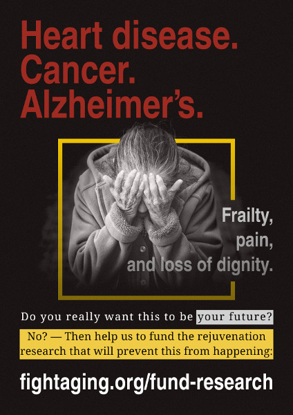
Doctors and scientists spent the last century struggling heroically against these diseases - and we invented antibiotics and vaccines in order to fight them. Those were the biggest challenges of the 20th century. We've been so successful fighting them that now we live in a world where the average life expectancy is 80. In developed countries all ten of the leading causes of death are caused by simply living long enough to not die of anything else. The 21st century will be defined by our struggle against these diseases of aging, and it is not going to be an easy one. For 75 years, these have been the leading causes of death for humanity, and everything we've done to fight them has barely made a dent in the number of people dying of any one of them. In fact, they are rising as a fraction of total deaths in the world, as we continue to make strides against infectious disease, malnutrition, and violence around the world.

You see, as a society we look to medicines to make us well when we're sick, and so far almost everything that we've designed to treat the diseases of aging has fit in that paradigm. We wait for someone to get cancer, to have a stroke, or start losing their memories, and then we try to do something about it. But this approach hasn't really been working. Since 2000, we've done 200 clinical trials in humans just for Alzheimer's disease, and 99% of those have failed. The two that succeeded haven't even given us a drug that does much to treat Alzheimer's disease. We spend over $20 billion a year on cancer research and trials, but most of the gains we've made against cancer since 1970 have come from better diagnosis of cancer, not from curing the disease.

This should tell us that we're doing something wrong in our approach to the diseases of aging, because, unlike infections, the diseases of aging are caused by the slow, gradual build up of damage to our bodies over a lifetime, before they ever cause enough of a problem for us to go see a doctor. And by the time that we go to see that doctor, so much has happened inside of our bodies, that there is not much that they can do to help us. So this is how I started my academic career. I was one of a small group of scientists, and we were all thinking the same thing: if we ever wanted to eliminate Alzheimer's and cancer, the way that we eliminated smallpox, we would have to take a different approach to healthcare. We would have to treat the diseases of aging by anticipating them, building medicines that could remove damage caused by getting old before it ever accumulated enough to make us sick.

And this kind of makes sense, right? Because we all feel the effects of getting old right now, even when we're not sick. I mean, who here can run as fast as they could when they were 18 years old? Or maybe bounce back up after falling out of a tree like they could when they were 12. I am by no means old, and even though my risk of osteoporosis or cancer is diminishingly small, I am getting older, just like all of you are. My blood vessels are hardening. My neurons are starting to get tired. My DNA is mutating. I'm losing the battle to keep my cells and tissues in good condition. Right now we only think of this progressive accumulation of damage as a problem when everything goes to hell, and it erupts as some kind of disease. If we want to stop this gradual build up of damage in our bodies, we're told that the old things we can do are eat better, exercise, avoid smoking, hope that we've gotten lucky with our genes. It's not exactly a hopeful message. We're not leveraging the power of modern medicine to prevent us from getting sick from the things that are killing us the most.

But that is all changing. Because for the first time in history, we understand what makes us get older. We've traced to the biochemical level the diseases of aging and what causes them, and we've been able to categorize the damage of aging into nine buckets. Things like the random mutations of DNA, or the exhaustion of our stem cells. Our understanding is now at the cellular and molecular level, which means that we can actually design medicines to target and treat these things. And those medicines actually exist. We have a repository of over 50 interventions, whether a small molecule drug or a genetic change, that can extend healthy life by as much as 50%. Think about that: 50% longer without getting Alzheimer's disease, or cancer, or having a stroke, or having our bones and muscles wear down. 50%! In mice. And so the mice are super-excited about this. But what does it mean for us humans?

Well, luckily this is how new medicines are usually born. We take a piece of research and test it in mice to see if it works, and if it does then we advance that to human trials. And the good news is that we have 50 things that are ready to test. But making the jump from mice to humans for these sorts of diseases won't exactly be straightforward. You see, a trial to prevent a disease instead of to treat it has some additional challenges. It is more time-consuming and more expensive, which means the companies that would have to pay the tens of millions of dollars for these trials are often hesitant to do so, when they are used to doing the more traditional reactive trials. However, we have a glimmer of hope here too, that may be able to fast-track some of these preventative medicines into the clinic. You see, if you build a medicine that does a good job preventing damage that could eventually cause disease, it turns out that the same medicine can stop a disease from getting any worse by halting that same damage. And sometimes we can even repair the damage, reversing the effects of a disease.

Now, if you caught yourself thinking "but wait, that sounds completely obvious!" I might forgive you for that. It does seem reasonable that if something is going wrong with my cells, and I fix that thing, then it would help whether or not I've labelled my cells as diseased. But until very recently we just didn't know that, because it hadn't really been tested. The people who are working on studying what goes wrong in an old mouse and the people who are giving treatments to Alzheimer's patients weren't really talking to each other. But now they are, and armed with this new knowledge of what makes us age, and what we can do about it, we're able to pursue two ambitious goals at the same time. First, we have the ability to create new medicines to treat patients suffering from diseases of aging. This is what motivates me and the people that I work with, every single day, using this new research to come up with a medicine that can impact millions of people who are sick right now.

But there's also a second thing we can be doing. As we create new medicines for these diseases, and test them in the traditional way, we have to remember that what we really want is a medicine that can prevent disease instead of just treating it. And to get there, we're going to need to have proven, safe, effective medicines targeted at treating the damage of aging, and there has been progress on this front as well. One of the 50 interventions I told you guys about before happens to be an approved drug that's already been used in humans for decades. So after results in mice came out showing that we could extend their healthspan, a group of researchers started combing through hundreds of thousands of patient records who had been taking this drug, and they found something incredible. This drug - that people didn't take to prevent the diseases of aging, they took to prevent their blood glucose from going up, because they had diabetes - but when then were on this drug, they had a lower incidence of both cancer and Alzheimer's disease. Even compared to healthy people that didn't have diabetes.

So this thing may actually be working. This drug, which is called metformin, can extend mouse life span on average by 5-10%, which, if it works the same in humans, would mean 4 to 8 extra healthy years. And that's a lot, because if we invented a pill that miraculously cured all cancer in all of humanity right now, we would expect an average life span gain of about three and a half years, because we would succumb to another disease as we got older. So based on this research, a new clinical trial has started to test whether people who are healthy can take metformin and avoid cancer and Alzheimer's disease. So you might want to wait for the results of that trial before you go and beg your doctor for diabetes meds.

So now you may be asking yourself, whether it's this trial or another medicine that gets approved, who is going to pay for these preventative medicines? And it's worth pausing here for a moment to reflect that insurance companies are actually already paying for something very similar. Many of us in this room may be taking medicines that lower our cholesterol, which reduce the chances of getting stroke. When we invent new medicines that can not just reduce your chances of getting a stroke, but also Alzheimer's disease and cancer, which are way more expensive for those insurance companies to treat, you can bet that they'll be lining up to pay for those drugs too. With insurance companies in the game, this means that pharma companies are going to pay top dollar for the rights to test and sell these medicines. And that means that scientists working at universities or at biotech companies are going to be competing with each other to create the next greatest preventative medicine. It's a positive feedback loop, and the cycle can be kicked off with just one victory. Even a drug that extends healthy life span by a year or two can start a cycle of investment and research and testing that can change the way we do healthcare forever.

And I have good news, because there are 50 interventions that we already have ready to go. We just need to get to work. As I close up here, I think it's worth addressing one little thing, which is that I get asked all the time if we even should be trying to treat aging or extend life. I think that this is absolutely the wrong question. I think that the question we should be asking ourselves is "when do you want to get Alzheimer's disease?" When do you want to have a stroke? When do you want your muscles to break down? For most of these, I think everyone that I know would say "never". It's like asking someone in 1900 at what age they'd like to get tuberculosis. Or polio. "No thank you!" The evidence we have suggests that we can make new medicines based on our understanding of aging that can help people who are suffering from today's biggest killers. Yes, this will change healthcare. It will redefine medicine in the 21st century in the same way that vaccines and antibiotics redefined medicine in the 20th. But that's good! That's progress. Because I want to be able to enter the 22nd century and face our newest medical challenges, whatever those may be.

We have the opportunity in our lifetimes to flip healthcare on its head, by wedding the power of modern medicine with our understanding of what makes us age. We're going to invent new medicines that can treat the damage caused by getting old before we ever get sick. And that? That's a future that I can look forward to.

Peyer's position on aging incorporates the views of the Hallmarks of Aging authors and the Longevity Dividend scientists, in that while he views aging as damage accumulation, and our responsibility as being to build the means to repair and prevent that damage, he has enthusiasm for moving ahead with approaches to slow aging that I consider to be largely a waste of time and effort. Take metformin, for example. To my eyes the animal data is shabby and unreliable, with studies showing all sorts of outcomes, and the effects in humans are too small to spend any time on in a world that includes the SENS rejuvenation research programs and the senolytic therapies to clear senescent cells currently under development.

For Peyer, the present TAME metformin trial is a useful step on the road towards obtaining more funding and attention in order to build better therapies: it is a wake up call. I'm dubious, however, that all it will take to start the avalanche is just a little success in the matter of life span, health, and mortality. We have plenty of examples from past years of what I would call a little success: the effects of statins on cardiovascular mortality, the bisphosphonate studies showing significant reductions in mortality, and so forth. The revolution hasn't happened in response to any of this; 99% of medical research and development is still business as usual, creating expensive and marginal patches that fail to address aging in any meaningful way. So why would it happen over a modest reduction in rates of age-related disease for people taking metformin?

I think we need bigger and better successes. Marginal improvement won't cut it. We need outright, obvious, sizable rejuvenation. Will senolytics wake the world if they produce a reliable five year gain in healthy life expectancy, as well as reversing numerous diseases and conditions of aging? I don't know. It may be that even that will just be absorbed into the current state of things, and 95% rather than 99% of medical research and development will continue to be business as usual. Inertia is an impressive thing in these large institutional scientific and regulatory communities. Nonetheless, we need to keep aiming high. If we aim low, then all we'll get in the end is poor results on the only metric that matters, the degree to which health is restored and extended.

Spurring Blood Vessel Growth via Signaling is Not as Simple as Hoped

One of the strategies under development to tackle age-related ischemia, in which blood flow to a limb becomes insufficient due to vascular damage or dysfunction, is to attempt to use signaling mechanisms to spur the development of new blood vessels that bypass the damaged area. Results to date have been mixed, and as the researchers here note, this is probably because the process of blood vessel growth is complex and staged. Simple treatments employing a single signal molecule are unlikely to make much headway.

A new study identifies a signaling pathway that is essential for angiogenesis, the growth of new blood vessels from pre-existing vessels. The findings may improve current strategies to improve blood flow in ischemic tissues. "Our research shows that the formation of fully functional blood vessels requires activation of protein kinase Akt by a protein called R-Ras, and this mechanism is necessary for the formation of the hallow structure, or lumen, of a blood vessel. The findings are important because they shed new light on the biological process needed to increase blood flow in ischemic tissues."

Previous efforts to treat ischemia by creating new blood vessels have focused on delivering angiogenic growth factors like vascular endothelial growth factor (VEGF) to ischemic sites. But all of these studies, including more than 25 phase II and III clinical trials, have failed to offer significant benefit to patients. The research team used a combination of 3D cell culture and living tissue to show that VEGF promotes vascularization, but the vessel structures formed are chaotic, unstable and non-functional. "Functional vessels need to have a lumen; a pipe-like opening that allows oxygenated blood and nutrients to travel through the body, and VEGF alone cannot fully support the formation of such a vessel structure."

"Generating new blood vessels is similar to the way trees grow; sprouts develop from existing vessels and then branch out further and further to restore vascularity. This study shows that there are distinct steps and signals that control the process. First, VEGF activates Akt to induce endothelial cells to sprout. Then, R-Ras activates Akt to induce lumen formation. The second step involving Akt activation by R-Ras stabilizes the microtubule cytoskeleton in endothelial cells, creating a steady architecture that promotes lumen formation. We propose that VEGF and R-Ras activation of Akt signaling are complementary to each other, both are necessary to generate fully functional blood vessels to repair ischemic tissue. Our next step is to work toward promoting the combined signaling of Akt in clinical studies; prompting R-Ras activation through either gene therapy or pharmacologically in parallel with VEGF therapy."

Link: https://www.eurekalert.org/pub_releases/2017-11/spmd-sfk112017.php

More Evidence Against a Late Life Mortality Plateau

It has been suggested that in very late life mortality rates flatten out and cease to increase. This effect has been observed in flies and other short-lived species, and insofar as aging is defined as an increase in mortality rate over time, it implies that old individuals cease to age. This isn't a desirable sort of agelessness, of course, as the plateaued mortality rates are very high; individuals are in poor health and do not live much longer. How might we interpret this? That all of the most harmful damage has already been done, and further accumulated damage doesn't much change the near future outcome?

In humans it is questionable as to whether there is enough data for people of 110 years and older to support any sort of rigorous conclusion about mortality rate trends in that sparse age group. The few researchers who have tried to crunch the numbers come away with quite different conclusions, depending on the details of their methodology, with the example here being one of those leaning towards an absence of a late life mortality plateau in our species.

Accurate estimates of mortality at advanced ages are essential for forecasts of population aging and for testing the predictions of competing theories of aging. They also contribute to more reliable forecasts of future longevity. Earlier studies suggest that exponential growth of mortality (Gompertz law) is followed by a period of deceleration, with slower rates of mortality increase at extreme old ages. This mortality deceleration eventually produces the "late-life mortality leveling-off" and "late-life mortality plateaus" at extreme old ages. Researchers have provided a detailed description of this phenomenon in humans and even made the first estimates for the asymptotic value of the upper limit to human mortality. The same phenomenon of "almost non-aging" survival dynamics at extreme old ages is detected in other biological species, and in some species the mortality plateau can occupy a sizable part of their life.

Studies of mortality after age 110 years are scarce because of difficulties in obtaining reliable age estimates. It was demonstrated that the age misreporting at older ages results in mortality underestimation. Also, it was found that mortality deceleration is more expressed in the case of poor-quality data than with data of better quality. Recent analysis of detailed records from the U.S. Social Security Administration Death Master File for several single-year extinct birth cohorts demonstrated that the Gompertz law fits mortality data better than other models up to ages 105-106 years. However, existing studies of mortality after age 110 years reported flat mortality, which does not grow with age.

In this paper, we analyze mortality trajectories for supercentenarians, using data on a sufficiently large sample of supercentenarians (aged 110 and older) available in the International Database on Longevity (IDL). All ages of supercentenarians in the database were subjected to careful validation. These results demonstrate that hazard rates after age 110 years do not stay constant and suggest that mortality deceleration at older ages is not a universal phenomenon. These findings may represent a challenge to the existing theories of aging and longevity, which predict constant mortality in the late stages of life. One possibility for reconciliation of the observed phenomenon and the existing theoretical consideration is a possibility of mortality deceleration and mortality plateau at very high yet unobservable ages.

Link: https://www.ncbi.nlm.nih.gov/pmc/articles/PMC5696798/

Recent Insight into the Processes of Rejuvenation that Act to Ensure the Offspring of Adult Parents are Born Young

Parents and their germline cells are biologically old, and yet developing offspring produced from the germline are biologically young. Therefore a form of cellular rejuvenation takes place somewhere between the start and the end of reproduction in multicellular organisms, whether they are nematode worms of a few hundred cells, or vastly larger and more complex species such as our own. New research on this topic from the usually secretive research groups at Calico was widely announced today; it is focused on the nematode Caenorhabditis elegans, but the findings are probably of relevance to the processes of rejuvenation that take place in mammalian reproduction. Aging is a matter of accumulated damage, of quite similar forms in nematodes and mammals: to make offspring young, all of this damage must be cleared away, or the germline shielded from it.

The rejuvenation that occurs in mammalian zygotes is not all that well characterized, though you'll find papers on the topic from recent years. It appears to overlap with processes observed to take place when cells are reprogrammed into a state of induced pluripotency: researchers have seen mitochondrial damage repaired, for example. This present work in nematodes is interesting for its focus on the lysosome and clearance of metabolic waste, as there isn't all that much work on what happens to such waste during induced pluripotency or in early mammalian embryonic development. Clearly it has to be successfully removed if present in order for offspring to be young, but this doesn't necessarily mean that the various mammalian processes of rejuvenation are anything like those of nematodes in their details and ordering, even if there are strong similarities at the high level.

The research here is intriguing for extending the findings in nematodes to frogs - give it a few years and we'll no doubt be seeing the study results for mammals. In mammals, early life rejuvenation must accomplish the same goal as it does in frogs and nematodes, regardless of how it is organized, which is to ensure that offspring are biologically young. Further, it must take place when those offspring are still a collection of just a few cells, as these processes would be highly disruptive and probably fatal if they took place throughout a more developed, complex organism. But perhaps such processes of rejuvenation could be selectively targeted to small and vital collections of cells. Perhaps it already takes place in some such cell populations as a way to maintain their function for a lifetime; consider stem cells, for example. This remains to be seen, as does how useful the rejuvenation processes that make offspring young might be as a starting point for the construction of therapies to slow aging.

Young Again: How One Cell Turns Back Time

None of us was made from scratch. Every human being develops from the fusion of two cells, an egg and a sperm, that are the descendants of other cells. The lineage of cells that joins one generation to the next - called the germline - is, in a sense, immortal. Over time, a cell's proteins become deformed and clump together. When cells divide, they pass that damage to their descendants. Over millions of years, the germline ought to become too devastated to produce healthy new life. "You take humans - they age two, three or four decades, and then they have a baby that's brand new. There's some interesting biology there we just don't understand."

Researchers have now reported the discovery of one way in which the germline stays young. Right before an egg is fertilized, it is swept clean of deformed proteins in a dramatic burst of housecleaning. The researchers discovered this process by studying a tiny worm called Caenorhabditis elegans. Most C. elegans are hermaphrodites, producing both eggs and sperm. As the eggs mature, they travel down a tube, at the end of which they encounter sperm. Researchers discovered that a worm's eggs carry a surprisingly heavy burden of damaged proteins, even more than in the surrounding cells. But in eggs that were nearing the worm's sperm, the researchers found far less damage. These experiments raised the possibility that the sperm were sending out a signal that somehow prompted the eggs to rid themselves of damaged proteins.

The researchers then created mutant "female" worms and observed that their eggs all became littered with protein clumps. When the researchers let them mate with males, however, the clumps disappeared from the eggs. They then carried out additional studies, such as looking for other mutant worms that could not clear out protein clumps even though they could make sperm. Combining these findings, the researchers worked out the chain of events by which the eggs rejuvenate themselves.

It begins with a chemical signal released by the sperm, which triggers drastic changes in the egg. The protein clumps within the egg "start to dance around." The clumps come into contact with little bubbles called lysosomes, which extend fingerlike projections that pull the clumps inside. The sperm signal causes the lysosomes to become acidic. That change switches on the enzymes inside the lysosomes, allowing them to swiftly shred the clumps. Researchers hypothesize that the worms normally keep their eggs in a dormant state. The eggs accumulate a lot of damage, but make little effort to repair it. Only in the last minutes before fertilization do they destroy protein clumps and damaged proteins, so that their offspring won't inherit that burden.

"The hypothesis is that it's not just a worm thing." In their new paper, the researchers reported that they had tested this hypothesis on frogs, which are much more closely related to humans than is C. elegans. The scientists exposed frog eggs to a hormone that signals them to mature. The lysosomes in the frog eggs became acidic, just as happens in worms. The germline may not be the only place where cells restore themselves in this way. Throughout our lives, we maintain a supply of stem cells that can rejuvenate our skin, guts and brains. It may be that stem cells also use lysosomes to eradicate damaged proteins. It might be possible, for example, to treat diseases by giving aging tissues a signal to clean house.

A lysosomal switch triggers proteostasis renewal in the immortal C. elegans germ lineage

Although individuals age and die with time, an animal species can continue indefinitely, because of its immortal germ-cell lineage. How the germline avoids transmitting damage from one generation to the next remains a fundamental question in biology. Here we identify a lysosomal switch that enhances germline proteostasis before fertilization. We find that Caenorhabditis elegans oocytes whose maturation is arrested by the absence of sperm exhibit hallmarks of proteostasis collapse, including protein aggregation. Remarkably, sperm-secreted hormones re-establish oocyte proteostasis once fertilization becomes imminent.

Key to this restoration is activation of the vacuolar H+-ATPase (V-ATPase), a proton pump that acidifies lysosomes. Sperm stimulate V-ATPase activity in oocytes by signalling the degradation of GLD-1, a translational repressor that blocks V-ATPase synthesis. Activated lysosomes, in turn, promote a metabolic shift that mobilizes protein aggregates for degradation, and reset proteostasis by enveloping and clearing the aggregates. Lysosome acidification also occurs during Xenopus oocyte maturation; thus, a lysosomal switch that enhances oocyte proteostasis in anticipation of fertilization may be conserved in other species.

Considering Age-Related Changes in Molecules in the Bloodstream in the Context of Cell Therapies for the Old

Parabiosis studies in which the circulatory systems of an old mouse and a young mouse are linked, and in which the old mouse shows a reversal of some measures of aging, have given rise to a broadening exploration of age-related changes in the molecules carried in the bloodstream. The high level picture of what is taking place here is this: reactions to rising levels of the forms of cell and tissue damage that cause aging include changes in the signal molecules released by cells into the surrounding environment. These are influential on stem cell function, chronic inflammation, and other line items known to be important in aging.

The paper here picks out a few such molecules of the many under study and discusses their likely roles and activities. The focus of these authors is on enhancing regenerative cell therapies by finding ways to make the tissue environment more receptive to transplanted cells and their ability to spur greater regeneration. That signaling changes in old tissues dampen stem cell activity is a major concern for the regenerative medicine community. This is one part of a field of research that includes numerous other efforts to try to adjust the circulating levels of these molecules, and thus to try to block some of the consequences of the underlying damage of aging. Like all similar efforts, I have to feel it will be much less effective than actually repairing that damage: in principle that should result in a reversal of the signaling changes.

It is undeniable that the incidence of cardiovascular diseases, mainly heart failure, increases in the elderly population. Global aging is a hallmark of our century: the eldery population comprise roughly 15% of the population, and this scenario will increase of an additional 25% on average by 2050. This unprecedented population profile will inevitably imply, among others, an increasing burden of cardiovascular events, some of which are directly linked to cellular senescence and dysfunction. Thus, increasing knowledge on the various mechanisms causing the progressive decline of cellular and tissue function may aid in developing therapies to delay or treat age-related conditions and diseases. Consequently, the discovery of pathways responsible for increasing life span and health span, as both potential biomarkers and targets, is currently of primary interest.

Endothelial progenitor cells (EPCs) are considered a main circulating stem cell population finely controlling vascular homeostasis and repair, therefore representing an interesting crossroad between circulating markers, regenerative cells, and aging mechanisms. Importantly, the demonstration that EPCs can be systemically recruited from the bone marrow-associated niche, and that after engraftment are able to replace old vasculature with new mature endothelial cells, has completely overturned the theory about aging and can be considered a significant reference for the relationship between progenitor cells and aging. To date, EPCs represent one of the most studied example tools to rejuvenate the vascular system or to potentially delay the damages induced by aging.

Notably, multiple studies suggest that, in the settings of cell transplantation for cardiovascular regenerative purposes, it is important not only to enhance intrinsic "young" properties of therapeutic cells, such as EPCs, but also to grant an ideal host microenvironment where engraftment can occur. Therefore, approaches able to rejuvenate regenerative cells and/or preserve tissue homeostasis and physiology (i.e., delaying overall aging) should be synergistically combined.

One of the main mechanisms affecting senescence and aging at multiple levels is oxidative stress, which originates from several biochemical pathways triggered, among others, by environmental factors, and overall imbalancing the final amount of reactive oxygen species. In this review, we will discuss few circulating molecules, proteins and microRNAs, selected among those whose levels and related signaling pathways have been correlated to life span and healthy aging. In particular, we will discuss pathways with specific biological and rejuvenating roles in cellular senescence, cardiovascular functions, and with a potential or known role in the control of regenerative cell populations.

Link: https://doi.org/10.3389/fcvm.2017.00062

More Evidence for Even Modest Levels of Physical Activity to be Beneficial

Quite a few studies on physical activity and mortality rate have been published in the past few weeks. They lean towards supporting the hypothesis that low levels of activity are still beneficial to some degree in older individuals. The benefits scale up as activity becomes more intense, but there isn't a threshold that must be hit in order to obtain at least some improvement in health and reduction in mortality rate. The research here is another example of this sort of study outcome. Note that human studies generally show correlations, not causation. Corresponding animal studies of exercise and health that do prove causation are the reason why we can be fairly confident that exercise causes better health.

This study compared the association between different levels of physical activity and the risk of cardiovascular disease in elderly to middle-aged individuals. "We know that regular physical activity has major health benefits. Healthy adults are advised to do at least 150 minutes a week of moderate intensity or 75 minutes a week of vigorous intensity aerobic exercise to reduce their risk of cardiovascular disease. These recommendations are based primarily on research in middle-aged adults and we wanted to know whether regular physical activity yields comparable cardiovascular health benefits in elderly people."

The study included 24,502 adults aged 39 to 79 years who participated in the European Prospective Investigation into Cancer (EPIC) Norfolk cohort, a prospective population study that is part of the ten-country collaboration EPIC study. The cohort was primarily designed to assess dietary and other determinants of cancer, but data were also collected on determinants of cardiovascular disease. Participants were recruited between 1993 and 1997 from registries of general practices in the county of Norfolk, UK. On enrollment into the study, participants completed a health and lifestyle questionnaire, underwent a standardised physical examination and gave blood samples. Physical activity during work and leisure time was assessed with a questionnaire and participants were categorised as active, moderately active, moderately inactive and inactive.

Patients were followed up until 31 March 2015 for hospitalisation or death from cardiovascular events (coronary heart disease or stroke). Physical activity levels and time to cardiovascular events were investigated in three age categories: less than 55, 55 to 65 (middle-aged), and over 65 years of age (elderly). During a median follow-up of 18 years there were 5,240 cardiovascular disease events. In elderly participants, hazard ratios for cardiovascular events were 0.86, 0.87, and 0.88 in moderately inactive, moderately active and active people, respectively, compared to inactive people. In those aged 55-65 and less than 55 years, the associations were directionally similar, but not statistically significant.

"We observed an inverse association between physical activity and the risk of cardiovascular disease in both elderly and middle-aged people. As expected, there were more cardiovascular events in elderly participants, which could explain why the association only reached significance in this age category. Elderly people who were moderately inactive had a 14% reduced risk of cardiovascular events compared to those who were completely inactive. This suggests that even modest levels of physical activity are beneficial to heart health. Elderly people should be encouraged to at least do low intensity physical activities such as walking, gardening, and housework."

Link: https://www.escardio.org/The-ESC/Press-Office/Press-releases/any-physical-activity-in-elderly-better-than-none-at-all-for-reducing-cardiovascular-risk

The Most Obvious Tau Aggregates in Tauopathies, the Neurofibrillary Tangles, are not the Primary Cause of Harm

Altered proteins build up in the aging brain, forming solid deposits. The most prominent of them are amyloid-β, altered forms of tau, and α-synuclein, giving rise to amyloidosis, tauopathies, and synucleinopathies respectively. Some conditions mix and match: Alzheimer's disease is both an amyloidosis and a tauopathy. To further muddy the waters, any aging brain far enough along in the process to exhibit full-blown neurodegeneration will also exhibit significant levels of all of the other forms of dysfunction caused by aging.

Present thinking on the roots of protein aggregation conditions is fairly diverse. Insofar as there is a consensus, the root causes are considered to include issues such as failing cellular maintenance processes, failure of the drainage of cerebrospinal fluid as a way to export waste to the rest of the body, infection by pathogens capable of generating more of these unwanted proteins, and failure of the immune system - in defending against those pathogens, in generating inflammation that causes all sorts of breakage and change in cellular behavior, and in cleaning up the waste and debris produced by other cells. Amyloid-β, altered tau, and α-synuclein are all produced in some amount by normal, healthy, young people, but clearly they do not suffer for it, and nor does it build up. Any hypothesis of disease progress must account for what changes in older individuals.

An interesting point of commonality between the various forms of aggregated protein in the brain is that the largest and most obvious deposits, neurofibrillary tangles in the case of tau, are not the worst of the problem. You might think of them as the result of our biology trying to build ever bigger middens to cope with the waste that piles up. Cells dump it into the surrounding environment, or become overridden with garbage that they sequester into lumps when they can't even keep up with that. This is harmful, but as it turns out not as harmful as the surrounding halo of related biochemistry: for the most part it isn't the garbage in the middens that causes cell death and dysfunction, but rather a collection of associated proteins and their subtle interactions with cells. This is well established for amyloid-β, and the paper noted here makes an argument for this to be the case for tau as well.

Researchers describe new biology of Alzheimer's disease

Scientists have known for a long time that two proteins, β-amyloid and tau, clump and accumulate in the brains of Alzheimer patients, and this accumulation is thought to cause nerve cell injury that results in dementia. Recent work by these researchers has shown that the clumping and accumulation of tau occurs as a normal response to stress, producing RNA/protein complexes termed "stress granules," which reflect the need for the brain to produce protective proteins. The persistence of this stress response leads to excessive stress, the accumulation of pathological stress granules, and the accumulation of clumped tau, which drives nerve cell injury and produces dementia.

In the current study, the researchers use this new model and show that reducing the level of stress granule proteins yields strong protection, possibly by reducing persistent pathological stress granules as well as changing the type of tau clumping that occurs. The team hypothesized that they could delay the disease process by reducing stress granules and decreasing this persistent stress response by genetically decreasing TIA1, which is a protein that is required for stress granule formation. Reducing TIA1 improved nerve cell health and produced striking improvements in memory and life expectancy in an experimental model of AD.

Although the experimental models had better memory and longer lives, the team observed more clumped tau in the form of neurofibrillary tangles. To explain how this might be associated with a better outcome, the researchers looked at the type of tau pathology and showed that reducing TIA1 dramatically lowered the amount of tiny clumps, which are termed tau oligomers and are particularly toxic. "Reducing TIA1 shifted tau accumulation from small to large clumps, decreasing the amount of small tau clumps and producing a proportional increase in the large tau clumps that generate neurofibrillary tangles and are less toxic."

Reducing the RNA binding protein TIA1 protects against tau-mediated neurodegeneration in vivo

Emerging studies suggest a role for tau in regulating the biology of RNA binding proteins (RBPs). We now show that reducing the RBP T-cell intracellular antigen 1 (TIA1) in vivo protects against neurodegeneration and prolongs survival in transgenic P301S Tau mice. Biochemical fractionation shows co-enrichment and co-localization of tau oligomers and RBPs in transgenic P301S Tau mice. Reducing TIA1 decreased the number and size of granules co-localizing with stress granule markers. Decreasing TIA1 also inhibited the accumulation of tau oligomers at the expense of increasing neurofibrillary tangles.

Despite the increase in neurofibrillary tangles, TIA1 reduction increased neuronal survival and rescued behavioral deficits and lifespan. These data provide in vivo evidence that TIA1 plays a key role in mediating toxicity and further suggest that RBPs direct the pathway of tau aggregation and the resulting neurodegeneration. We propose a model in which dysfunction of the translational stress response leads to tau-mediated pathology.

Recent Research Implicates Astrocytes in the Progression of Alzheimer's Disease

Astrocytes are one of a number of different classes of supporting cells of the brain, and researchers here investigate how they might be involved in the progression of Alzheimer's disease - though with the caution they they are looking at early-onset Alzheimer's linked to specific mutations. These variants of the condition may be accelerated by processes that are not relevant in the more common form. Either way, Alzheimer's disease is an enormously complex condition; all cell types in the brain change their behavior or are impacted in some way by inflammation, rising levels of protein aggregates such as amyloid-β, or other aspects of aging. Separating cause and effect of the disease state from everything else is a challenging undertaking, not least because the animal species used in the laboratory do not naturally suffer any sort of condition resembling Alzheimer's. So there is always the question of whether or not the very artificial animal models of the disease are close enough to the human condition to steer research in the right direction. This is the case for the biology of astrocytes in particular, and so the researchers here adopt a more modern approach of generating cells for study from human patients.

Alzheimer's disease (AD) is the most common dementia type, with no treatment to slow down the progression of the disease currently available. The mechanisms of AD are poorly understood, and drug therapy has focused on restoring the normal function of neurons and microglia, i.e. cells mediating brain inflammation. The new study shows that astrocytes, also known as the housekeeping cells of the brain, promote the decline of neuron function in AD. The findings suggest that at least some familial forms of AD are strongly associated with irregular astrocyte function, which promotes brain inflammation and weakens neurons' energy production and signalling.

Astrocytes are important brain cells, as they support neurons in many different ways. Astrocytes are responsible, for example, for the energy production of the brain, ion and pH balance, and they regulate synapse formation, the connections between neurons. Recent evidence suggests that human astrocytes are very different from their rodent counterparts and thus, it would be essential to use human cells to study human diseases. However, the availability of human astrocytes for research has been very limited. The study used the induced pluripotent stem cell technology, which enables the generation of pluripotent stem cells from human skin fibroblasts. These induced stem cells can then be further differentiated to brain cells, e.g. neurons and astrocytes, with the same genetic background as the donor had. The study compared astrocytes from familial AD patients carrying a mutation in the presenilin 1 gene to astrocytes from healthy donors, and the effects of these cells on healthy neurons were also analysed.

The researchers found out that astrocytes in patients with Alzheimer's disease produced significantly more beta-amyloid than astrocytes in persons without AD. Beta-amyloid is a toxic protein that is known to accumulate in the brains of AD patients. In addition, AD astrocytes secreted more cytokines, which are thought to mediate inflammation. AD astrocytes also showed alterations in their energy metabolism which likely led to increased production of reactive oxygen species and reduced production of lactate, an important energy substrate for neurons. Finally, when astrocytes were co-cultured with healthy neurons, AD astrocytes caused significant changes on the signaling activity of neurons when compared to healthy astrocytes.

Link: http://www.uef.fi/-/aivojen-astrosyyteilla-havaittiin-yhteys-alzheimerin-tautiin

Failing Mitochondria and Cellular Senescence in the Aging Lung

Mitochondrial dysfunction and cellular senescence are two of the root causes of aging targeted by the SENS rejuvenation research programs. They overlap at least a little, in that one might cause the other, but it is unclear as to whether this is significant for the specific types of mitochondrial damage considered important in the SENS view of aging. The open access paper here walks through this territory in the case of the aging lung; in recent years, it has become clear that senescent cells are important in the development of fibrosis in lungs and other organs, as well as in other aspects of aging in lung tissue. The present development of various forms of senolytic therapies to remove these cells should result in treatments capable of turning back lung aging to some degree, as well as treating presently intractable lung conditions such as idiopathic pulmonary fibrosis.

Cellular senescence is generally defined as irreversible cell-cycle arrest. Importantly, senescence is characterised by the development of a pro-inflammatory secretory phenotype, termed the senescence-associated secretory phenotype (SASP). The SASP is thought to be important for the immune-mediated clearance of senescent cells, however, may also be a contributor to tissue dysfunction. Evidence suggests that accumulation of senescent cells with time, leads to age-related loss of tissue function. Accordingly, senescent cells are found at sites of chronic age-related disease and have been causally implicated in the development of osteoarthritis, atherosclerosis, liver steatosis and pulmonary fibrosis.

The lung is particularly affected by the ageing process, showing clear decline in structure and function with age. Moreover, the ageing lung is characterised by the presence of senescent cells and several respiratory diseases have been identified as diseases of accelerated lung ageing. Chronic obstructive pulmonary disease (COPD) and idiopathic pulmonary fibrosis (IPF) are classic examples of respiratory diseases that increase in prevalence with age and have been associated with senescence.

The mitochondria can impact on aspects of the senescence phenotype in a number of possible ways and it has been suggested that dysfunctional mitochondria are an additional feature of senescent cells that enable them to mediate paracrine effects. Mitophagy, the selective degradation of defective mitochondria by autophagy, is reduced in senescent cells. This could, in part, be responsible for the increase in mitochondrial mass that has been described in senescence. The accumulation of the mitochondrial compartment and of dysfunctional mitochondria in particular, may be an important contributor to the pro-inflammatory aspects of cellular senescence.

It has been shown that mitochondrial dysfunction induced by mitochondrial DNA depletion, knockdown of mitochondrial sirtuin 3 (SIRT3), or through inhibition of the electron transport chain (ETC) induces senescence with a distinct phenotype, termed MiDAS (mitochondrial dysfunction-associated senescence). Our group recently designed a proof-of-principle experiment, which interrogated whether mitochondria are truly necessary for senescence. Utilising the parkin-mediated mitophagy system to completely remove mitochondria upon their depolarisation, we found that following a variety of senescence triggers (e.g. oxidative stress and oncogene activation) features of cellular senescence, including Sen-β-Gal activity and the SASP, were suppressed. The mitochondria may therefore be key to the regulation of some aspects of cellular senescence, such as the pro-inflammatory phenotype, and may be promising targets for SASP modulation.

Link: https://doi.org/10.1016/j.pharmthera.2017.10.005

Are Low Levels of Physical Activity Significant in Health and Longevity?

Mapping the dose-response curve for exercise, its effects on health and life expectancy, is of great interest to the research community. Given the significant time and effort required to make progress via epidemiological studies, this mapping will no doubt still be an ongoing concern even after the first rejuvenation therapies are widely available. The best we can expect from present day data on physical activity in humans are broad conclusions, such as that regular moderate exercise is good for you, while being sedentary is not, and a highlighting of areas of uncertainty.

One of these areas of uncertainty is the question of low level activity: walking around the house, gardening, shopping, and so forth. Things that don't rise to the level of deliberate physical exercise. Do these activities have a noticeable impact on health and longevity? Is it a case of more is better? Prior to the creation of small accelerometers, of the sort found inside every mobile device these days, there was simply no way to tell. Studies used self-reported data, which is unreliable enough to obscure small differences. With accelerometers, the first studies appeared to suggest that yes, low levels of exercise do correlate with better health in later life. Human epidemiology can rarely do more than point out correlations, but animal studies of exercise definitively show causation of improved health. There is every reason to believe that the observed human data is due to exercise causing improved health.

Not all accelerometer studies produce results that support the hypothesis that benefits arise from low levels of physical activity, however. A paper from earlier this year reported finding no association between low level physical activity and mortality rate, for example. This is a slow-moving part of the field, in which one has to weigh the balance of many studies carried out over a decade or more. At the present time the scales tip towards casual activity providing a modest benefit; more papers arrive with conclusions akin to the one noted here. Still, by the time all is said and done, a couple of decades from now at the present pace, degree of exercise will be nowhere near as influential on your health as whether or not you have access to rejuvenation therapies after the SENS model of periodic damage repair. It is still a good plan to exercise, as it would be foolish to turn down highly reliable, free benefits to health, even if they are modest in comparison to the rewards the future will bring.

For older women, every movement counts, new study finds

Folding your laundry or doing the dishes might not be the most enjoyable parts of your day. But simple activities like these may help prolong your life, according to the findings of a new study in older women. In the U.S. study of more than 6,000 white, African-American and Hispanic women ages 63 to 99, researchers reported significantly lower risk of death in those who were active at levels only slightly higher than what defines being sedentary. Women who engaged in 30 minutes per day of light physical activity - as measured by an accelerometer instead of a questionnaire - had a 12 percent lower risk of death. Women who were able to do a half-hour each day of moderate to vigorous activity had a 39 percent lower mortality risk.

For the age group in this study, light physical activities include regular chores such as folding clothes, sweeping the floor or washing the windows. Activities like these account for more than 55 percent of how older people spend their daily activity. Moderate to vigorous activities would be brisk walking or bicycling at a leisurely pace. The bottom line? "Doing something is better than nothing, even when at lower-than-guideline recommended levels of physical activity."

Even when researchers simultaneously accounted for the amount of each type of activity (light and moderate-to-vigorous) a woman did, they still observed significantly lower mortality associated with each time, independently of the other. "Current public health guidelines require that physical activity be of at least moderate or higher intensity to confer health benefits. Our study shows, for the first time in older women, that health is benefitted even at physical activity levels below the guideline recommendations. The mortality benefit of light intensity activity extended to all subgroups that we examined, and was similar for women younger than 80 compared to women over the age of 80. It was similar across racial/ethnic backgrounds, and among obese and non-obese women. Perhaps most importantly for this population, the mortality benefit was similar among women with high and low functional ability."

Accelerometer-Measured Physical Activity and Mortality in Women Aged 63 to 99

Age-related deterioration in health is associated with a reduction in physical activity (PA). U.S. and international guidelines on PA and public health recommend that healthy older adults perform at least 2.5 hours/week of moderate-intensity or 1.25 hours/week of vigorous-intensity aerobic PA for health benefits, a target that few older U.S. adults meet, often because they are not capable of engaging in moderate- to vigorous-intensity PA (MVPA). Substantially lower all-cause mortality risk is associated with relatively high MVPA levels (3-5 times guideline recommended) assessed using questionnaires. The extent to which this extends to older adults is unclear.

Typically, self-reported activity explains only 10% to 20% of the variance in device-measured PA. PA misclassification is large in older adults, especially for light-intensity PA, which these individuals commonly perform but is currently not recommended for public health. Use of accelerometers to measure PA is novel in prospective studies on older adults and provides the ability to calibrate the effect of PA much better than with self-report, especially for light-intensity PA. We examined associations between mortality and accelerometer-measured PA using age-relevant intensity cutpoints in older women of various ethnicities.

The results support the hypothesis that higher levels of accelerometer-measured PA, even when below the moderate-intensity threshold recommended in current guidelines, are associated with lower all-cause and CVD mortality in women aged 63 to 99. Our findings expand on previous studies showing that higher self-reported PA reduces mortality in adults aged 60 and older, specifically in older women, and at less than recommended amounts. Moreover, our findings challenge the conclusion of recent meta-analyses that MVPA, measured by to self-report, is required to offset mortality risk in adults.

First, absolute rates of all-cause and cardiovascular disease mortality were at least 50% lower in cohort members in the middle tertile of each PA exposure than in those in the lowest tertile. This is particularly impressive when considering the small mean differences between these tertiles of 50 minutes/day for low light-intensity PA, 33 minutes/day for high light-intensity PA, and 20 minutes/day for MVPA. Use of accelerometers enhanced accurate quantification of such small differences in usual daily PA, which is not possible using questionnaire assessments. Small increases in daily PA, which older adults can achieve, could have a substantial effect on mortality in later life. Even in the oldest cohort members, ages 80-89 and ≥90 years, absolute rates of all-cause mortality were 44% and 15% lower, respectively, when comparing the middle and lowest total PA tertile.

Bisphosphonates May Act to Reduce Mortality through Vascular Mechanisms

Bisphosphonates are used as a treatment for osteoporosis. Like most pharmaceutical therapies for age-related disease, they have a set of unpleasant side-effects, but a couple of studies have found evidence for long-term bisphosphonate use to reduce mortality in older individuals. In one case the effect was quite large, a dramatic decrease in mortality versus the expected rates. I think there remains some skepticism about an effect of that size resulting from commonly used medications, versus it being an accident of the data or the study group or some other correlated but unrecorded difference, at least until further studies with larger patient groups take place.

What might the mechanism be, however? Past work suggests that bisphosphonates have some beneficial effect on stem cell activity, which might be a viable explanation, given better evidence in patients. The paper here is focused instead on cardiovascular issues, such as (a) the calcification of blood vessels that contributes to hypertension, and (b) the development of atherosclerosis, in which fatty plaques form to narrow and weaken blood vessels, ultimately causing death when one of these weak points ruptures. These are prominent issues in aging, and given strong evidence for bisphosphonates to produce benefits on this front, it would be a plausible mechanism for reduced mortality. The open access review paper here walks through the current evidence for this hypothesis.

In the past, osteoporosis and atherosclerosis were considered as separate entities with a similar increasing prevalence with aging. Recently, studies have outlined that patients with low bone mineral density (BMD) are at significantly greater risk of developing cardiovascular disease (CVD) as well as unexpected cardiovascular events, more severe coronary atherosclerosis and vascular calcification. In addition, it is known that postmenopausal women with osteoporosis have an increased risk of developing cardiovascular events and that the increased risk is proportional to the severity of osteoporosis. These data have also suggested a possible influence of drugs affecting bone metabolism on lipid and atherosclerosis mechanisms, or that drugs effective on the atherosclerosis process could also be efficacious in fracture prevention.

An initial interesting theory was that CVD and osteoporosis were linked by a common denominator, such as serum lipid profile, which could act in parallel on both vascular and bone cells. However, an interesting observational study showed that in a multiple regression analysis, lipid profile did not predict osteoporosis or fracture risk, whereas aortic calcification severity significantly explained BMD at the hip. On the other hand, low BMD at the distal radius was found to be associated with increased risk of stroke and CVD mortality.

The common finding of simultaneous vascular calcification and osteoporosis in individual patients suggests that local tissue factors could have a crucial role in the regulation of mineralization and cell differentiation. Cardiovascular calcification was conventionally viewed as an inevitable consequence of aging, but some landmark studies have demonstrated that it is a highly regulated process of mineralization which involves cellular and molecular signaling processes similar to those found in normal osteogenesis. The similarity of the molecular mechanisms in osteogenesis and vascular calcification has led to the knowledge that atherosclerotic calcification is an actively regulated process, not a passive mineralization.

The growing evidence that atherosclerosis and osteoporosis share several pathophysiologic mechanisms reinforces the interest in pharmacologic agents which could inhibit bone loss and also provide benefits in terms of slowing the progression of atherosclerosis. At present, only bisphosphonates (BPs), currently considered the drug of choice for the prevention and treatment of osteoporosis, could have this potential.

The interest in the relationships between BPs and atherosclerosis has recently shown a further increase after the publication of the results of the HORIZON study which reported a 28% reduction in mortality in hip fracture patients treated with an annual i.v. dose of zoledronic acid. In another study, it was revealed that patients who received BP therapy for osteoporotic fracture had a lower hazard of myocardial infarction during the 2-year follow-up period with respect to controls. Moreover, two recent studies have reported that oral BPs reduce mortality in osteoporotic patients and that the reduction in mortality could be mainly due to cardiovascular and cerebrovascular deaths.

To sum up, the BPs seem to have the potential of influencing atherosclerosis and calcium homeostasis at the level of vascular walls with several possible mechanisms which may differ according to the type, potency, dosage and administration route of BPs. However, until the present time, it is not yet clear which of these above-mentioned mechanisms may be the most important in humans and additional studies are needed to specifically address the mechanism by which BPs use could influence cardiovascular morbidity and mortality.

Link: https://doi.org/10.2147/CIA.S138002

To What Extent are Gut Bacteria Involved in the Benefits of Fasting?

Calorie restriction improves health and extends life in most species and lineages tested, while both Protein restriction and intermittent fasting can provide similar but usually lesser packages of benefits. Once delving into the details of the biochemistry involved, however, the picture becomes very complex, and is still quite uncertain. These strategies probably work through overlapping collections of mechanisms that in turn interact with one another. Intermittent fasting and protein restriction still provide some benefits even when calorie level is kept constant, for example, and assays of epigenetic changes look fairly different for each of these dietary strategies.

Part of the challenge inherent in investigating calorie restriction, protein restriction, and intermittent fasting lies in the fact that near everything in the operation of metabolism changes in response. To the degree that these approaches modestly slow aging, near every measure of aging is affected. How to pinpoint root causes, or important causes, or chains of cause and effect? It isn't easy, as demonstrated by the very slow progress on this front despite a great deal of investment in time and effort over the past three decades.

The scope of "near everything" certainly includes the behavior and distribution of gut bacteria, and in recent years researchers have devoted increasing attention to their role in health and aging. That may well turn out to be in the same ballpark of importance to life expectancy as, say, exercise, but the degree to which it is entirely secondary to dietary choice or other factors in aging - such as immune dysfunction - is an interesting question. Certainly in the case of calorie restriction there is strong evidence for the benefits to be near-completely a function of increased autophagy, and thus there is little room for gut bacteria in that picture.

What about intermittent fasting, however? Researchers here demonstrate the ability to replicate at least some measures observed in intermittent fasting in mice by transplanting gut microbiota from fasting mice into non-fasting mice. This is quite interesting as a point of comparison for what we think we know about how calorie restriction works. It suggests that intermittent fasting with overall calorie restriction is probably quite a different beast from intermittent fasting without overall calorie restriction.

Obesity and related metabolic disorders are growing health challenges; they mainly result from an imbalance between energy intake and energy expenditure. Emerging evidence suggests that non-shivering thermogenesis can re-establish energy balance and therefore counter the effects of elevated energy intake. This process is mediated primarily by the thermogenic activity of uncoupling protein 1 (UCP1), mainly in brown and beige fat cells. In this context, activating brown adipose tissue (BAT) or browning of white adipose tissue (WAT) could be a promising therapy for obesity and related metabolic diseases.

Recently, intermittent fasting was demonstrated to optimize energy metabolism and promote health. However, the mechanism for these benefits is unclear. Notably, one study found that time-restricted feeding can counteract obesity without reducing energy intake. Although perturbation of circadian rhythm was considered as a significant contributor to the increased energy expenditure, the possibility exists that white adipose browning would be a more direct mechanism. Therefore, in the current study, mice were placed on an every-other-day fasting (EODF) regimen to explore its effect on white adipose beiging and metabolic disorders. Evidence suggests that EODF selectively activates beige fat thermogenesis and ameliorates obesity-related metabolic diseases, probably via a microbiota-beige fat axis.

Gut microbiota play a critical role in energy metabolism and lipid homeostasis, and germfree or microbiota-depleted rodents have decreased susceptibility to diet-induced obesity and metabolic syndrome. Based on the above findings, EODF treatment could alter the microbiota compositions and prevent high-fat-diet-induced obesity and metabolic disorders. To further clarify the role of gut microbiota in mediating the beneficial effects of EODF regimen on metabolic diseases, the effect of EODF in control and microbiota-depleted high-fat-diet-induced obesity mice was compared. EODF treatment significantly reduced obesity and hepatic steatosis and improved insulin sensitivity in control mice, but not in microbiota-depleted mice, indicating that the effects of EODF depend on gut microbiota.

To examine whether gut microbiota are sufficient to replicate the effects of EODF, microbiota-depleted mice with high-fat-diet-induced obesity were transplanted with microbiota from ad libitum (AL) feeding and EODF mice, respectively. Compared with the AL microbiota-transplanted group, EODF microbiota transplantation did mimic all the beneficial effects of EODF treatment on metabolic dysfunctions.

In summary, the present work uncovered novel perspectives on beige-fat development in white adipose tissue. EODF was shown to selectively activate beige fat, probably by re-shaping the gut microbiota, which led to increases in the beiging stimuli acetate and lactate. EODF also dramatically ameliorated metabolic syndrome in a mouse model of obesity. This alternative beige fat activation by EODF offers new insights into the microbiotabeige fat axis and provides a novel therapeutic approach for the treatment of obesity-related metabolic disorders.

Link: http://dx.doi.org/10.1016/j.cmet.2017.08.019

Defenestration and the Roots of Age-Related Insulin Resistance

Defenestration is apparently a word with two meanings. The second, a scientific term, is the removal or loss of fenestrations. Let it never be said that this is not a place of learning. What, one might ask, are fenestrations? This is another word adopted by the scientific community and given an additional meaning: it refers to a collection of small openings or pores in our biology. The particular small openings or pores that concern us today are those found in the blood vessels of the liver, one of the organs involved in the development and progression of type 2 diabetes.

While we might tend to think of type 2 diabetes as a disease caused by excess fat tissue, and for more than 90% of patients in our modern era of cheap calories this is entirely true, it is also the case that the damage of aging ultimately leads to a similar dysfunction in insulin metabolism. The path to the same end is quite different, however. While even the comparatively late stages of visceral-fat-induced diabetes can be reversed through a sustained low-calorie diet and loss of that fat, there is nothing much that can yet be done to effectively deal with purely age-related diabetes. This is just one of the many age-related conditions we'd like to reverse through rejuvenation therapies based on the SENS research programs.

The short open access commentary below summarizes some of the mechanisms involved in loss of insulin sensitivity in the old, distinct from those losses caused by fat tissue. This is where the fenestrations of blood vessels in the liver enter the picture. The authors present evidence to suggest the loss of fenestrations - defenestration - increasingly blocks the passage of insulin to where it is needed, producing what is in effect insulin resistance and all of its secondary consequences. To me the interesting questions attend the cause of this change: is it a form of dysfunction in tissue maintenance of the sort that arises due to growing inflammation in aging tissues? Is it some other secondary effect, a change in signaling that disrupts whatever cellular coordination is needed to form fenestrations? Further research is needed.

It's the holes that matter

Before circulating insulin can interact with membrane bound insulin receptors and trigger downstream signalling it must first cross the endothelium of the blood vessels in the target tissue. This transfer across the endothelium from the blood is recognised as a rate limiting step in insulin action in muscle and fat in humans, but the role of the liver endothelium in insulin uptake has not been examined previously. Recent research explores the contribution of insulin transfer from the blood, across the liver sinusoidal endothelium and to the insulin receptors on the hepatocytes as a mechanism for the development of hyperinsulineamia and insulin resistance, as identified as a major risk factor for the development of age-related disease in humans.

The sinusoids, or blood vessels of the liver are lined by specialized endothelial cells that are very thin and perforated with transcellular holes or pores that traverse the entire cell. These pores, known as fenestrations, have no diaphragm and are patent passages through the cell. The fenestrations provide efficient ultrafiltration of small material from the blood into the liver. Coupled with very little extracellular matrix and a highly adapted hepatocyte membrane, uptake of substrates, such as nutrients, toxins, and insulin into the liver for metabolism, detoxification, and signalling is rapid and regularly overlooked. However, in older age, the morphology of the liver sinusoids and the endothelium changes significantly. The cells become thicker, and the diameter and number of fenestrations is reduced by up to 50% (known as defenestration), there is extracellular matrix deposition and evidence of loss of hepatocyte microvilli. Collectively, these changes have been called pseudocapillarization. It has previously been shown that these changes reduce hepatocyte uptake of lipoproteins and some drugs.

In the current work, the hepatic and systemic disposition of insulin was explored in young and old animals and insulin resistance was confirmed to be present in the older animals. Critically, using multiple indicator techniques insulin transfer across the liver endothelium was shown to be significantly impaired. The 20% reduction in insulin's volume of distribution in the liver was consistent with limited transfer across the sinusoidal endothelium and retention of insulin in the sinusoid. In concordance with these changes, there were very high circulating insulin levels indicative of both increased secretion and impaired clearance. Despite normal glucose tolerance tests in the older animals, insulin resistance was present. Of key importance, insulin and glucose uptake into muscle and fat was shown to be unchanged with age, suggesting age related insulin resistance was most likely being driven by impaired hepatic uptake and clearance.

This work suggests that defenestration and pseudocapillarization of the liver sinusoidal endothelium seen in aging prevents the access of insulin to the insulin receptor on the hepatocyte membrane through impaired transfer across the endothelium. This results in hyperinsulinemia, impaired hepatic insulin signalling and insulin resistance. Further the work demonstrates that the liver endothelium does not provide a barrier for the uptake of insulin under normal conditions. In summary, patent fenestrations are required for hepatic insulin uptake, clearance, and signalling and loss of fenestrations is a probable causative mechanism for insulin resistance and diabetes seen with aging. This work provides evidence that maintaining the integrity of the liver sinusoidal endothelium into old age may prevent age-related insulin resistance and excitingly, introduces a novel therapeutic target.

Towards Better Artificial Alternatives to Cartilage Tissue

It will be interesting to watch the accelerating development of biological versus non-biological replacements for damaged tissue over the next few decades. Both are improving at a fair pace, and there is a sizable area of overlap between the two sides of the field. If a nonbiological alternative gets the job done, then why not use it in place of engineered tissue? At the moment, new patient-matched engineered tissue would be a better long term alternative, considering the various challenges that result from introducing long-term implants into the body, but in near all cases that is not yet an option. Twenty years from now, however, many forms of replacement will have competing tissue engineered and wholly artificial alternatives available in the market, and the trade-offs will be more subtle.

The liquid strength of cartilage, which is about 80 percent water, withstands some of the toughest forces on our bodies. Synthetic materials couldn't match it until "Kevlartilage" was developed. Many people with joint injuries would benefit from a good replacement for cartilage, such as the 850,000 patients in the U.S. who undergo surgeries removing or replacing cartilage in the knee. While other varieties of synthetic cartilage are already undergoing clinical trials, these materials fall into two camps that choose between cartilage attributes, unable to achieve that unlikely combination of strength and water content.

The other synthetic materials that mimic the physical properties of cartilage don't contain enough water to transport the nutrients that cells need to thrive. Meanwhile, hydrogels - which incorporate water into a network of long, flexible molecules - can be designed with enough water to support the growth of the chondrocytes cells that build up natural cartilage. Yet those hydrogels aren't especially strong. They tear under strains a fraction of what cartilage can handle.

The new Kevlar-based hydrogel recreates the magic of cartilage by combining a network of tough nanofibers from Kevlar with a material commonly used in hydrogel cartilage replacements, called polyvinyl alcohol, or PVA. In natural cartilage, the network of proteins and other biomolecules gets its strength by resisting the flow of water among its chambers. The pressure from the water reconfigures the network, enabling it to deform without breaking. Water is released in the process, and the network recovers by absorbing water later. This mechanism enables high impact joints, such as knees, to stand up to punishing forces. Running repeatedly pounds the cartilage between the bones, forcing water out and making the cartilage more pliable as a result. Then, when the runner rests, the cartilage absorbs water so that it provides strong resistance to compression again.

The synthetic cartilage boasts the same mechanism, releasing water under stress and later recovering by absorbing water like a sponge. The nanofibers build the framework of the material, while the PVA traps water inside the network when the material is exposed to stretching or compression. Even versions of the material that were 92 percent water were comparable in strength to cartilage, with the 70-percent version achieving the resilience of rubber. As the nanofibers and PVA don't harm adjacent cells, researchers anticipate that this synthetic cartilage may be a suitable implant for some situations, such as the deeper parts of the knee.

Link: http://ns.umich.edu/new/releases/25262-kevlar-based-artificial-cartilage-mimics-the-magic-of-the-real-thing

Stem Cell Therapy Partially, Unreliably Repairs Spinal Cord Injuries in Rats

Engineering regeneration of an injured spinal cord is one of the fields to watch as a marker of capabilities in stem cell medicine. There is a fair amount of funding and effort directed towards this goal, and it requires overcoming a number of issues that are relevant to other types of regenerative medicine. These include overcoming scarring, inducing healing in tissues that normally do not regenerate in adults, ensuring the reliability of the outcome, and so forth. As the study here indicates, reliability remains a challenge. In all stem cell therapies, the factors that affect patient outcomes are still poorly understood.

Engineered tissue containing human stem cells has allowed paraplegic rats to walk independently and regain sensory perception. The implanted rats also show some degree of healing in their spinal cords. Spinal cord injuries often lead to paraplegia. Achieving substantial recovery following a complete spinal cord tear, or transection, is an as-yet unmet challenge. The researchers implanted human stem cells into rats with a complete spinal cord transection. The stem cells, which were derived from the membrane lining of the mouth, were induced to differentiate into support cells that secrete factors for neural growth and survival.

The work involved more than simply inserting stem cells at various intervals along the spinal cord. The research team also built a three-dimensional scaffold that provided an environment in which the stem cells could attach, grow and differentiate into support cells. This engineered tissue was also seeded with human thrombin and fibrinogen, which served to stabilize and support neurons in the rat's spinal cord.

Rats treated with the engineered tissue containing stem cells showed higher motor and sensory recovery compared to control rats. Three weeks after introduction of the stem cells, 42% of the implanted paraplegic rats showed a markedly improved ability to support weight on their hind limbs and walk. 75% of the treated rats also responded to gross stimuli to the hind limbs and tail. In addition, the lesions in the spinal cords of the treated rats subsided to some extent. This indicates that their spinal cords were healing. In contrast, control paraplegic rats that did not receive stem cells showed no improved mobility or sensory responses. While the results are promising, the technique did not work for all implanted rats. An important area for further research will be to determine why stem cell implantation worked in some cases but not others.

Link: https://www.eurekalert.org/pub_releases/2017-11/f-prw111617.php

Mild Mitochondrial Stress Found to Prevent Some of the Age-Related Declines in Cellular Maintenance in Nematodes

Hormesis is a near ubiquitous phenomenon in living organisms and their component parts: a little damage, a short or mild exposure to damaging circumstances, can result in a net benefit to health and longevity. Cells respond to damage or stress by increasing their self-repair efforts for some period of time, maintaining their function more effectively than would otherwise have been the case. At the high level, the outcomes of hormesis have been measured for a wide variety of stresses and systems, from individual cells to entire organisms. At the low level of specific biochemical processes and interaction of components inside the cell, there is a lot more mapping and cataloging to be accomplished, however.

The research noted below is an example of the this sort of exploration. It is an interesting study for demonstrating that some forms of stress response can turn back a fraction of the age-related decline in cellular maintenance processes, at least temporarily. It is well known that cellular maintenance falters in later life. This is in some cases a form of unhelpful reaction or side-effect caused by rising levels of damage and dysfunction, and in others it is a direct consequence of damage to the systems responsible for maintenance and repair. As an example of the second type, the lysosomes responsible for recycling broken molecules and structures in the cell can become clogged with rare, resilient waste compounds that they cannot process. The whole process of repair runs down when that happens.

The research here appears to touch on the first type of decline, demonstrating that controlling signals can be overridden to turn on the repair machinery once more. In the nematode worms the researchers work with, the species Caenorhabditis elegans, the result is a fair-sized increase in life span. Based on the results of numerous other interventions that increase the activities of cellular maintenance processes, this sort of outcome is expected. It is worth noting that very large increases of this nature in nematode life span - or indeed in any short-lived species - do not map to noteworthy increases in human life span. Our life spans are far less plastic in response to circumstances, despite benefiting from similar types of intervention. Calorie restriction is one of the better known ways to spur greater cellular maintenance activity, and while it certainly improves human health, it doesn't make us live significantly longer, as is the case in short-lived species.

Mitochondrial stress enhances resilience, protects aging cells and delays risk for disease

In a genetic study of the transparent roundworm C. elegans, a research team found that signals from mildly stressed mitochondria (the cellular source of energy) prevent the failure of protein-folding quality-control (proteostasis) machinery in the cytoplasm that comes with age. This, in turn, suppresses the accumulation of damaged proteins that can occur in degenerative diseases, such as Alzheimer's, Huntington's and Parkinson's diseases and amyotrophic lateral sclerosis (ALS).

"People have always known that prolonged mitochondrial stress can be deleterious. But we discovered that when you stress mitochondria just a little, the mitochondrial stress signal is actually interpreted by the cell and animal as a survival strategy. It makes the animals completely stress-resistant and doubles their lifespan. It's like magic. Our findings offer us a strategy for looking at aging in humans and how we might prevent or stabilize against molecular decline as we age. Our goal is not trying to find ways to make people live longer but rather to increase health at the cellular and molecular levels, so that a person's span of good health matches their lifespan."

The study builds on earlier work in which the researchers reported that the molecular decline leading to aging begins at reproductive maturity due to inhibitory signals from the germ line cells to other tissues to prevent induction of protective cell stress responses. In C. elegans, this is between eight and 12 hours of adulthood, yet the animal will typically live another three weeks. The researchers screened the roundworm's approximately 22,000 genes and identified a set of genes, called the mitochondrial electron transport chain (ETC), as a central regulator of age-related decline. Mild downregulation of ETC activity, small doses of xenobiotics and exposure to pathogens resulted in healthier animals, the researchers found.

Mitochondrial Stress Restores the Heat Shock Response and Prevents Proteostasis Collapse during Aging

Old age is the primary risk factor for many human diseases, but the overarching principles and molecular mechanisms that drive aging remain poorly understood. Aging has long been thought of as a stochastic process that is characterized by the gradual accumulation of cell damage. However, recent evidence suggests that aging arises, at least in part, from programmed events early in life that promote reproduction. In the nematode Caenorhabditis elegans, the ability to prevent metastable proteins from misfolding and aggregating fails early in adulthood, resulting in the appearance and persistence of protein aggregates in multiple tissues before animals have ceased reproduction.

Proteostasis is routinely maintained through the activity of constitutive and inducible stress response pathways. Among these, the transcription factor HSF-1 promotes the expression of molecular chaperones and enhances protein-folding capacity in the cytosol and nucleus through the heat shock response (HSR). During C. elegans adulthood, the HSR undergoes rapid repression as animals commence reproduction, thereby leaving cells vulnerable to environmental stress and proteostasis collapse well before overt signs of aging are distinguishable. This suggests that precise regulatory switches actively repress the HSR early in life as part of programs that promote reproduction at the cost of proteostasis.

To this end, we performed an unbiased genetic screen to identify genes whose knockdown maintains resistance to thermal stress and prevents repression of the HSR in reproductively active adults. We identified the mitochondrial electron transport chain (ETC) as a robust determinant of the timing and severity of the decline in the HSR and show that mild mitochondrial stress increases HSF-1 binding at target promoters, maintains the HSR, and preserves proteostasis in reproductively active animals. These beneficial effects were achieved without the severe physiological defects typically associated with impaired mitochondrial function, suggesting that modulation of mitochondrial activity is a physiologically relevant determinant of the timing of repression of the HSR and cytosolic proteostasis collapse with age.

The Results of Most Potential Biomarkers of Aging Vary Considerably

As expected, a study finds that the numerous candidate biomarkers of aging vary widely in their assessments of biological age. This makes complete sense, as (a) aging is caused by a number of distinct processes of damage accumulation, and (b) most of the assessments measure one or more metrics that are more influenced by some forms of damage than by others. To pick an easy example, when measuring aging by skin-related metrics such as wrinkles, appearance, and elasticity, what is seen is primarily the consequences of cross-linking. If measuring fibrosis in organs, then that is primarily cellular senescence and immune system dysfunction. If measuring grip strength, falling numbers here are caused by the contributions to sarcopenia, which so far appears to be caused primarily by failing stem cell activity.

Of all of the potential biomarkers of aging, I would hypothesize that those based on patterns of DNA methylation are the best to date, as they likely measure blended cellular responses to all of the forms of damage that cause aging. That said, it is thought-provoking to see the evidence here suggest that a suitable combination of simple measures such as grip strength and bloodwork is more effective. The conclusion that biomarkers of aging are still a work in progress is no doubt an accurate one.

A head-to-head comparison of 11 different measures of aging, including blood and chromosome tests like those being sold commercially, has found that they don't agree with one another on how fast a given person is growing older. This comparison is based on a life-long study of nearly 1,000 people in Dunedin, New Zealand who have been studied extensively from birth to age 38. Researchers working with this study cohort had earlier reported that a panel of 18 biological measures might be used to predict the pace of aging, based on how these markers had changed from age 26 to 38 in a given individual. But when they expanded their analysis to look at whether these measures and others all pointed in the same direction at age 38, the picture was much less clear.

"People age at different rates and geriatric medicine needs a way to measure that, but when measuring all sorts of different aspects of a person's physiology, from genes to blood markers to balance and grip strength, you see a lot of disagreement. Based on these results, I'd say it's premature to market aging tests to the public."

For comparisons, the researchers drew on physical measures of aging collected from the Dunedin study group, including balance, grip, motor coordination, physical limitations, cognitive function and decline, self-reported health and facial aging as judged by others. Measuring the length of telomeres, protective caps of DNA at the end of chromosomes that unravel as we age, turned up no evidence of the ability to predict physical or cognitive changes, except possibly facial aging. "Telomeres are a fundamental mechanism of aging and cancer prevention, that's true. But saying it's useful to measure in a 50-year-old to see whether they're aging is a different matter."

The team also examined hundreds of locations in the genome to see changes in the patterns of DNA methylation, molecular controls that govern whether a gene is active or not. These epigenetic patterns have been studied by other researchers as clocks thought to measure the aging rate. The researchers measured the clocks when people were 26 and again when they were 38 and found the expected 12 years of progress. The good news is that the three different epigenetic clocks they tested seem to keep time pretty well. "But the clocks were less clearly related to changes in people's physiology or problems with physical or cognitive performance. That raises questions about whether they could be used to survey patients or populations to predict health span."

The team also applied algorithms developed by other teams to analyze a large collection of physiological measures, including blood markers and tests of heart and lung function, and found a somewhat stronger signal. When they statistically examined all of their tests against each other to see whether biological aging measures could predict physical changes or mental changes, they found that the physiological measures performed somewhat better than telomeres or epigenetic clocks. But none of the measures performed well enough to argue for including them in an annual physical exam. The search will continue. As scientists investigate therapies to slow aging, "we'd like to know in less than 30 years whether the treatment works." Ideally, such a measure would be related to chronological age and would be inexpensive and non-invasive so it could be given to people before and after testing an anti-aging therapy to see whether it's working.

Link: https://today.duke.edu/2017/11/aging-tests-yield-varying-results

A Profile of James Clement's Supercentenarian Research

Should James Clement's name remain well-known in association any of the present day work on human longevity, one would hope it will be as one of the pioneers to first organize trials of senolytic therapies in humans, via his Betterhumans organization. This is far from the only research interest of this citizen scientist, however, and in past years he has put in a great deal of time and effort to expand what is known of the genetics and biochemistry of supercentenarians, rare individuals who survive past the age of 110. That is the focus of the article here.

For my part I think that the genetics of supercentenarians are not the place to look for meaningful therapies to lengthen life. After all, these individuals are still very frail, enormously impacted by the damage of aging. So far as past genetic assessments have shown, there isn't much of a difference between the survivors and the dead in any given birth year. A tiny fraction of people beat the odds even when the odds are long, and that may well be all there is to it: chance in complex system. Still, rare discoveries such as that announced yesterday keep the hope alive that there is some genetic rarity in supercentenarians that might be more relevant to future medicine. Regardless, I see the path forward as something other than genetic mapping. Instead it is that of senolytics and other forms of therapy that aim to periodically repair the damage that causes aging before it rises to pathological levels, to prevent and turn back aging, not just slow it a little.

The full genetic sequences of some three dozen genomes of North American, Caribbean, and European supercentenarians being made available this week by a nonprofit called Betterhumans to any researcher who wants to dive in. A few additional genomes come from people who died at 107, 108 or 109. If unusual patterns in their three billion pairs of A's, C's, G's and T's - the nucleobases that make up all genomes - can be shown to have prolonged their lives and protected their health, the logic goes, it is conceivable that a drug or gene therapy could be devised to replicate the effects in the rest of us.

The rare cache of supercentenarian genomes, the largest yet to be sequenced and made public, comes as studies of garden-variety longevity have yielded few solid clues to healthy aging. Lifestyle and luck, it seems, still factor heavily into why people live into their 90s and 100s. To the extent that they have a genetic advantage, it appears to come partly from having inherited fewer than usual DNA variations known to raise the risk of heart disease, Alzheimer's disease and other afflictions.

That is not enough, some researchers say, to explain what they call "truly rare survival," or why supercentenarians are more uniformly healthy than centenarians in their final months and years. Rather than having won dozens of hereditary coin tosses with DNA variations that are less bad, scientists suggest, supercentenarians may possess genetic code that actively protects them from aging. But the effort to find that code has been "challenged," as a group of leading longevity researchers put it in a recent academic paper, in part by the difficulties in acquiring supercentenarian DNA.

The DNA sequences being released this week were acquired almost single-handedly by James Clement, 61. A professed citizen-scientist, Mr. Clement collected blood, skin, or saliva from supercentenarians in 14 states and seven countries over a six-year period. The usefulness of such a small group for a genetic study is unclear, which is one reason Mr. Clement's company Androcyte, now defunct, has turned into a crowdsourcing project. So despite the limitations of Mr. Clement's database, several prominent researchers have already expressed interest in it. "This could show the utility of starting a bigger collection."

There was, nominally, the prospect of making money. But with a business plan that, even to some of his investors, sounded more like a research project, Mr. Clement seems to have undertaken the task largely because it provided the chance to act on a longstanding interest in human longevity, including his own. A self-described transhumanist who eats mostly low-glycemic vegetables and nuts and walks seven miles a day, Mr. Clement has accumulated an eclectic résumé that includes starting a brew pub, practicing international tax law, and cofounding a futurist magazine. He harbors what he prefers to call a "healthy love of life," rather than an aversion to death, and he is possessed of an apparently genuine conviction that longer lives would make humans more humane.

"My hat was off to someone who was willing to take the time out of his life to go get these precious specimens," said Dr. George Church, the Harvard geneticist, who has devoted a portion of his laboratory to research into the reversal of aging. The kind of ultrarare mutations that supercentenarians might harbor, Dr. Church believed, were not likely to be detected with standard techniques, which scan only the places in the genome where DNA is already known to vary between individuals.

To look for as-yet-uncataloged variations would require sequencing all of the supercentenarians' six billion genetic letters, a far more expensive procedure. When he and Mr. Clement first discussed the idea in 2010, the cost was about $50,000 per genome. But the price was falling. And with the financial support of a handful of like-minded wealthy individuals who agreed to invest in the exploratory phase of the project, "it just seemed," Mr. Clement said, "like something I could do."

Link: https://www.nytimes.com/2017/11/13/health/supercentenarians-genetics-longevity.html

Human PAI-1 Loss of Function Mutants Found to Live Seven Years Longer than Peers

Researchers have found a noteworthy effect on longevity in a small study population that includes the only known individuals with a loss of function mutation in plasminogen activator inhibitor-1 (PAI-1). Individuals with the mutation live seven years longer on average than near relatives without it. Repeating the study with larger groups of people obviously isn't a practical option in the case of rare mutations - we're stuck with the family trees that the research community is fortunate enough to identify - but one nonetheless has to wish for more individuals, in order to obtain a more reliable confirmation, when an effect of this size is reported. It means taking a step back to revisit questions we've asked ourselves about the odds of finding significant longevity-enhancing mutations in our species, based upon the absence of results for the past twenty years of searching.

This is also a finding that can and probably should be taken as support for current work on elimination of senescent cells as a potential rejuvenation therapy. PAI-1 isn't a gene pulled from thin air in this context. It is well studied for its influence on aging, and appears to be one of the driving regulators of the harmful effects of cellular senescence. Lingering senescent cells accumulate with age, and secrete a mix of damaging signal molecules that produce chronic inflammation, damage tissue structure, and alter the behavior of nearby cells for the worse. This is known as the senescence-associated secretory phenotype (SASP), and PAI-1 is involved in both the SASP and in some of the processes by which cells become senescent. Studies show that inhibition or loss of PAI-1 reduces some of the harms now known to be associated with senescent cell presence, and in doing so slows measures of aging.

There is all sorts of past research into PAI-1 and senescent cells that we might choose to draw lines between. To pick one example, PAI-1 inhibition can slow atherosclerosis, just as can removal of senescent foam cells in atherosclerotic plaque. There are no doubt overlapping mechanisms here, though it seems clear that reducing PAI-1 levels has a variety of other effects as well. Those effects can't be all that terrible given the existence of a lineage of thriving human mutants lacking PAI-1, something that is always a good demonstration to have in hand. There are a few other beneficial mutations with a small human population to examine, such as those related to reduced blood lipids; we may see many of these lines of research result in therapies in the years ahead. And yet! While there will no doubt be an avalanche of funding into bringing PAI-1 inhibitors to the clinic, ask yourself this: if tinkering with a fraction of the harmful secretions of senescent cells is this beneficial, how much better will it be to remove these damaging cells entirely via senolytic therapies? All of those involved in this field should spend more time than they do on work with a higher expectation value, I believe.

Genetic mutation in extended Amish family in Indiana protects against aging and increases longevity

The first genetic mutation that appears to protect against multiple aspects of biological aging in humans has been discovered in an extended family of Old Order Amish. An experimental "longevity" drug that recreates the effect of the mutation is now being tested in human trials to see if it provides protection against some aging-related illnesses. Indiana Amish kindred (immediate family and relatives) with the mutation live more than 10 percent longer and have 10 percent longer telomeres (a protective cap at the end of our chromosomes that is a biological marker of aging) compared to Amish kindred members who don't have the mutation.

Amish with this mutation also have significantly less diabetes and lower fasting insulin levels. A composite measure that reflects vascular age also is lower - indicative of retained flexibility in blood vessels in the carriers of the mutation - than those who don't have the mutation. These Amish individuals have very low levels of PAI-1 (plasminogen activator inhibitor,) a protein that comprises part of a "molecular fingerprint" related to aging or senescence of cells. It was previously known that PAI-1 was related to aging in animals but unclear how it affected aging in humans.

"For the first time we are seeing a molecular marker of aging (telomere length), a metabolic marker of aging (fasting insulin levels) and a cardiovascular marker of aging (blood pressure and blood vessel stiffness) all tracking in the same direction in that these individuals were generally protected from age-related changes. That played out in them having a longer lifespan. Not only do they live longer, they live healthier. It's a desirable form of longevity. It's their 'health span.'"

The researchers have partnered with another group in the development and testing of an oral drug, TM5614, that inhibits the action of PAI-1. The drug has already been tested in a phase 1 trial in Japan and is now in phase 2 trials there. The team will apply for FDA approval to start an early phase trial in the U.S., possibly to begin within the next six months. The proposed trial will investigate the effects of the new drug on insulin sensitivity on individuals with type 2 diabetes and obesity because of the mutation's effect on insulin levels in the Amish.

A null mutation in SERPINE1 protects against biological aging in humans

Aging remains one of the most challenging biological processes to unravel, with coordinated and interrelated molecular and cellular changes. Humans exhibit clear differential trajectories of age-related decline on a cellular level with telomere attrition across various somatic tissues and on a physiological level across multiple organ systems. In addition to telomere length, researchers have proposed several molecular drivers of aging, including genomic instability, epigenetic alterations, loss of proteostasis, deregulated nutrient sensing, mitochondrial dysfunction, cellular senescence, stem cell exhaustion, and altered intercellular communication. Despite knowledge of these potential molecular causes of aging, no targeted interventions currently exist to delay the aging process and to promote healthy longevity.

In the United States, cardiometabolic disease influences life span as a leading cause of death and disability in adult men and women. Cardiometabolic disease is associated with a shorter leukocyte telomere length (LTL). Telomere shortening, which results from replication of somatic cells in vitro and in vivo, may cause replicative senescence. Senescent cells and tissues exhibit a distinctive pattern of protein expression, including increased plasminogen activator inhibitor-1 (PAI-1) as a part of the senescence-associated secretory phenotype (SASP).

PAI-1, which is encoded by the SERPINE1 gene, is the primary inhibitor of endogenous plasminogen activators and is synthesized in the liver and fat tissue. In addition to its role in regulating fibrinolysis, PAI-1 also contributes directly to cellular senescence in vitro. Genetic absence or pharmacologic inhibition of PAI-1 in murine models of accelerated aging provides protection from aging-like pathology, prevents telomere shortening, and prolongs life span. Cross-sectional human studies have demonstrated an association of plasma levels of PAI-1 with insulin resistance. Large genome-wide association studies (GWAS) provide an additional supportive evidence for a casual effect of PAI-1 on insulin resistance and coronary heart disease.

The role of the SASP, in general, and specifically PAI-1 in longevity in humans is uncertain. We have previously reported the identification of a rare frameshift mutation in the SERPINE1 gene in the Old Order Amish (OOA), living in relative geographic and genetic isolation; this mutation results in a lifelong reduction in PAI-1 levels. Therefore, we tested the association of carrier status for the null SERPINE1 mutation with LTL as the prespecified primary end point in the only known cohort with a SERPINE1 null mutation. The central findings of our study are that heterozygosity for the null SERPINE1 gene encoding PAI-1, which is associated with a lifelong reduction in PAI-1, is associated with longer LTL, a healthier metabolic profile with lower prevalence of diabetes, and a longer life span. The Amish kindred provide an unprecedented opportunity to study the biological effects of a private loss-of-function mutation with a large effect on circulating PAI-1 on longevity in humans.

The current study builds upon the available cellular and animal evidence supporting the role of PAI-1, the product of SERPINE1, as an important contributor to aging. PAI-1 expression is increased in senescent cells and tissues and is a fundamental component of the SASP. There is a compelling evidence that senescent cells accumulate in the tissues and contribute to the aging process. In addition to contributing to the molecular fingerprint of senescence, PAI-1 is necessary and sufficient for the induction of replicative senescence in vitro and is a critical downstream target of the tumor suppressor p53. The contribution of PAI-1 to cellular senescence is broadly relevant in the organism as a whole.

The Limits to Human Longevity, or Lack Thereof

This open access paper is a good resource if you happen to want a list of references to the mainstream scientific discussion of the past twenty years regarding trends in human life expectancy, and the predicted future of those trends. It is somewhat myopic beyond that in the sense that it gives little credit to the idea that the trend might continue or increase, as a result of future technological progress in medicine. The trend is an artifact of human efforts, and as such the size of the trend is entirely dependent on how well medicine can be made to address the causes of aging.

In the past, no effort at all was directed towards treating the causes of aging, and the small degree of extended healthy life with each passing year was an entirely accidental benefit. We are now at a point in time in which the scientific community is transitioning into making deliberate efforts to treat the causes of aging, with increasing enthusiasm and funding. Therefore expecting the future trend to look like the past trend, or even slow down, or thinking that we are in any way approaching a limit to human life span, appears to me to be a nonsensical position. We can understand why human life span is limited today, and why it was limited in the past: it does not follow that it will be limited in the future, because medical science will address the biological mechanisms involved, the accumulation of cell and tissue damage that causes aging.

How long can we live? How fast can we run or swim? Demographers disagree about the lifespan trend and its potential limit, while sports scientists discuss the frontiers of maximal physical performance. Such questions stimulate large and passionate debates about the potential of Homo sapiens and its biological upper limits. Historical series, defined as the measurable data collected since the nineteenth century for lifespan, sport, or height provide crucial information to understand human physiology and the form and nature of our progression over the last 10 generations.

Recent studies about lifespan trends increased interest about the possible ceilings in longevity for humans. This long-lasting debate increased in strength at the beginning of the 1990s. Using biological and evolutionary arguments, the first leading opinion postulated an upper limit for life expectancy at birth and maximal longevity. These limits may have already been approached: around 85-95 years for life-expectancy and 115-125 years for maximal longevity, as a result of nutritional, medical, societal, and technological progress. A second school of thought considered that life expectancy may continue to progress indefinitely at a pace of 2 to 3 added years per decade. They claim that most of the babies born during the 2000s, "if the present yearly growth in life expectancy continues through the twenty-first century," will celebrate their 100th birthday or, potentially reach physical immortality due to undefined scientific breakthroughs.

Human life-expectancy and maximal lifespan trends provide long historical series. Similar to sport achievements, though somewhat less precisely measured, it followed an unprecedented progression during the twentieth century supported by major nutritional, scientific, technological, societal, and medical innovations. From 1900 to 2000 in the majority of high-income countries, life expectancy at birth increased by ~30 years, mostly due to a reduction of child mortality through nutrition, hygiene, vaccination, and other medical improvements.

Concerning the future, trends oscillate, from pessimistic to optimistic views, but recent data suggest a slow-down in the progress of life-expectancy related to the stabilization of a very low level of infant mortality (0.2-1% of births in the healthiest countries in the world). The present slow progress in high-income countries is mostly due to reduced mortality rates of chronic non-communicable diseases, principally among cardiovascular diseases and cancers. However, those advancements have a much lower impact on life-expectancy as compared to vaccination campaigns.

Predicting a continuous linear growth of life-expectancy in the long term may probably not be relevant if the major progresses have already been accomplished. Beyond the fittest mathematical model for estimating future trends, we need to carefully examine the consistency with structural and functional limits determining maximal lifespan related to life-history strategies and evolutionary and environmental constraints. For example, aging is an irreversible process: it is complex as it concerns all physiological functions, organs, and maintenance systems. But, it also has universal characteristics, showing a continuous exponential decline starting in the third decade for all maximal indicators with an accelerated loss of physical performance until death. No escape from decline is observed, despite the best efforts of the oldest old.

Similarly, maximal lifespan increased slightly during the last two centuries, but since 1997, nobody has lived for more than 120 years. Surpassing mathematical models, projecting 300 years into the future without biological considerations, most recent data showed evidence of a lifespan plateau around 115-120 years, despite a sharp increase in the number of centenarians and supercentenarians. Jeanne Calment with 122.4 years has certainly come close to the potential biological limit of our species in term of longevity, at the benefit of an extremely rare long-lived phenotype supported by a specific lifestyle and chance.

Link: https://doi.org/10.3389/fphys.2017.00812

Libella Gene Therapeutics Plans Human Telomerase Gene Therapy Trial

My attention was recently directed to another new group planning patient paid human trials of telomerase gene therapy. This is a company associated with Sierra Sciences and the RAAD Festival crowd, meaning the Life Extension Foundation principals. These folk have of late started to fund a number of interesting efforts, such as the Betterhumans senolytics trials. This is another in that series.

Is telomerase gene therapy a useful treatment for aging? In mice it extends life span, most likely through effects such as greater immune activity and greater stem cell activity, but possibly also via other mechanisms. Telomerase acts to lengthen telomeres at the ends of chromosomes, but it also has a range of other functions, some of which might positively impact mitochondrial function. Average telomere length in tissues falls with age: it is a function of the rate of cell division, as telomeres shorten every time a cell divides, and stem cell activity, as stem cells produce daughter cells with long telomeres. So telomere length is very much an assessment of some of the processes of aging, not a cause of aging. In turn, telomerase gene therapy is not a means of targeting the causes of aging - rather, it is one of the more effective classes of compensatory treatment identified to date, alongside forms of stem cell therapy.

Whether telomerase gene therapy will have the same sort of risk and benefit profile in humans as it does in mice is something of an open question. Mice have very different telomere dynamics in comparison to humans, and the risk of cancer may well be quite different as well. Counterintuitively, in mice that risk actually appears to be reduced by introduction of telomerase, though the mechanisms involved are not well understood. We might hypothesize that increased immune system efficiency in removing potentially cancerous cells counterbalances the telomerase-induced tendency for those cells to become more active. Still, how do you find out other than by trying? Making the attempt is the most cost-effective means of obtaining human data.

Our mission is to reverse aging and cure all age-related diseases, starting with Alzheimer's. Libella Gene Therapeutics has exclusively licensed the technology of Sierra Sciences to conduct a human research project. We believe we have the scientist, the technology, the physicians, and the lab partners, all of which are necessary to get this done. By activating telomerase, we hope to lengthen telomeres in the body's cells. To have an effective delivery system for the telomerase to reach every cell in the body, quadrillions of gene therapy particles must be produced for each test subject. The production of enough gene therapy particles to treat one person takes anywhere from four months to a year to complete. Because of the demands on production, we will have a limited number of tests available. We anticipate having around 50 spots over the next 12 months.

We believe the most expedient way to test revolutionary evidence-based technology, such as gene therapy, is a pay to play model. The FDA passed legislation in 2009 allowing for patients to pay for their care when other viable options are not available. Libella Gene Therapeutics (LGT) strongly believes an informed choice is a right, not a privilege. LGT believes that "pay to play" is ethical. The data has continued to mount that telomerase activation and lengthening of telomeres may be the most exciting and disruptional breakthrough in the history of medicine. LGT is committed to bringing telomerase therapy to the world.

Today the majority of human clinical studies are performed outside of the United States. 65% of clinical studies are performed off shore. Typically it is cheaper, quicker, and involves less regulation. LGT believes it is most ethical to conduct our studies outside of the United States where we can move faster, and at a lower cost, as long as there is no reduction in quality or safety for our study participants.

Link: http://www.libellagenetherapeutics.com/mission/

A Review of the Recent History of Parabiosis Research

Today, a history of parabiosis studies, albeit one rather biased towards the idea that signals present in young blood might be used to produce benefits in the old. It is possibly a little early to be taking firm sides on that question given the contradictory research results to date. Parabiosis is the name given to linking the circulatory systems of two individuals in order to compare the effects on both sides. Of late it has been used in aging research, joining a young mouse and an old mouse in search of answers regarding the degree to which aging is influenced by a changing balance of signals in the bloodstream. This influence should be a secondary or later consequence in the chain of cause and effect that drives aging: signal and other molecules are secreted by cells, and changes in the mix of these molecules are a reaction to the current state of the cells and their tissues. In the case of age-related change, it is a reaction to underlying molecular damage in cells and their surroundings.

Changing the levels of various signaling molecules carried in the fluids suffusing tissues can have potent effects. Think of most present stem cell therapies, for example: comparatively small numbers of transplanted cells can produce a period of enhanced regeneration and reduced inflammation simply by changing the balance of signals for a short time before they die. This is an essentially compensatory strategy, one that doesn't address root causes, but tries to ameliorate some of their consequences for at least a little while. Far more members of the research community work on this sort of approach than are striking at the root, more is the pity.

Parabiosis is a starting point on the road to identifying which of the signaling changes in blood and tissues are most important, or at the very least, most easily mapped and manipulated. Goals include most of the same outcomes found in stem cell therapies: dampening chronic inflammation; increasing stem cell activity and tissue regeneration; boosting organ function. One might think of parabiosis, stem cell therapy, and a few other related lines of research as parallel roads heading at some pace to the same future destination, which is the ability to directly deliver or block signal molecules to produce the same or greater benefits presently observed in simple stem cell transplants. To adjust the operation of metabolism to disable as much as possible of the harmful further reaction to initial age-related damage. This will probably be a diverse and widespread form of medicine two decades from now, but it is nonetheless second fiddle to the primary goal of repairing the root cause damage of aging. Fix the root cause, and much of the rest of the problem fixes itself.

The Fountain of Youth: A tale of parabiosis, stem cells, and rejuvenation

The claim that blood can rejuvenate our organs has been revitalized by one research group at the Stanford University School of Medicine in 2005 and 2010. These studies stemmed out from observations which show that tissue regenerative capacity declines with age. In tissues such as muscle, blood, liver, and brain this decline has been attributed to a diminished responsiveness of tissue-specific stem and progenitor cells. However, aged muscle successfully regenerates when grafted into muscle in a young host, but young muscle displays impaired regeneration when grafted into an aged host.

Either local or systemic factors could be responsible for these reciprocal effects. In order to test whether systemic factors can support the regeneration of tissues in young animals and/or inhibit regeneration in old animals, the the paper by Conboy and colleagues of 2005 reported an experimental setup in which - in contrast to transplantation - regenerating tissues in aged animals are exposed only to circulating factors of young animals, and vice versa. Thus, they established parabiotic pairings between young and old mice (heterochronic parabioses), with parabiotic pairings between two young mice or two old mice (isochronic parabioses) serving as controls. In parabiosis, two mice are surgically joined, such that they develop a shared blood circulation with rapid and continuous exchange of cells and soluble factors at physiological levels through their common circulatory system.

Parabiosis was invented in 1864 by the physiologist Paul Bert in order to see whether a shared circulatory system was created. Clive McCay, a biochemist and gerontologist at Cornell University in Ithaca, New York, was the first to apply parabiosis to the study of ageing, but this technique fell out of favour after the 1970s, likely because many rats died from a mysterious condition termed parabiotic disease, which occurs approximately one to two weeks after partners are joined, and may be a form of tissue rejection. Only at the beginning of the 21st century, Irving Weissman and Thomas A. Rando at the Stanford University brought parabiosis back to life, to study the movement and fate of blood stem cells.

The Stanford group investigated muscle regeneration and liver cell proliferation in the parabiosis setting. Notably, parabiosis with young mice significantly enhanced the regeneration of muscle in old partners. The regeneration of aged muscle was almost exclusively due to the activation of resident, aged progenitor cells, and not to the engraftment of circulating progenitor cells from young partners. In the case of liver studies, and as in muscle, parabiosis to a young partner significantly increased hepatocyte proliferation in aged mice. As also in muscle, the enhancement of hepatocyte proliferation in aged mice was due to resident cells and not the engraftment of circulating cells from young partners.

From that start, the paper walks through more recent years of work, including the ongoing debate over whether GDF11 is or is not important in the effects of parabiosis, and the beginning of human trials of blood transfusion from young donors. It omits last year's findings that suggest dilution of harmful factors in old blood is the more important mechanism in parabiosis studies, possibly because it was written prior to that point. Papers can take a long time to make it through peer review to publication. Results from human transfusion studies are so far entirely unspectacular, which at the outset seemed to me a likely outcome given disappointing results in mice. Transfusion is quite different from parabiosis, but we should at least think that this might be telling us something about which mechanisms are more plausible.

A Klotho Gene Therapy Produces Long-Lasting Cognitive Enhancement in Mice

Klotho is a longevity-associated gene in mice and humans, but in recent years researchers have seemed more interested in delving into its effects on cognitive function. Now a team has demonstrated that one of the various forms of klotho protein can be increased via gene therapy in order to produce long-lasting cognitive enhancement following a single treatment. This is somewhat more interesting than earlier work involving genetic manipulation of klotho levels, and similar to another study that used a different protein derived from the klotho gene. It remains to be seen as to whether this sort of approach will hold up for human subjects, though some of the evidence for human cognitive function to associate with klotho levels is intriguing.

αKlotho is a gene regulator of aging, increasing life expectancy when overexpressed and accelerating the development of aging phenotypes when inhibited. Research has shown that elevating Klotho levels have beneficial effects on synaptic and cognitive functions through a mechanism involving the NMDA receptor (NMDAR). Moreover, studies in three independent human cohorts showed that human carriers of the klotho KL-VS allele, which increases secretion of Klotho in vitro, obtained better results in various cognitive tests.

To date, all studies have focused on the transmembrane and the processed forms of Klotho (named m-KL and p-KL). In pioneering work, it was recently demonstrated that alternative splicing of Klotho (s-KL) produces a stable truncated isoform. This work also shows a strong correlation between high expression levels of the two klotho transcripts in brain and healthy status while aging. Significantly, the secreted s-KL isoform is almost exclusively found in brain, while m-KL is mostly expressed in kidney and to a lesser extent in brain. This suggests s-KL may have an important role in the brain.

More detailed study revealed that the s-KL protein could be detected in different murine brain regions involved in learning and memory processes, such as prefrontal cortex, motor cortex, and hippocampus. Conceivably both isoforms may have similar roles, but as they are transcribed differently, they may have distinct functions. Here we study the role of s-KL in cognitive processes. We hypothesise it is a neuroprotective protein involved in the onset and/or progression of cognitive deficits associated with aging. To explore its effects, we modified s-KL levels in the brains of adult wild-type C57Bl/6J mice using AAVrh10 gene therapy vectors.

This study demonstrates for we believe the first time in vivo that 6 months after a single injection of s-KL into the central nervous system, long-lasting and quantifiable enhancement of learning and memory capabilities are found. More importantly, cognitive improvement is also observable in 18-month-old mice treated once, at 12 months of age. These findings demonstrate the therapeutic potential of s-KL as a treatment for cognitive decline associated with aging.

Link: https://doi.org/10.1038/mp.2017.211

Inhibiting Interleukin 11 can Suppress Fibrosis

The ability to reverse fibrosis would turn back some fraction of the progression of age-related failure in heart, kidney, lungs, and other organs. Fibrosis is a form of scarring in tissue that forms in place of functional structures, and appears to caused by a chronic inflammation state of the immune system, as well as by the growing number of senescent cells found in older tissues. Normal regeneration and tissue maintenance is a complicated, coordinated process involving stem cells, transient senescent cells, immune cells, somatic cells in the vicinity, and a whole lot of signaling back and forth. So it is perhaps understandable that lingering senescent cells, altered signaling, and a dysfunctional immune system could cause it to run awry.

Removal of senescent cells reverses fibrosis to some degree in studies where it has been attempted. Senescent cells cause chronic inflammation through the mix of signals they generate, known to include the inflammatory cytokine interleukin 6 (IL-6), among many others. IL-6 and interleukin 11 (IL-11) are known to share many commonalities. The latter is not among the interleukins so far found to be secreted by senescent cells, however. So given this, and that senescent cells are now so strongly tied by evidence to fibrosis, it is somewhat interesting see compelling evidence for IL-11 to be a driver of fibrosis. It suggests that it is simplistic to blame direct signaling from senescent cells for all inflammatory issues: a great deal of secondary signaling and activity is no doubt taking place as well, not to mention other possible independent causes of inflammation in aging.

Researchers have discovered that a critical protein, known as interleukin 11 (IL11) is responsible for fibrosis and causes organ damage. While it is surprising that the importance of IL11 has been overlooked and misunderstood for so long, it has now been very clearly demonstrated by this work. A protein known as transforming growth factor beta 12 (TGFB1) has long been known as the major cause of fibrosis and scarring of body organs, but treatments based on switching off the protein have severe side effects. The scientists discovered that IL11, is even more important than TGFB1 for fibrosis and that IL11 is a much better drug target than TGFB1.

Fibrosis is the formation of excessive connective tissue, causing scarring and failure of bodily organs and the skin. It is a very common cause of cardiovascular and renal disease, where excessive connective tissue destroys the structure and function of the organ with scar tissue. Fibrosis of the heart and kidney eventually leads to heart and kidney failure, thus this breakthrough discovery - that inhibiting IL11 can prevent heart and kidney fibrosis - has the potential to transform the treatment of millions of people around the world.

"The team is at the stage of developing first-in-class therapies to inhibit IL11 and this offers hope to patients with heart and kidney disease. This therapeutic target for fibrotic diseases of the heart, kidney and other organs may be exactly what we need to fill the unmet pressing clinical gap for preventing fibrosis in patients. We are proud to announce that the suite of intellectual property arising from this research has been licensed to a newly launched Singapore-funded biotechnology start-up Enleofen Bio."

Link: https://www.eurekalert.org/pub_releases/2017-11/dms-sma111317.php

Giving Tuesday Approaches: Help us to Fund SENS Rejuvenation Research

Giving Tuesday is two weeks away, on November 28th. As manufactured celebrations go, I think we could do far worse than a holiday that encourages philanthropy. While most people are basically well-meaning, and I think would agree in principle that support for medical research is to the common good, we all lead busy lives and need prompting.

So here is a prompt, to remind you that we are all still aging, that aging causes an enormous toll of suffering and death, and that, absent progress, you too will be one of the victims. To offer material support to the research groups that are working to treat the causes of aging is not just the most compassionate thing you can do for the millions suffering today, it is also in your self-interest for tomorrow. If you are organized enough to save for retirement, because it will make your life easier decades from now, then you should also be organized enough to help establish the new medical technologies that will reduce or eliminate the age-related disease that also lies ahead, waiting.

The most effective way to help make progress through charitable contributions is to give to the SENS Research Foundation or their allies such as the Methuselah Foundation. This year we have put out a call for SENS Patrons, people willing to pledge a monthly contribution to the SENS Research Foundation. Josh Triplett, Christophe and Dominique Cornuejols, and Fight Aging! have put up a $36,000 challenge fund to encourage new supporters, and we will match the next full year of your donations if you sign up before the end of 2017.

There is a growing movement that calls itself Effective Altruism, a reaction to the haphazard nature of most philanthropy, both at the small scale and the large scale. The core of the argument is focused on the individual choice, and suggests that instead of giving to charity on impulse - obtaining a short-term flush, the feeling of having done something, but with the high likelihood of actually having achieved nothing - we are better served by taking the small amount of time needed to choose high impact causes and organizations. To have greater certainty that our contributions do the most amount of good, in other words.

Obviously this is not a binary choice, rather a spectrum of effort, but the central point is that it doesn't in fact take a great deal of effort to prevent a donation from being largely wasted. It also doesn't take a great deal of effort to move a fair way upwards on the curve of utility, the measure of just how much good is achieved for a given amount. What causes the greatest degree of suffering and death in the world today? Aging. Hundreds of millions must live with age-related disease and declines for which there are no effective therapies, and more than 100,000 lives are lost each day, most of those struggling, painful endings. Enormous sums are spent on trying and failing to cope with the consequences of aging. The burden falls most heavily on the poor, as in all such matters. Thus if you identify competent organizations working on treatment of the causes of aging, a donation given should be far more effective when it comes to improving the human condition than is the case for near any other cause.

When it comes to competent organizations working on the treatment of aging, the SENS Research Foundation shines. It is a part of a network, including the Methuselah Foundation, that has made enormous strides over the past fifteen years towards launching a rejuvenation research and development industry. You might take a look at the summary of progress in the Fight Aging! FAQ for a sense of just how much has been accomplished with the charitable donations of past years. There is so much left to achieve, however! The SENS Research Foundation is powered by our donations, and that has proven to be a very effective vehicle for progress. This is why I ask you all to help keep this wheel turning, and why I devote significant amounts of my own resources to this organization and its allies.

An end to the disease and frailty and pain and death of aging can be engineered, and indeed the first rejuvenation therapies are edging their way towards the clinic. But comprehensive, significant success in our lifetimes is only possible given widespread support and far more funding. We lead the way towards making that happen.

A Study of Early Life Adversity versus Later Immune Aging Points to Cytomegalovirus as the Problem

A fair number of research groups study psychological stress and aging, and investigations of early life adversity versus later risk of age-related disease fall into this category. The paper here finds that persistent cytomegalovirus infection is likely the mediating mechanism linking early life stress and later increased risk of age-related disease, acting through accelerated immune system dysfunction. This implies that the early stress may or may not be all that important, as - for whatever reason - the groups selected as examples of stress in early life are also more likely to be infected. That might be the short-term detrimental effects of stress on immune function, or it could be a matter of being in close contact with more distinct groups of people during childhood, as is the case for the adopted individuals in the study here.

Cytomegalovirus is a persistent herpesvirus that the immune system cannot effectively clear from the body. Near everyone becomes infected at some point in life, and extensive evidence links this infection with immune system dysfunction. Increasing numbers of immune cells become dedicated to uselessly fighting cytomegalovirus, and ever fewer are left for everything else the immune system must accomplish. Other than this long-term corrosion, cytomegalovirus doesn't cause obvious symptoms in the vast majority of people - few notice the initial infection. Removing cytomegalovirus isn't that helpful, as the damage is already done in the old, and the young will be reinfected. A more useful approach might be to selectively target and destroy cytomegalovirus-specific immune cells to free up space for their replacement.

Adverse and stressful events in childhood, such as parental loss, low childhood socioeconomic status, or institutionalization, have been associated with elevated levels of inflammation and an increased risk for multiple age-related diseases, such as cardiovascular disease. Many efforts have been made to understand the mechanisms underlying long-term effects of ELA. One of the mechanisms proposed is accelerated aging of the immune system, also known as immunosenescence. Immunosenescence refers to the process of progressive deterioration of immune functions that go hand in hand with normal aging. If ELA affects the rate of immunosenescence, this may explain an increased risk and earlier onset of age-related disorders.

It remains an open question as to what drives ELA-associated immunosenescence. Besides ELA, several other environmental factors have been found to modulate the rate of immunosenescence, such as persistent viral infections. Herpes simplex virus (HSV), Epstein-Barr virus (EBV), and cytomegalovirus (CMV) are among the most prevalent viral infections that establish latency after primary infection and reactivate when the immune system is compromised. Latent infections with CMV in particular are believed to play an important role in immunosenescence and are associated with age-related alterations of T cell immunity.

In this study, we investigated T cell-specific immunosenescence (T cell differentiation and CD57 expression) in participants with and without a history of ELA. Participants in the ELA group had experienced separation from their parents in early childhood and were subsequently adopted, which is a standard model of ELA. This study cohort is a healthy subset of the EpiPath cohort, excluding all participants with acute or chronic diseases. With a mediation analysis we examined whether CMV titers may account for immunosenescence observed in ELA.

In this study, we have shown that ELA is associated with higher levels of T cell senescence in healthy participants. Not only did we find a higher number of senescent cells (CD57+), these cells also expressed higher levels of CD57, a cell surface marker for senescence, and were more cytotoxic in ELA compared to controls. Control participants with high CMV titers showed a higher number of senescent cells, compared to controls with low titers. Importantly, we found that the effect of ELA on immunosenescence was associated with CMV infection specifically, rather than being the consequence of continued reactivation of latent viruses in general.

Our findings have important implications for this literature on senescence in ELA. Most evidence for accelerated immunosenescence in ELA comes from telomere length, but none of these studies have accounted for CMV infections. Our results suggest that the association between ELA and shorter telomeres - or immunosenescence in general - may have been largely mediated by CMV infection. First of all, because there is a clear link between CMV infection and immunosenescence. CMV infection is related to expanding populations of specific memory T cells, and a shrinking population of naïve T cells, similar to what is observed in aging. CMV seropositivity has been shown to reduce life expectancy by almost 4 years in an elderly population, especially due to an increase in cardiovascular deaths.

Second, there is reason to believe that children in adverse circumstances are at higher risk for CMV infection. For instance, the likelihood of CMV infection is higher in children raised in poverty and low socioeconomic status. There is no clear epidemiological data on the prevalence of infection in international adoptees, as were included in this study. However, most adopted children have been institutionalized prior to adoption, which arguably increases the risk for CMV infection, as is the case for day-care center attendance.

Link: https://doi.org/10.3389/fimmu.2017.01263

Why is Sepsis a Condition of the Elderly?

Sepsis and consequent septic shock occur more frequently in the old and cause greater harm and mortality in older individuals. The condition occurs when an infection spurs the immune system into a state of runaway inflammation and then shutdown, sufficient to disrupt or permanently damage metabolism and organ function. The open access paper here dives into the details of age-related immune system dysfunction, with an eye to explaining why exactly these failures cause sepsis to be both worse and more prevalent in the elderly. As for so many of the specific frailties of old age, the best solution is to repair the immune system - to address the specific, most important root causes of its decline, such as failing blood stem cells, atrophy of the thymus, and accumulation of malfunctioning or overspecialized immune cells.

The treatment of critically ill aged patients is challenging. Older people frequently exhibit atypical symptomatology, due to comorbidities and dysfunctions throughout all body systems that are related to the aging process. Sepsis is a disease of the elderly. The incidence of sepsis increases exponentially with age, and sepsis-associated long-term sequelae particularly affect older patients. Sepsis survivors are at substantial risk for poor quality of life, functional disability, and cognitive impairment. As advances in medicine and quality of life extend the life expectancy worldwide, a growing number of aged patients need critical care. A recent study demonstrated a significant rise in survivorship after sepsis in the United States, caused by a rising incidence of sepsis rather than improvements in its case fatality rate, generating a substantial population burden of aged patients with disabilities.

The reason for the higher susceptibility to infection and increased mortality in older adults remains in debate. The basal inflammatory state found in healthy seniors suggests that aged people possess a limited capacity to control inflammation. Similarly, the critically ill are frequently affected by overwhelming inflammatory syndromes, where the host response is the major cause of damage. The chronic low-grade inflammation in the elderly and the explosive inflammation in the critically ill share several commonalities. We propose that, together, these processes may have synergistic effects, leading to a worse outcome.

Notably, these synergistic effects have interesting peculiarities. A study performed by our group found that older people are as immunocompetent as young individuals regarding the cytokines, chemokines, and growth factors produced in response to devastating infections. After our analysis of several inflammatory mediators in the plasma of critically ill individuals, we were unable to find any reason that could serve to better explain why the aged show an increased susceptibility and mortality to septic shock. This phenomenon can be partially explained by the fact that aged people probably display a prolonged inflammatory systemic response under acute stress conditions, when compared with the systemic response of the young, even though both groups share the same ability to trigger and sustain the same intensity of inflammatory signaling in the acute phase.

The intestinal mucosal barrier is a fundamental line of defense against undesirable microorganisms, toxins, and antigens, preventing their entrance into the bloodstream. Aged people are in a persistent systemic inflammatory state that may be partially attributed to increased bacterial translocation, secondary to intestinal barrier dysfunction. As people age, the intestinal barrier weakens, partially due to decreased levels of tight junctions connecting epithelial cells, and the enteric immune system becomes ineffective. These observations suggest that the increased mortality of aged patients in critical care conditions is probably due to a prolonged systemic inflammatory response, at least partially caused by increased bacterial translocation and defective bacterial clearance.

Link: https://doi.org/10.3389/fimmu.2017.01389

The Microbes of Periodontitis as a Contributing Cause of Alzheimer's Disease

The open access review paper I'll point out today is good overview of current thinking on the microbial contribution to Alzheimer's disease, with a particular focus on the microorganisms involved in gum disease, or periodontitis. The past century has seen huge strides in our control over the worst of the microbial life that caused so much suffering and death to our ancestors. Nowadays, of that worst, what is left uncontrolled are those microbes whose impact is more subtle and slow, or where it is inherently challenging to intervene. Tooth decay and gum disease remain widespread because these problems typically do not kill people rapidly, and because none of the simple approaches to unwanted microbes work when it comes to removing problem bacteria from the mouth.

In Alzheimer's disease, the dominant theme for research is the aggregation of harmful proteins in the brain, and how exactly it is that these aggregates and their consequences kill cells. The dominant theme for the development of therapies is a focus on removing protein aggregates. This is a good thing for the field of medicine as a whole, as it is the case that protein aggregation is one of the causes of aging, and success in for any one such unwanted protein should eventually lead to technologies to address them all. Unfortunately, large-scale investment in this plan for Alzheimer's disease has produced only very limited positive outcomes over the last decade: many clinical trials have launched and failed. This may well be because it is intrinsically challenging to safely intervene in the brain, since our understand is still very incomplete, and the primary choice of approach, meaning forms of immunotherapy, is still a comparatively young and developing technology.

As protein aggregate clearance has progressed without any attempt reaching the clinic, a great deal of reexamination of assumptions and theorizing has taken place. In the course of this, newfound support has emerged for the role of microbes in the development of Alzheimer's. There is a solid foundation of evidence to support the view that lingering infection by microbial life capable of disrupting the biochemistry of the brain is one of the important causes of this and other neurodegenerative conditions. The bacteria of the mouth, those involved in gum disease, are a good candidate. This is particularly true given the range of evidence gathered over the years to link periodontitis to chronic inflammation, heart disease, and neurodegenerative conditions such as Alzheimer's.

Periodontitis, Microbiomes and their Role in Alzheimer's Disease

As far back as the eighteenth and early nineteenth centuries, microbial infections were responsible for vast numbers of deaths. The trend reversed with the introduction of antibiotics coinciding with longer life. Increased life expectancy, however, accompanied the emergence of age related chronic inflammatory states including the sporadic form of Alzheimer's disease (AD). Taken together, the true challenge of retaining health into later years of life now appears to lie in delaying and/or preventing the progression of chronic inflammatory diseases, through identifying and influencing modifiable risk factors.

Diverse pathogens, including periodontal bacteria have been associated with AD brains. Amyloid-beta (Aβ) hallmark protein of AD may be a consequence of infection, called upon due to its antimicrobial properties. Up to this moment in time, a lack of understanding and knowledge of a microbiome associated with AD brain has ensured that the role pathogens may play in this neurodegenerative disease remains unresolved. The oral microbiome embraces a range of diverse bacterial phylotypes, which especially in vulnerable individuals, will excite and perpetuate a range of inflammatory conditions, to a wide range of extra-oral body tissues and organs specific to their developing pathophysiology, including the brain.

This offers the tantalizing opportunity that by controlling the oral-specific microbiome; clinicians may treat or prevent a range of chronic inflammatory diseases orally. Evolution has equipped the human host to combat infection/disease by providing an immune system, but Porphyromonas gingivalis and selective spirochetes, have developed immune avoidance strategies threatening the host-microbe homeostasis. It is clear from longitudinal monitoring of patients that chronic periodontitis contributes to declining cognition.

Undoubtedly, a complex etiology underlies the clinical manifestations seen in AD. Candidate microbes conforming to the AD microbiome would be those that induce immunosuppression, are pathogenic, are able to evade the innate and adaptive immune recognition, incite local inflammation and are incapable of allowing entry of activated peripheral blood myeloid cells in the brain. The periodontal microbiome does concur with the type of expected bacteria in AD brains. As an analogy to the dysbiotic periodontal microbial communities driving periodontal disease, the AD microbiome may reflect similar traits.

One such example is the keystone periodontal pathogen P. gingivalis, which is a master immune evader and an immunosuppressor of the host through IL-2 suppression. Although P. gingivalis lacks the curli gene, it has alternative inflammatory mechanisms to indirectly activate β secretases and contribute to host derived Aβ as well as correlate with loss of mental function. A recent systematic review and a 16-year follow-up retrospective cohort study significantly link 10-year exposure to chronic periodontitis as a risk factor for AD. These reports, together with effort from other researchers firmly places periodontitis as a risk factor for AD.

Evidence for Aging of the Thymus to have a More Subtle Detrimental Effect on the Immune System than Thought

The T cells of the adaptive immune system are created in the bone marrow by hematopoietic stem cells, but migrate to the thymus to mature. Both the stem cell population and the thymus decline with age, reducing the rate at which new immune cells arrive to take up the fight against pathogens and potentially cancerous cells. This reduced rate contributes to the age-related failure of the immune system, as misconfigured and damaged cells start to accumulate more rapidly than they can be replaced with fresh, functional cells. The open access paper here presents evidence to suggest that the effects of thymic decline are more subtle than simply an across the board reduction in the rate at which new T cells are supplied, however.

Both of these proximate causes of immune system aging might be addressed in the years ahead. There are several lines of research into thymic regrowth, such as through tissue engineering or delivery of FOXN1. Meanwhile the field of stem cell research should arrive at ways to invigorate old and declining stem cell populations, both replacing damaged cells with new cells, and reverting the stem cell niche changes that cause stem cells to become less active in later life.

Chronic inflammation in the elderly is partially attributed to atrophy of the thymus - an organ that regulates the immune system - and in particular the ability of organisms to recognize their own cells-a phenomenon known as central tolerance. Immune central tolerance is established by two processes: first, immune cells that react strongly to self are eliminated in a process called negative selection, and second, thymic regulatory T (tTreg) cells are generated to suppress self-reactive immune reactions. The former has already been reported to be defective in the aged thymus, but whether the generation of new tTreg cells is also impaired has remained unclear.

Here, we analyzed the effect of aging on tTreg cell generation and found that the atrophied thymus is still able to make new tTreg cells; indeed, we show that tTreg cell generation capacity is enhanced when compared with other naïve T cells from the same thymus. We conclude that the balance of defective negative selection with enhanced tTreg cell generation may be necessary to avoid autoimmune diseases during aging.

Both negative selection and tTreg cell generation are critically dependent on medullary thymic epithelial cell (mTEC)-presentation (promiscuous expression) of self-antigens/peptides. Changes with age are potentially attributed to decreased T cell receptor (TCR) signaling strength due to inefficiency in promiscuous expression of self-antigens or presenting a neo-self-antigen by medullary thymic epithelial cells, displaying decreased negative selection-related marker genes (Nur77 and CD5high) in CD4 single positive (SP) thymocytes. Our results provide evidence that the atrophied thymus attempts to balance the defective negative selection by enhancing tTreg cell generation to maintain central T-cell tolerance in the elderly. Once the balance is broken, age-related diseases could take place.

Link: https://doi.org/10.1371/journal.pbio.2003352

More Support for Impaired Drainage Theories of Neurodegenerative Disease

There is increasing evidence to suggest that one of the contributing causes of neurodegenerative conditions such as Alzheimer's disease is the failure of drainage of cerebrospinal fluid. Metabolic wastes such as misfolded amyloid and hyperphosphorylated tau are no longer carried from the brain rapidly enough as drainage becomes impaired, and thus build up to cause harm. Leucadia Therapeutics, shepherded by the Methuselah Fund, is focused on relieving the progressive failure of a path through the cribiform plate in the roof of the mouth. Other researchers, such as those here, are investigating drainage through the lymphatic system, which as we well know becomes impaired in a number of characteristic ways with advancing age. These are most likely two ways of looking at the same primary drainage paths.

Our brain swims. It is fully immersed in an aqueous liquid known as cerebrospinal fluid. Every day, the human body produces about half a litre of new cerebrospinal fluid in the cerebral ventricles; this liquid originates from the blood. This same quantity then has to exit the cranial cavity again every day. Researchers have now shown that in mice, the cerebrospinal fluid exits the cranial cavity through the lymph vessels. Past research has inadequately explained how cerebrospinal fluid exits the cranial cavity. Scientists knew that two paths were available - blood vessels and lymphatic vessels, but for a long time, and due due to insufficient research tools, they had assumed that drainage through the veins was by far the predominant pathway.

The researchers have now been able to refute this assumption. They injected tiny fluorescent dye molecules into the ventricles (cavities) of the brain in mice and observed how these molecules exited the cranial cavity. They used a sensitive non-invasive imaging technique to examine the blood vessels in the periphery of the animals' bodies, as well as the lymphatic and blood vessels directly draining the skull. It turned out that the dye molecules appeared after just a few minutes in the lymphatic vessels and lymph nodes outside the brain. The researchers were unable to find any molecules in blood vessels so quickly after the injection.

They were also able to determine the exact path of the dye molecules and thus the cerebrospinal fluid: it leaves the skull along cranial nerve sheaths - in particular around the olfactory and optic nerves. Once in tissue outside the brain, it is removed by the lymphatic vessels. The scientists are not entirely able to rule out whether a small portion of the cerebrospinal fluid also leaves the brain as previously assumed - through the veins. However, based on their research findings, they are convinced that the lion's share travels through the lymphatic system, and that the anatomy textbooks will have to be rewritten.

"The immune system eliminates toxins elsewhere in the body, but the brain is considered to be largely disconnected from this system. Only a few immune cells have access under normal conditions. The cerebrospinal fluid steps into the breach here. By constantly circulating, it flushes the brain and removes unwanted substances." This flushing function could offer a starting point for treatment of neurodegenerative diseases such as Alzheimer's. Alzheimer's is caused by misfolded proteins that accumulate in the brain. Researchers speculate that these misfolded proteins could be eliminated by, for example, drugs that induce lymphatic flow.

Link: https://www.ethz.ch/en/news-and-events/eth-news/news/2017/11/exit-through-the-lymphatic-system.html

Two Studies Showing Exercise to Correlate with Reduced Mortality in Old Age

Today, I'll point out two studies that explore the relationship between exercise and mortality. It should be no surprise to hear that regular exercise is a good thing, even (or perhaps especially) in later life. The overwhelming weight of evidence demonstrates that maintaining an exercise program over the years is, alongside the practice of calorie restriction, the most reliable and effective approach to modestly slow the consequences of aging. That statement will not continue to be true for many more years, but even as the first rejuvenation therapies arrive, those based on clearance of senescent cells, it will remain the case that exercise delivers some degree of benefits - and for free. Perusing numerous studies of exercise and life span conducted over the years, the difference in life expectancy between a sedentary lifestyle and a moderately active lifestyle is probably in somewhere in the lower end of the five to ten year range. The quality of health in the last decades of life is also notably different between the two extremes.

Most human studies only show correlations. It is the animal studies that prove causation - that it is the exercise producing the difference in health and longevity, not a matter of those in better shape who were going to live longer anyway also being more likely to exercise. As the use of cheap, lightweight accelerometers to measure activity has spread, and research groups are becoming better at mapping the dose-response curve for exercise, it is beginning to appear to be the case that even those of us who are moderately active - say, by following the long-standing 150-210 minutes per week guideline - are probably exercising too little to come close to the 80/20 point. Double that might be more on the mark. But of course, the current consensus is a moving target, and one should be wary of any attempt to extract pinpoint accuracy from epidemiology. It is better mined for rough guidelines, and in the studies here those rough guidelines tend towards a recommendation for more vigorous activity and more strength training.

Accelerometer-Measured Physical Activity and Sedentary Behavior in Relation to All-Cause Mortality: The Women's Health Study

Physical inactivity is estimated to cause as many deaths globally each year as smoking. Current guidelines recommend ≥150 minutes per week of moderate-intensity aerobic physical activity (PA) and muscle-strengthening exercises on ≥2 days per week. These guidelines are based primarily on studies using self reported moderate-to vigorous-intensity PA (MVPA). Technological developments now enable device assessments of light-intensity PA (LPA) and sedentary behavior, and well-designed studies with such assessments that investigate clinical outcomes are needed for updating current guidelines. Here, we present data from the WHS (Women's Health Study).

Women were mailed a triaxial accelerometer and asked to wear it on the hip for 7 days (except during sleep and water-based activities) and then to mail back the device. A total of 17,708 women wore their devices. Of 17,466 devices recording data (242 devices failed), 16,741 (96%) had data from ≥10 hours/day on ≥4 days. Women were followed up through December 31, 2015, for mortality, with deaths confirmed with medical records, death certificates, or the National Death Index. We examined the associations of total volume of PA (total accelerometer counts per day), MVPA (minutes per day), LPA (minutes per day), and sedentary behavior (minutes per day) with mortality using proportional hazards regression.

At baseline, the mean age was 72.0 years, and mean wear time was 14.9 hours/day. The median times of MVPA, LPA, and sedentary behavior were 28, 351, and 503 minutes/day, respectively. During an average follow-up of 2.3 years, 207 women died. Total volume of PA was inversely related to mortality after adjustment for potential confounders. For MVPA, there also was a strong inverse association. This association persisted in sensitivity analyses that excluded women with cardiovascular disease and cancer and those rating their health as fair/poor or deaths in the first year.

Three main findings emerged. First, we observed a strong inverse association between overall volume of PA and all-cause mortality. Although this inverse relation is not novel, the magnitude of risk reduction (≈60%-70%, comparing extreme quartiles) was far larger than that estimated from meta-analyses of studies using self-reported PA (≈20%-30%). Second, the strong inverse association for overall volume of activity was attributable primarily to the strong inverse association between MVPA and mortality. Third, we did not find any associations of LPA or sedentary behavior with mortality after accounting for MVPA.

This study is one of the first investigations of PA and a clinical outcome using newer-generation accelerometers capable of measuring activity along 3 planes. Using triaxial instead of uniaxial data increases the sensitivity for recognizing PA, detecting more time in LPA and less time in sedentary behavior. This study provides support for the 2008 federal guideline recommendation of MVPA, but it does not support either increasing LPA or decreasing sedentary behavior for mortality risk reduction.

Does strength promoting exercise confer unique health benefits? A pooled analysis of eleven population cohorts with all-cause, cancer, and cardiovascular mortality endpoints

The largest study to compare the mortality outcomes of different types of exercise found people who did strength-based exercise had a 23 percent reduction in risk of premature death by any means, and a 31 percent reduction in cancer-related death. "The study shows exercise that promotes muscular strength may be just as important for health as aerobic activities like jogging or cycling."

Public health guidance includes strength-promoting exercise (SPE) but there is little evidence on its links with mortality. Using data from the Health Survey for England (HSE) and Scottish Health Survey (SHS) from 1994-2008 we examined the associations between SPE and all-cause, cancer, and cardiovascular disease mortality. The core sample comprised 80,306 adults aged ≥30 years corresponding to 5,763 any cause deaths (681,790 person years).

Following exclusions for prevalent disease/events in the first 24 months, participation in any SPE was favorably associated with all cause (0.77) and cancer mortality (0.69). Adhering only to the SPE guideline of (≥2 sessions/week) was associated with all-cause (0.79) and cancer (0.66) mortality; adhering only to the aerobic guideline (equivalent to 150 minutes/week of moderate intensity activity) was associated with all-cause (0.84) and cardiovascular disease (0.78) mortality. Adherence to both guidelines was associated with all-cause (0.71), and cancer (0.70) mortality. Our results support promoting adherence to the strength exercise guidelines over and above the generic physical activity targets.

Early Life Protein Restriction can Extend Fly Lifespan by Reducing Levels of Late Life Metabolic Waste

Calorie restriction, reducing the intake of calories while maintaining an optimal intake of micronutrients, slows aging in near all species and lineages tested to date. The effects are much larger in short-lived species, however, despite short-term health benefits observed in calorie restricted humans in studies conducted over the past decade. Protein restriction, in which only dietary protein (or just one type of protein, usually methionine) is reduced, produces similar results to overall calorie restriction. The precise balance of low-level mechanisms involved is somewhat different, however, judging by evaluation of epigenetic and gene expression changes. In the open access paper noted here, researchers investigate some of the metabolic changes brought on by protein restriction in early life in flies, finding once more that the outcomes are similar to calorie restriction or lifelong protein restriction, but the fine details of how those outcomes are achieved are different. Metabolism is complex.

There is now substantial evidence from human and rodent studies that early-life nutrition can have a long-term effect (often termed nutritional programming) upon the risks of coronary heart disease, stroke, hypertension, obesity, type 2 diabetes and osteoporosis during adulthood. Similarly, developmental nutrition has been shown to regulate lifespan, increasing or decreasing it depending upon the particular dietary alteration and when it was experienced. For example, a maternal low protein diet during suckling increases the longevity of male mice.

Drosophila has proved a useful genetic and physiological model for studying nutrition, growth, and metabolism. Developmental nutrition is known to influence several aspects of adult metabolism in Drosophila. Lifespan in Drosophila and in other species can be extended by dietary restriction (DR) during adulthood. In contrast to studies of adult diet, much less is known about how developmental diet regulates Drosophila lifespan. Depriving larvae of dietary yeast, the major protein source, during only the last (third) instar is known to produce adults with a small body size without significantly altering lifespan. It has also been reported that diet or yeast dilution throughout larval development can lead respectively to minor (~7%) or moderate (~25%) increases in lifespan. Hence, there is some evidence that developmental dietary history influences Drosophila lifespan but the regimes tested thus far have only generated modest effects and the underlying mechanisms have not yet been identified.

This study shows that dietary yeast restriction during Drosophila development can induce long-term changes in adult triglyceride storage, xenobiotic resistance, and lifespan. It can also extend lifespan even when adults are switched to a high yeast diet. In contrast, longevity obtained via adult-onset dietary restriction (DR) is largely reversible upon switching to a non-restricted diet. Developmental-diet induced extensions of median lifespan can be as large or larger than those observed with adult DR but this depends strongly upon the adult environment. We found that yeast restricted males reproducibly lived longer than controls, with median lifespan increases ranging from 20% up to a striking 145%, varying with adult diet. Hence, it is the combination of developmental and adult environments that determines survival outcomes, not one or the other.

We explored the possibility that flies themselves might condition the environment with endogenously produced substances detrimental to survival (hereafter called autotoxins). A differential production and/or response to these autotoxins could then contribute to lifespan regulation by developmental diet. A major finding of this study is that male and female flies condition their environment with alkene autotoxins that decrease the survival of both adult sexes. Developmental yeast restriction influences adult oenocytes to synthesise a hydrocarbon blend that contains a lower proportion of alkene autotoxins. In turn, this promotes increased longevity in many adult environments but not those where lifespan is limited by other toxic factors, such as paraquat or a high glucose-to-yeast ratio diet.

Alkenes appear to have a selective mechanism-of-action as physiological amounts of tricosene kill adult flies but not larvae. Their influence upon Drosophila adults is far-reaching and affects not only how survival is regulated by developmental dietary history but also by population density, sex, and insulin signalling. This has important implications for laboratory lifespan studies, with our results suggesting that the autotoxin contribution can be teased apart from other mechanisms by measuring survival curves on a case-by-case basis. During evolution, the selective advantages conferred by alkenes as sex pheromones, barrier lipids, and/or mediators of other beneficial activities are likely to have outweighed any disadvantages due to decreased longevity.

It is surprising that a major class of Drosophila autotoxins corresponds to lipids on the body surface, some of which are known to act as sex pheromones. This link is also emerging in Caenorhabditis elegans, where recent work shows that male pheromone contains ascaroside lipids that mediate the density-dependent mortality of grouped males and that shorten the survival of both sexes. Future studies will be needed to determine whether physiological amounts of skin-derived lipids can influence longevity in mammals, as they do in Drosophila.

Link: https://doi.org/10.1038/s41467-017-01740-9

Preventing the Death of Neurons in Alzheimer's Disease without Clearing Amyloid and Tau Aggregates

This is an excellent example of what I consider to the wrong high-level strategy for medical research, particularly so given that the results appear promising. Rather than attacking the root causes of an age-related condition, scientists search for ways to block one or more of the consequences of those root causes, a much narrower set of potential benefits. Here, in the context of Alzheimer's disease, the root causes include aggregation of misfolded or otherwise problematic proteins - amyloid-β and tau. The biochemistry surrounding these aggregates causes cell dysfunction and death. Researchers have now found a way to block much of the resulting cell death without actually removing the aggregates, and this also prevents much of the cognitive decline, at least in an animal model of the condition.

These results represent an unusually effective outcome for this approach to therapy. We should consider that since it fails to clear aggregates, any and all of the other effects that might occur as a result of their presence are still in fact taking place. For example, amyloid is thought to negatively impact vascular function. In general this is the problem with blocking consequences rather than removing causes: because cellular biochemistry is so very complex, it is very hard to block more than a narrow slice of the consequences of any given set of the causes of aging. It is hard to even effectively map and catalog all of those consequences. And yet sometimes very promising results are produced, amidst the myriad failures and marginal outcomes, and that encourages people to continue trying this development strategy rather than to work on addressing the root causes of the problem. Nonetheless, it remains the case that if the root cause can be addressed, then all of its consequences are also addressed, whether or not they are known and mapped. It is a much more efficient way forward, on balance.

A soon-to-be-published study indicates that protecting nerve cells with a specific compound helps prevent memory and learning problems in lab animals. Although the treatment protects the animals from Alzheimer's-type symptoms, it does not alter the buildup of amyloid plaques and neurofibrillary tangles in the rat brains. "We have known for a long time that the brains of people with Alzheimer's disease have amyloid plaques and neurofibrillary tangles of abnormal tau protein, but it isn't completely understood what is cause or effect in the disease process. Our study shows that keeping neurons alive in the brain helps animals maintain normal neurologic function, regardless of earlier pathological events in the disease."

Researchers used an experimental compound called P7C3-S243 to prevent brain cells from dying in a rat model of Alzheimer's disease. P7C3-based compounds have been shown to protect newborn neurons and mature neurons from cell death in animal models of many neurodegenerative diseases. P7C3 compounds also have been shown to protect animals from developing depression-like behavior in response to stress-induced killing of nerve cells in the hippocampus, a brain region critical to mood regulation and cognition.

The researchers tested the P7C3 compound in a well-established rat model of Alzheimer's disease. As these rats age, they develop learning and memory problems that resemble the cognitive impairment seen in people with Alzheimer's disease. The new study, however, revealed another similarity with Alzheimer's patients. By 15 months of age, before the onset of memory problems, the rats developed depression-like symptoms. Developing depression for the first time late in life is associated with a significantly increased risk for developing Alzheimer's disease, but this symptom has not been previously seen in animal models of the disease.

Over a three-year period, researchers tested a large number of male and female Alzheimer's and wild type rats that were divided into two groups. One group received the P7C3 compound on a daily basis starting at six months of age, and the other group received a placebo. The rats were tested at 15 months and 24 months of age for depressive-type behavior and learning and memory abilities. At 15 months of age, all the rats - both Alzheimer's model and wild type, treated and untreated - had normal learning and memory abilities. However, the untreated Alzheimer's rats exhibited pronounced depression-type behavior, while the Alzheimer's rats that had been treated with the neuroprotective P7C3 compound behaved like the control rats and did not show depressive-type behavior.

At 24 months of age (very old for rats), untreated Alzheimer's rats had learning and memory deficits compared to control rats. In contrast, the P7C3-treated Alzheimer's rats were protected and had similar cognitive abilities to the control rats. The team also examined the brains of the rats at the 15-month and 24-month time points. They found the traditional hallmarks of Alzheimer's disease - amyloid plaques, tau tangles, and neuroinflammation - were dramatically increased in the Alzheimer's rats regardless of whether they were treated with P7C3 or not. However, significantly more neurons survived in the brains of Alzheimer's rats that had received the P7C3 treatment.

Link: https://medicine.uiowa.edu/content/saving-neurons-may-offer-new-approach-treating-alzheimer%E2%80%99s-disease-0

Geroscience Interviews Michael Fossel on the Subject of Telomeres and Aging

Geroscience recently published a long two part discussion with Michael Fossel. He is among the more prominent advocates for treating aging as a medical condition from the past few decades, and has written a couple of books on the topic. As a very brief summary of his views, I'd say he is fairly narrowly focused on telomerase therapies and telomere lengthening as a mode of treatment. This isn't because he sees telomere erosion, the reduction in average telomere length in tissues over the course of a life, to be a cause of aging. Rather he sees it as a convenient point of intervention that might at least partially reverse many of the epigenetic changes that occur with aging.

Epigenetic decorations to DNA adjust the pace at which specific proteins are produced from their genetic blueprints. Cellular machinery is controlled by the amounts of various proteins that are present in the cell: more or less of a given protein and the machinery acts in different ways. The internal activity of a cell is a highly dynamic feedback loop running from protein production to protein activity to epigenetic change to protein production again, with thousands of proteins participating and interacting with one another. It is enormously complex, and patterns of epigenetic markers are constantly changing in response to the circumstances a cell finds itself in. Some of these changes are reactions to the rising levels of metabolic waste and molecular damage that cause aging, and can in and of themselves be either helpful or cause further harm.

A number of factions within the research community are interested in trying force a reversal of age-related epigenetic changes: to make cells act in more youthful ways, overriding their reaction to damage and dysfunction in tissues. The fact that stem cell therapies can work even when the delivered cells die, and the only outcome is signaling that alters native cell behavior for some period of time, demonstrates that there are gains to be obtained in this sort of approach. It is nonetheless not really rejuvenation. It doesn't address the causes of aging, it is not repair in that sense even if can spur greater tissue regeneration and stem cell activity. It is instead something more akin to revving up a damaged engine - with all of the obvious downsides even if goals are achieved in the short-term.

Fossel isn't the only one advocating telomerase therapies. Maria Blasco's group is very much in favor of this path to treatment of aging, and accordingly telomeres and telomerase are found in the Hallmarks of Aging. Thinking of telomere erosion as a cause of aging and acting accordingly is, I think, the wrong path, however. Average telomere length in a tissue is a function of (a) the rate at which somatic cells divide, losing a little of their telomere length each time until they self-destruct or become senescent, and (b) the rate at which the stem cells supporting that tissue provide fresh somatic cells with long telomeres. So average telomere length is clearly secondary to declining stem cell activity, and it is well known that stem cell populations decline and falter with age.

Più mosso, Maestro! An interview in the key of telomere with Dr. Michael Fossel

When a lot of people look it aging, they view it in a very simplistic way: "Well, things fall apart, what do you expect?" You're accumulating amyloid, tau tangles, your collagen and elastin break down. But they're thinking about it mechanically, not biologically. I'll give you an analogy: I have a beautiful picture of a 1930 Duesenberg, and the car looks absolutely gorgeous - spot free, runs smoothly. If compare that to my five-year-old car, mine is in much worse shape. But the reason the 1930 Duesenberg looks fantastic is that five generations of absolute fanatics took care of it.

What happens with humans is that our rate of turnover comes down with age. If you look at beta-amyloid in regard to Alzheimer's disease, for example, you find that the pool of beta-amyloid is dynamic. It's continually being picked up, brought through the cell membrane, broken down, reconstituted, rebuilt, and put out again. But if you measure the rate of turnover in, for example, microglial cells with age, you find that the more senescent a cell is, the slower all of these turnover processes are - the rate of capture, the transmembrane translation, the rate of degradation. It's not that beta-amyloid denatures and therefore you get plaques. Instead, as the rate of turnover goes down, the percentage of denatured molecules goes up.

This is true throughout the entire human body. Everything that you think of as aging or age-related disease is a dynamic process, and all of those processes slow with age. The Duesenberg doesn't do well because it was well-made or because it had "great genes". It's the epigenetics, the turnover rate, that counts. And that's why telomeres matter - telomeres per se aren't important, but they modulate a slew of genes controlling turnover rate.

The mechanism of aging is a cascade of changes. Let's take Alzheimer's, for example. Why does Alzheimer's occur? Well, it occurs because the neurons die. Why does that happen? Well, because of the beta amyloid, and the tau tangles, and the changes in mitochondria and the oxidative damage. Well, what's upstream of that? I would argue it's because the microglial cells have changed their behavior. And why did that happen? Because the telomeres were shortened and now the pattern of epigenetic expression is playing a different tune. Why did that happen? Well, because the cell divided.

Then it gets messier and brings you back to clinical medicine. For example, we know that the rate of microglial cell senescence - that is, microglial cell divisions - goes up in patients with closed head injury and infection. So is that why you find that some patients are people with viral infection, bacterial infection, fungal infection, closed head injury? Well, again you have to go back and ask yourself what you're exposing for underlying genetic risks. You could also ask why the cells are dividing in the first place, if that's how far up you want to trace it.

I would never say that telomeres cause aging - they don't! The question I'm asking is, out of this whole cascade of changes, where's the single most effective point of intervention? I don't think it's preventing infection or preventing closed head injuries. I think the more effective point of intervention has to do with changing the pattern of gene expression. But rather than going after gene by gene by gene, rather than approaching an orchestra instrument by instrument, I would rather go to the conductor and say, "Play this tune." And that's where the telomere comes in.

Michael Fossel on telomerase therapy in cancer, Alzheimer's, and more

If you asked me when we would first able to reverse human aging, technically I'd have to say it already happened back in 1999. That was when we showed in the lab that when you reset the telomere length in individual human cells like fibroblasts, you reset the pattern of gene expression, and then they act like young cells. Alright, but that's cells. Let's get a little more realistic: what about human tissue? There, the answer is the year 2000, when someone showed that you could grow young human skin cells. And likewise you can do the same thing with endothelial cells, vascular structures, bone, and a number of other tissues. But if you look at the data on the supplement TA-65 and a number of other things, it's just not impressive. It is suggestive and intriguing, though.

There are at least four ways, probably five, that we can reset telomeres in patients. The problem is that we need techniques that allow us to actually do that. Ronald DePinho did some really nice work seven years ago, but what he'd done was to alter the germ cell line so that he could turn telomerase on and off. I can't do that to you! Then Maria Blasco did the same thing with gene therapy. And the viral vector she used has been used in humans already, so we can actually do this now.

There are a couple of odd variables. Let's say I put a telomerase gene into one of your cells and it resets your telomeres. The first question is, how long does it stay there before the cell tears up the little plasmid that I put in there, because it's not on your chromosome? The answer is that it gets torn down at a certain rate that's a little hard to predict, since it depends on which cells and which species you're looking at. But it also depends on how fast your cells divide. If I put one little plasmid into a microglial cell and it divides, now I've got one cell with the plasmid and one without, or two with half a plasmid. So if this happens every time your cells divide, the more rapidly they divide, the less they have the telomerase. It's not like I've made you immortal - all I've done is reset your telomeres and gene expression, and they will un-reset again over time.

I actually see this as an advantage in several ways. One of the academic fads in the last twenty years (that's not well-substantiated) is that telomerase causes cancer. It really doesn't, but it is permissive of cancer. Even then, telomerase's effect on DNA repair means it's a genomic stabilizer which decreases the rate of new mutations. That doesn't mean telomerase is totally safe though. I think of it as three different zones a cell can be in. If you have long telomeres, you repair DNA really quickly. If your telomeres are short enough, the cell can no longer divide, so it's damaged, but it's not a complex problem. But if they're a bit less short, your cells are still dividing but you're not repairing damage - a cancer disaster. Most cancers maintain their telomeres just long enough that they remain unstable from a genetic standpoint, but not long enough that they can repair. So if I give you telomerase, I want to make sure that I either give you a lot, enough to get through that risk zone, or none at all.

Anxieties over Individual and Societal Stasis are a Displacement Activity

People express all sorts of strange objections to bringing an end to the disability, pain, suffering, and death caused by aging. I think one possible reason for these objections is that they are a form of displacement activity, a way to put off engaging with the uncomfortable topic of declining health and death. So there are complaints about unimportant things such as the possibility of boredom, possible changes in the arrangement of society, and whether or not an individual would keep the same job for a century - whether our society would become absurdly static. As these silly little debates take place, more than 100,000 people die of aging each and every day. Were that the result of natural disaster or plague, you can be certain that it would dominate the headlines, and there would be an outpouring of support for efforts to address the issue. Few voices would be suggesting a halt to this work because it might alter international trade, or because the focus of some institutions changed as a result. We all know where the importance lies here: it is a matter of life and death. So too with aging.

Rejuvenation biotechnology would allow older adults to continue working and producing wealth for much longer than they can today, thus benefiting society in many ways. However, some people are concerned that this might do more harm than good; imagine all those rejuvenated elders holding onto their jobs forever, preventing the young from getting jobs themselves! Not to mention the risk of a gerontocratic world, where powerful older people get a touch too attached to their chairs, never allowing younger people a chance!

Quite frankly, what's wrong with that? Just because someone has been in charge of the same position for long, it doesn't mean that it's necessarily a bad thing. If you think otherwise, you might be making the incorrect assumption that, rejuvenated or not, older people will always tend to do things in old ways, eventually making them a worse choice than younger people. On the contrary, their long experience might make them more fit than others, especially if we're talking about chronologically older but open-minded people who keep up to date. Personally, I think what matters is that people in certain positions, whether within government or a company, are the right people for the job. If they aren't, old or young, they should be replaced by other people who are more fit, and, generally, there are more efficient and humane ways to do so than letting them get age-related diseases - for example, voting for someone else or hiring a different person.

It's easy to hypothesize that a generation of rejuvenated 200-year-olds could end up becoming a gerontocratic elite that maintains power over younger people, but how would this be accomplished, exactly? Maybe the older generation is rich and powerful, but unless we're talking about a totalitarian world in which the masses are intentionally kept ignorant and poor, younger generations do have fair chances to make positions for themselves. Power and wealth come from knowledge, and, these days, knowledge is more freely and widely available than ever before.

Yet power and wealth don't come only from knowledge; they also come from powerful and wealthy ancestors. If we didn't develop rejuvenation, certainly all the Scrooge McDucks of the world would die sooner than they would otherwise, but their power and wealth would go to their heirs, and so on over the generations, which wouldn't do much to prevent the creation of an elite. So, no, old age is not an easy way out of the problem of powerful elites ruling the world, and its absence wouldn't make the problem any worse, really. The only possible way out is giving everyone equal access to knowledge and equal opportunities. Inevitably, some will end up being more successful and thus more powerful than others anyway; however, if this allows them to become an oppressive force on the rest of us, I think this is a problem with our socio-economic system, not with the existence of lifesaving medical technology. I don't know about you, but I'm not very keen on waiting until the "perfect" society or "perfect" economic system are built before we decide to cure the diseases of old age.

I think fears of a society where rejuvenated elderly make younger people's lives more difficult are misplaced in that they assume present-day scenarios will exist in the far future. Take the concern about jobs, for example, rejuvenated old people would stick to their jobs forever and make it harder for young people to enter the workforce. It sounds bad, but there are a few assumptions behind it that we should question. First, would rejuvenated old people actually stick to their jobs forever? Why? You hardly hear of a professional who was in the exact same job for forty years these days. More broadly, career change is a thing already. After all, after forty years in the same line of work, it's conceivable you might want to try something else, thus making room for others to take your place. Will rejuvenated old people be allowed to stick to their jobs forever? Not everyone is a manager in charge of decisions, and your boss may well decide to lay you off, rejuvenated or not, and hire someone else.

I'd say it's rather silly to oppose rejuvenation today for the reason that, in a century or two, it might cause an unemployment problem due to too many people being alive. It's simply too long a time to make any even remotely accurate predictions on what the job market will be like or if there even will be any. In all honesty, I think it makes more sense to worry about a concrete problem that we already have today - the ill health of old age - than worry about a hypothetical one that might or might not happen in a hundred years' time. As time goes by, we'll have a better picture of potential future problems lying ahead, and we'll be in a better position than we are in today to do something about them.

Link: https://www.leafscience.org/gerontocracy/

The Playboy Interview with Ray Kurzweil

Ray Kurzweil is an entrepreneur and futurist who sees the upward curve of technology continuing to physical immortality in the decades ahead, and the transformation of humanity into something greater. He has said comparatively little about SENS rejuvenation biotechnology over the years, however. One way to look at his thinking on the matter, I believe, is to consider him fairly uninterested in implementation details. They are just color painted atop fundamental capabilities such as computational power. He has amassed considerable data on and studied the shape of trends in these fundamental capabilities, and predicts based on those trends - "The Singularity is Near" is still the definitive form of his arguments.

I think this a defensible methodology over the average and in the long term, but one that doesn't allow you to say much about short-term futures or specifics. When he does put dates on the table, most of us believe they are too early. So I'll advance the argument that Kurzweil's writings, even Fantastic Voyage on actuarial escape velocity, don't really intersect strongly with the work of advocates and biotechnologists who are currently trying to raise funding and build the first rejuvenation therapies. We are very interested in short-term futures and specific implementation details, and much less interested in trends, since we're about to disrupt them. Kurzweil's visions form a part of the zeitgeist, the background of persuasion and aspiration against which this work takes place.

When people talk about the future of technology, especially artificial intelligence, they very often have the common dystopian Hollywood-movie model of us versus the machines. My view is that we will use these tools as we've used all other tools - to broaden our reach. And in this case, we'll be extending the most important attribute we have, which is our intelligence.

How will all this help us live longer? Let's start with genetics. It's beginning to revolutionize clinical practice and will completely transform medicine within one to two decades. We're starting to reprogram the outdated software of life - the 23,000 little programs we have in our bodies, called genes. We're programming them away from disease and away from aging. We can subtract genes. We can modify stem cells to have desirable effects such as rejuvenating the heart if it's been damaged in a heart attack, which is true of half of all heart attack survivors. The point is health care is now an information technology subject to the same laws of acceleration and progress we see with other technologies. We'll soon have the ability to rejuvenate all the body's tissues and organs and develop drugs targeted specifically at the underlying metabolic process of a disease rather than taking a hit-or-miss approach. But nanotechnology is where we really move beyond biology.

By the 2020s we'll start using nanobots to complete the job of the immune system. Our immune system is great, but it evolved thousands of years ago when conditions were different. It was not in the interest of the human species for individuals to live very long, so people typically died in their 20s. The life expectancy was 19. Your immune system, for example, does a poor job on cancer. It thinks cancer is you. It doesn't treat cancer as an enemy. It also doesn't work well on retroviruses. It doesn't work well on things that tend to affect us later in life, because it didn't select for longevity. We can finish the job nature started with a nonbiological T cell. T cells are, in fact, nanobots - natural ones. We could have one programmed to deal with all pathogens and could download new software from the internet if a new type of enemy such as a new biological virus emerged.

I believe we will reach a point around 2029 when medical technologies will add one additional year every year to your life expectancy. By that I don't mean life expectancy based on your birthdate but rather your remaining life expectancy. People say they don't want to live forever. Often their objection is that they don't want to live hundreds of years the way the quintessential 99-year-old is perceived to be living - frail or ill and on life support. First of all, that's not what we're talking about. We're talking about remaining healthy and young, actually reversing aging and being an ideal form of yourself for a long time. They also don't see how many incredible things they would witness over time - the changes, the innovations. Me, I'd like to stick around.

I regard death as the greatest tragedy. People talk about getting used to death and accepting it, but the end of each life is a terrible loss, like the Library of Alexandria burning down. All that information, all their skills, their personality, their memories are gone. The people who loved that person also suffer. A significant portion of their neocortex had evolved to understand the person and interact with them, and then suddenly that person is no longer there for them to use that part of their brain, which leads to the shock of mourning. But I think it's humanity's mission to transcend our limitations, and the most profound limitation we have is that of our life span. That's the hardest thing for people to accept, because birth and life and death have been with us since the beginning of recorded history. But I can see a path that's not far off where we can indefinitely extend our lives.

Link: https://www.playboy.com/articles/playboy-interview-ray-kurzweil

A Demonstration in which Cellular Senescence is Reversed

In principle any cell state can be reprogrammed into another cell state - it is a matter of figuring out the machinery involved, which remains no small task even now in this age of revolutionary progress in the tools of biotechnology. Some cell state changes are more plausible and easily discovered since they correspond, nearly or exactly, to transitions that already take place in at least some circumstances and species. So skin cells can be turned into the induced pluripotent stem cells that are near identical to embryonic stem cells, and which can then differentiate into another cell type, such as a neuron. Alternatively those skin cells can be converted directly to entirely different cell types without going through the pluripotent stage, via forms of transdifferentiation.

Senescent cells are those that have entered a state of growth arrest in response to damage, a toxic environment, or hitting the replication limit that exists for all somatic cells. Senescent cells do not replicate, and they either remain in this state indefinitely, in a tiny minority of cases, or self-destruct, in the vast majority of cases. They never return to replication. But when we say that the state of cellular senescence is irreversible, we mean that it is observed to be irreversible in the normal run of things in our tissues, just as skin cells don't randomly turn into induced pluripotent stem cells in the normal run of things in our tissues. Once researchers can start tinkering with cell programming and the controlling levers of cell state, however, all the rules are there to be broken. Senescent cells can be made to replicate once more, given the right modification.

The presence of growing numbers of lingering senescent cells is one of the root causes of aging. In recent years there has been an explosion of interest in developing therapies to prevent the contribution of cellular senescence to aging - and to turn it back to generate rejuvenation in the old. This is enormously gratifying to advocacy groups such as the SENS Research Foundation and Methuselah Foundation, and advocates such as Aubrey de Grey, who have been trying to create this surge of investment and progress since just after the turn of the century. The primary therapeutic approach to senescent cells is to selectively destroy them. It is simple, it absolutely gets rid of all the problems, whether known or yet to be cataloged, and it is shown to extend life and reverse measures of aging in mice.

Should we be interested in reversal of senescence as an approach, however? Senescent cells are generally senescent for a reason, and that reason either involves their age and amount of replication, or it involves internal damage that can be harmful to the surrounding tissues. That includes cells with DNA damage that causes them to be potentially cancerous. The relationship of cell damage to outcomes such as cancer is a numbers game: simply re-enabling replication in all senescent cells will probably raise the risk of issues down the line. However, the harms done by senescent cells due to the characteristics of their state are also significant. These cells cause harm through their signaling profile, a mix of secreted molecules that generate chronic inflammation, fibrosis, and all sorts of other woes in surrounding tissue. Turning off senescence and enabling replication in senescent cells should be a considerable improvement over leaving them as they are, provided that it does in fact prevent their damaging signaling. This is true, at least, in the short term, but I think it a poor second best to their destruction over the long term. These are not high-quality cells; on average they will bear a burden of damage and dysfunction that is distinct from whether or not they are senescent. Cancer is definitely one of the concerns.

Old human cells rejuvenated in breakthrough discovery on ageing

A new way to rejuvenate old cells in the laboratory, making them not only look younger, but start to behave more like young cells, has been discovered. A team has discovered a new way to rejuvenate inactive senescent cells. Within hours of treatment the older cells started to divide, and had longer telomeres - the 'caps' on the chromosomes which shorten as we age. This discovery builds on earlier findings showed that a class of genes called splicing factors are progressively switched off as we age. The team found that splicing factors can be switched back on with chemicals called resveratrol analogues. The chemicals caused splicing factors, which are progressively switched off as we age to be switched back on, making senescent cells not only look physically younger, but start to behave more like young cells and start dividing.

The discovery has the potential to lead to therapies which could help people age better, without experiencing some of the degenerative effects of getting old. Most people by the age of 85 have experienced some kind of chronic illness, and as people get older they are more prone to stroke, heart disease, and cancer. "This is a first step in trying to make people live normal lifespans, but with health for their entire life. Our data suggests that using chemicals to switch back on the major class of genes that are switched off as we age might provide a means to restore function to old cells. When I saw some of the cells in the culture dish rejuvenating I couldn't believe it. These old cells were looking like young cells. It was like magic. I repeated the experiments several times and in each case the cells rejuvenated. I am very excited by the implications and potential for this research."

As we age, our tissues accumulate senescent cells which are alive but do not grow or function as they should. These old cells lose the ability to correctly regulate the output of their genes. This is one reason why tissues and organs become susceptible to disease as we age. When activated, genes make a message that gives the instructions for the cell to behave in a certain way. Most genes can make more than one message, which determines how the cell acts. Splicing factors are crucial in ensuring that genes can perform their full range of functions. One gene can send out several messages to the body to perform a function - such as the decision whether or not to grow new blood vessels - and the splicing factors make the decision about which message to make. As people age, the splicing factors tend to work less efficiently or not at all, restricting the ability of cells to respond to challenges in their environment. Senescent cells, which can be found in most organs from older people, also have fewer splicing factors.

"This demonstrates that when you treat old cells with molecules that restore the levels of the splicing factors, the cells regain some features of youth. They are able to grow, and their telomeres are now longer, as they are in young cells. Far more research is needed now to establish the true potential for these sort of approaches to address the degenerative effects of ageing."

Small molecule modulation of splicing factor expression is associated with rescue from cellular senescence

Messenger RNA (mRNA) processing has been implicated as a key determinant of lifespan. Splicing factor expression is dysregulated in the peripheral blood of aging humans, where they are the major functional gene ontology class whose transcript patterns alter with advancing age and in senescent primary human cells of multiple lineages. Splicing factor expression is also an early determinant of longevity in mouse and man, and in both species these changes are likely to be functional, since they are associated with alterations in splice site usage for many genes. Recent data suggests that modification of the levels of SFA-1, a core component of the spliceosome, influences lifespan in C. elegans through interaction with TORC1 machinery.

The splicing process is regulated on two levels. Firstly, constitutive splicing is carried out by the core spliceosome, which recognises splice donor and acceptor sites that define introns and exons. However, fine control of splice site usage is orchestrated by a complex interplay between splicing regulator proteins such as the Serine Arginine (SR) class of splicing activators and the heterogeneous ribonucleoprotein (hnRNP) class of splicing repressors. Other aspects of information transfer from DNA to protein, such as RNA export and mRNA stability are also influenced by splicing factors. Intriguingly, in addition to their splicing roles, many splicing factors have non-canonical additional functions regulating processes relevant to ageing. For example, hnRNPK, hnRNPD and hnRNPA1 have been shown to have roles in telomere maintenance and hnRNPA2/B1 is involved in maintenance of stem cell populations.

Splicing factor expression is known to be dysregulated in senescent cells of multiple lineages and it is now well established that the accumulation of senescent cells is a direct cause of multiple aspects of both ageing and age-related disease in mammals. These observations suggest that an interrelationship may exist between well characterised mechanisms of ageing, such as cellular senescence, and the RNA splicing machinery where the mechanistic relationship to ageing remains largely correlational.

In contrast to the situation with core spliceosomal proteins such as SFA-1, perturbation of a single splicing regulator by standard molecular techniques such as knockdown or overexpression is unlikely to be informative for assessment of effects on ageing and cell senescence, since ageing is characterised by co-ordinate dysregulation of large modules of splicing factors. Thus experimental tools capable of co-ordinately modulating the expression of multiple components simultaneously are required to address the potential effects of the dysregulation of large numbers of splicing factors that we note during the ageing process. Small molecules such as resveratrol have been reported to influence splicing regulatory factor expression. Unfortunately, resveratrol has multiple biological effects, and thus a 'clean' assessment of the effects of moderation of splicing factor levels on cell physiology cannot be achieved using this compound alone.

We have overcome this limitation through development of a novel library of resveratrol-related compounds (resveralogues) which are all capable of either directly or indirectly influencing the expression of multiple splicing factors of both SRSF and HNRNP subtypes. Treatment of senescent human fibroblasts from different developmental lineages with any of these novel molecules shifts expression patterns of multiple splicing factors to those characteristic of much younger cells. This change occurs regardless of cell cycle traverse and is associated with a marked decrease in key biochemical and molecular biomarkers of senescence without any significant alteration in levels of apoptosis. Elevated splicing factor expression is also associated with elongation of telomeres, and in growth permissive conditions, these previously senescent populations show significant increases in growth fraction and in absolute cell number, indicating cell cycle re-entry.

Investigating the Cellular Biochemistry of Spinal Regeneration in Geckos

A broadening collection of research groups are investigating various highly regenerative species - zebrafish, salamanders, spiny mice, and in this case geckos - in order to understand what exactly how they achieve regrowth of lost limbs and organs. The answers will probably be at least slightly different in each case. It remains to be seen as to whether or not the basis for a near-term therapy for human medicine is there to be uncovered, a way to make a comparatively small adjustment to our biochemistry that leads to similar outcomes. Maybe so, maybe not.

Many lizards can detach a portion of their tail to avoid a predator and then regenerate a new one. Unlike in mammals, the lizard tail includes part of the spinal cord. Researchers have found that the spinal cord in the tail contained a large number of stem cells and proteins known to support stem cell growth. "We knew the gecko's spinal cord could regenerate, but we didn't know which cells were playing a key role. Humans are notoriously bad at dealing with spinal cord injuries, so I'm hoping we can use what we learn from geckos to coax human spinal cord injuries into repairing themselves."

Geckos are able to regrow a new tail within 30 days - faster than any other type of lizard. In the wild, they detach their tails when grabbed by a predator. The severed tail continues to wiggle, distracting the predator long enough for the reptile to escape. In the lab, researchers simulate this by pinching the gecko's tail, causing the tail to drop. Once detached, the site of the tail loss begins to repair itself, eventually leading to new tissue formation and a new spinal cord. For this study, the team investigated what happens at the cellular level before and after detachment.

They discovered that the spinal cord houses a special type of stem cell known as the radial glia. These stem cells are normally fairly quiet. "But when the tail comes off, everything temporarily changes. The cells make different proteins and begin proliferating more in response to the injury. Ultimately, they make a brand new spinal cord. Once the injury is healed and the spinal cord is restored, the cells return to a resting state." Humans, on the other hand, respond to a spinal cord injury by making scar tissue rather than new tissue, he added. The scar tissue seals the wound quickly, but sealing the injury prevents regeneration. "It's a quick fix, but in the long term it's a problem. This may play a role in why we have a limited ability to repair our spinal cords. We are missing the key cell types required."

Link: https://news.uoguelph.ca/2017/11/geckostail/

A Focus on Amyloid-β Outside the Brain in Alzheimer's Research

A few studies provide evidence to suggest that the levels of amyloid-β in the brains of Alzheimer's patients are influenced by the levels of amyloid-β outside the brain. These are based on parabiosis, the process of joining the circulatory systems of two mice for an extended period of time, in this case one engineered to accumulate amyloid-β and exhibit the symptoms of Alzheimer's disease and the other normal. Given the additional capacity of the normal mouse to clear amyloid-β outside the brain, the engineered mouse improves, and researchers observed reduced levels of amyloid-β in the brain.

The research results noted here illustrate the opposite effect, that a mouse engineered to accumulate amyloid-β and exhibit the signs of Alzheimer's disease can export those symptoms to a normal mouse through their shared circulatory systems. Given everything else that is exchanged and mixed in the course of parabiosis, it is far from certain that an interpretation focused on transport of amyloid-β between mice is the correct one, however. Any number of other, intermediary proteins and mechanisms could be involved. Nonetheless, it is an interesting demonstration.

Alzheimer's disease, the leading cause of dementia, has long been assumed to originate in the brain. But new research indicates that it could be triggered by breakdowns elsewhere in the body. The findings offer hope that future drug therapies might be able to stop or slow the disease without acting directly on the brain, which is a complex, sensitive and often hard-to-reach target. Instead, such drugs could target the kidney or liver, ridding the blood of a toxic protein before it ever reaches the brain.

The scientists demonstrated this mobility through a technique called parabiosis: surgically attaching two specimens together so they share the same blood supply for several months. The team attached normal mice, which don't naturally develop Alzheimer's disease, to mice modified to carry a mutant human gene that produces high levels of a protein called amyloid-β. In people with Alzheimer's disease, that protein ultimately forms clumps, or "plaques," that smother brain cells. Normal mice that had been joined to genetically modified partners for a year "contracted" Alzheimer's disease. The amyloid-β traveled from the genetically-modified mice to the brains of their normal partners, where it accumulated and began to inflict damage.

Not only did the normal mice develop plaques, but also a pathology similar to "tangles" - twisted protein strands that form inside brain cells, disrupting their function and eventually killing them from the inside-out. Other signs of Alzheimer's-like damage included brain cell degeneration, inflammation, and microbleeds. In addition, the ability to transmit electrical signals involved in learning and memory - a sign of a healthy brain - was impaired, even in mice that had been joined for just four months. Besides the brain, amyloid-β is produced in blood platelets, blood vessels and muscles, and its precursor protein is found in several other organs. But until these experiments, it was unclear if amyloid-β from outside the brain could contribute to Alzheimer's disease. "The blood-brain barrier weakens as we age. That might allow more amyloid-β to infiltrate the brain, supplementing what is produced by the brain itself and accelerating the deterioration."

Link: http://www.med.ubc.ca/alzheimers-disease-might-be-a-whole-body-problem/

Evidence for 7-Ketocholesterol Accumulation to Contribute to Heart Failure

The processes of cellular maintenance decline in effectiveness and activity with age, and this leads to a form of garbage catastrophe, a feedback loop of dysfunction and failure that starts with recycling systems. Metabolic waste accumulates constantly in cells, but is also cleared out constantly. Unfortunately, some fraction of that waste is made up of compounds that our biochemistry is not well equipped to handle. The maintenance process of interest here is autophagy, in which unwanted cell structures and other molecules are tagged and delivered to one of the cell's lysosomes to be broken down and recycled. Resilient forms of unwanted compound still end up in the lysosomes, and there they accumulate because they cannot be effectively broken down. As a result, the lysosomes in old tissues become bloated and dysfunctional, and this is particularly noteworthy in tissues with comparatively little cell replication and turnover, such as the nervous system and heart muscle. In turn, this means that recycling of other garbage declines.

What I have just described is one of the root causes of aging: a process that operates in a normal, youthful metabolism and acts to gradually destroy its function. There are other root causes of aging, but in this case the best way forward to rejuvenation therapies is to identify the problem metabolic waste compounds and then develop therapies to safely break them down. Periodic application of these therapies would hold back this contribution to the aging process indefinitely. Unfortunately there are a sizable number of these compounds, and so this task will keep the research community busy for a while, assuming they ever get around to getting started in a meaningful way. For now, progress is carried forward by just a few researchers through philanthropic funding, led by the SENS Research Foundation and a couple of allied research groups. We can hope that the compounds they have identified - and found candidate drugs to clear - are among the more important.

One of these compounds is 7-ketocholesterol, a form of cholesterol damaged by being oxidized. Oxidization is a common theme among the problem compounds that show up in old lysosomes. If you look at the literature, you will find that 7-ketocholesterol is implicated in all sorts of dysfunction in aged tissues. One of the most prominent conditions in which it plays a part is atherosclerosis, the irritation of blood vessel walls that grows inexorably into inflammatory, fatty plaques, and eventually causes death due to blood vessel or plaque rupture. The SENS Research Foundation uncovered potential drug candidates for 7-ketocholesterol a few years ago, and that work is being carried forward by human.bio, though with no public indications of progress since then. In the open access paper below, the authors provide evidence linking the presence of 7-ketocholesterol and other oxidized metabolic waste compounds to heart failure. This is yet another reason, atop all of the existing data, to support greater efforts to develop a means to safely break down these unwanted, harmful compounds.

Lipidomics reveals accumulation of the oxidized cholesterol in erythrocytes of heart failure patients

Cardiovascular disease is a major health problem and the leading cause of death globally. Cardiac function deterioration hampers the ability of the heart to support blood circulation, resulting in heart failure (HF). The pathogenic mechanism leading to this end stage is complicated. Myocardial infarction, hypertension, cardiomyopathy, valvular heart disease, and inflammation-induced oxidative stress are known risk factors for disease progression.

Changes in metabolites have been identified in plasma and are associated with clinical outcomes in patients with HF. These findings suggest that metabolic remodeling in patients may occur during HF progression, and the metabolite profile can thus be used as a biomarker panel for a variety of assessment purposes. Lipid metabolism alterations have been increasingly demonstrated to underlie the pathogenesis of cardiovascular disease. Currently, research on lipids has focused on the analysis of plasma lipids such as cholesterol, triacylglyceride, and phospholipids. Reports seldom indicate specific fatty acids and total cholesterol in erythrocytes (red blood cells) as a predictor of cardiovascular events. Given the relatively long life (approximately 120 days) of erythrocytes, any change in the lipid profile of erythrocyte membrane may reflect pathophysiologic changes associated with disease progression.

Few studies have reported on the comprehensive assessment of the metabolome and lipidome of erythrocytes, especially in the scenario of HF. The aim of this study was to identify lipid profiles of HF erythrocytes using high-throughput liquid chromatography time-of-flight mass spectrometry. Our findings suggested that the erythrocyte lipid profiles of patients with HF were significantly different from those of normal subjects. The levels of phosphatidylcholines (PCs), phosphatidylethanolamines (PEs), and sphingomyelins (SMs) decreased in HF erythrocytes. However, the levels of lysoPCs, lysoPEs, and ceramides increased in these cells. Of these lipids, 7-ketocholesterol (7KCh) accumulated in the erythrocytes of patients with HF. This accumulation may be of significance as a potential discriminator and as a player in the pathogenesis of HF. At molecular level, we demonstrated that intracellular 7KCh accumulation caused reactive oxygen species (ROS) formation and cardiomyocyte death.

Chronic inflammation is associated with HF progression. A number of proinflammatory cytokines, such as tumor necrosis factor α, interleukin (IL)-1, and IL-6, were implicated in this process. In general, chronic inflammation leads to increased oxidative stress and damage and probably accounts for some of the observed changes in HF erythrocytes. Oxidative stress induces phospholipase activity, which leads to a decline in phospholipid levels and an increase in lysophospholipids levels. Moreover, 7KCh, an oxidation product of cholesterol, accumulates as a consequence of oxidative stress. Previous studies have revealed that oxidative damage products, such as oxidized LDL and oxysterols, are found in patients with cardiovascular disease. 7KCh is considered an important metabolite for monitoring cardiovascular disease outcomes and mortality as well as for predicting the incidence of cardiovascular disease events in general population. Accumulation of 7KCh in HF erythrocytes suggests that 7KCh is a risk factor for HF, with a potential for clinical applications.

Considering the Evidence for Vascular Amyloidosis as a Cause of Aging

The balance of evidence for the aging of the cardiovascular system suggests the following view. It starts off in the blood vessels, with the accumulation of senescent cells and cross-links. Cross-links directly stiffen these tissues, while senescent cells produce inflammation and changes in cell behavior that promote calcification - again leading to stiffness. These and other processes also disrupt the delicate balance of cell signaling responsible for blood vessel constriction and relaxation. All of this combines to degrade the feedback system controlling pressure in the cardiovascular system, and blood pressure rises as a result. In turn, the heart remodels itself, becoming larger and weaker.

At the same time as this is going on, increased oxidation in the lipids carried by the bloodstream is produced as a result of greater inflammation, or via processes such as cells becoming taken over by damaged mitochondria. Blood vessel walls become irritated by oxidized lipids, and that produces a feedback loop in which inflammatory signaling draws in cells that attempt to clean up the problem compounds, but fail and die, adding their remains to a growing fatty plaque that narrows and weakens the blood vessel wall - the condition known as atherosclerosis. The combination of weakened blood vessels and rising blood pressure is ultimately fatal: a large vessel ruptures, producing a heart attack or stroke.

This lightly sketched overview touches on a number of the root causes of aging outlined in the SENS rejuvenation research portfolio. It doesn't, however, mention amyloid, the solid deposits of misfolded or damaged proteins that appear in old tissues, and which are known to contribute to a range of age-related conditions. Yet we now know that transthyretin amyloid is implicated in some fraction of cardiovascular mortality, and appears to be the majority cause of death in supercentenarians, their circulatory systems and heart tissue clogged with the stuff. So where does amyloid fit in to vascular aging? Is it mixed in with cross-links and senescent cells from the start, causing stiffening and failure of vascular contraction? Or does it only arise in significant amounts later, enabled by earlier forms of damage? This open access paper looks over some of what is known on this topic.

Amyloid is found in the aortic walls of almost 100% of the population above 50 years of age, and also aged people are susceptible to hypertension and atherosclerosis, which indicates that vascular amyloidosis (VA), hypertension, and atherosclerosis are highly associated with aging. However, few studies have focused on the relationship between amyloidosis and arterial diseases. Amyloidosis is a disorder of protein metabolism characterized by extracellular accumulation of abnormal insoluble amyloid fibrils. About 30 proteins are known to form pathogenic amyloid or amyloid-like fibrillary networks in a wide range of human tissues which are associated with diseases having high morbidity and mortality rates.

However, there are only four kinds of amyloid proteins which are mainly associated with VA. In general, these four amyloid proteins TTR (Transthyretin), Apo1 (Apolipoprotein A-1), immunoglobin γ, and medin are susceptible to deposit, respectively at cerebral artery, coronary artery and aorta. If amyloid proteins deposit within the walls of the cerebral vasculature with subsequent aggressive vascular inflammation, it will lead to recurrent hemorrhagic strokes; If they deposit within the walls of the coronary artery, they will lead to angina pectoris, even ischemic cardiomyopathy; If they deposit within the wall of aorta, they will lead to hypertension, atherosclerosis, and even dissecting aneurysm eventually.

Growing evidence has indicated that MFG-E8 is a secreted inflammatory mediator that orchestrates diverse cellular interactions involved in the pathogenesis of various diseases, including vascular aging and amyloidosis. During aging, both MFG-E8 transcription and translation increase within the arterial walls of various species. Many inflammatory molecules within the Ang II signal pathway are induced by MFG-E8. During amyloidosis, as the origin of amyloid protein, MFG-E8 cleaves into medin which increases the stiffness of vascular wall through the binding to tropoelastin. These medin amyloids have been observed within arterial walls, including that of both aorta and temporal artery.

Endothelial integrity is important to vascular health, with endothelial cells (ECs) building the frontline cells of the arterial wall. It is suggested that the amyloidosis associated protein medin is toxic to aortic ECs in vitro and may underlie the pathogenesis of aortic aneurysm in vivo through a weakening of the aortic wall. In addition, the increased inflammatory load, such as elevated MFG-E8 in the old endothelia may damage endothelial mitochondrial DNA and interfere with the mitochondria life cycle via enhanced reactive oxygen species generation, which consequently initiates and promotes EC senescence and apoptosis. These cellular events and micro-environments lead to endothelia dysfunction which renders the arterial wall a fertile soil in which amyloidosis and atherosclerosis may flourish. Interestingly, endothelial dysfunction also occurs with aging even in healthy adults, and collectively, endothelial dysfunction can be viewed as a prelude for arterial disease.

Link: https://doi.org/10.3389/fgene.2017.00126

Delivering AUF1 to Decrease Vascular Inflammation

Vascular inflammation is of note in aging because it speeds up the various processes that cause stiffening and dysfunction in blood vessels, which in turn leads to the spectrum of debilitating and fatal cardiovascular diseases that are collectively responsible for a sizable fraction of human mortality. Senescent cells appear to be a major cause of this rising inflammation, and targeted destruction of these harmful cells is proving beneficial in animal studies, but most scientists interested in blood vessel inflammation are instead looking for ways to interfere in inflammatory signaling. Adjusting cellular reactions to the root causes of aging is far more popular as a strategy than repairing those root causes, such as by removing senescent cells, sad to say. Similarly, slowing the progression of root causes is far more popular than reversing or removing them. Until this changes progress towards increased healthy longevity will remain frustratingly slow.

The research noted here is an example of the type. AUF1 has been found to be involved in muscle stem cell activity, among other items, but more pertinently also appears to control inflammatory signaling. Mice lacking AUF1 suffer accelerated aging, while the presence of more AUF1 acts to dampen inflammation. Thus the authors of this paper have packaged a therapy that delivers AUF1 to vascular tissues, and tested it in mice in an effort to block some of the secondary inflammatory consequences that arise from the root cause cell and tissue damage of aging.

Currently, aging and anti-aging research has become a focus worldwide. Living standards and quality of life will continue to improve in the 21st century as scientific countermeasures to aging progress. With increasing age and cell degeneration, vascular endothelial cells (VEC) renew very slowly and show manifestations of aging. Long-term stimulation by pro-inflammatory cytokines, such as tumor necrosis factor-α (TNF-α), interleukin-1β (IL-1β), and interleukin-6 (IL-6), leads to chronic and low-grade microinflammation of VECs, which leads to age-related degenerative diseases. Many studies have shown close correlations between inflammation and DNA damage and between cell senescence and aging. Animal experiments have found that inhibition of VEC inflammation could delay aging and prolong life, thus it is imperative that we further investigate inhibition of VEC senescence.

Studies have shown that the AU-rich region connecting factor 1 (AUF1) gene controls the inflammatory response and maintains chromosome integrity by activating telomerase to repair the ends of chromosomes, thus AUF1 reduces inflammation and prevents rapid aging. By delivering AUF1 to VECs, we may be able to weaken the inflammatory cytokine response. Platelet endothelial cell adhesion molecule-1, which is also called cluster of differentiation 31 (CD31), is a member of the immunoglobulin superfamily and is expressed on endothelial cells, platelets, macrophages, and neutrophils and is involved in inflammatory angiogenesis. Inflammation, cell adhesion, and migration of endothelial cells play important roles in inflammatory angiogenesis, thus it is feasible to target CD31 to modulate VECs.

By inhibiting VEC inflammation, aging may be delayed and life may be prolonged. However, there is no targeted therapy for the aging of vascular endothelial cells. To enhance the effects of anti-aging treatments, we constructed a drug delivery system using liposomes conjugated with anti-CD31 monoclonal antibody (CD31-PILs) because anti-CD31 monoclonal antibody targets VECS. This CD31-PILs delivery system was able to encapsulate the AUF1 plasmid and to deliver it to VECs. A decline in cell proliferation ability is one of the biological signs of aging, and cell cycle changes can reflect the ability of a cell to proliferate. Analysis of cell cycle distributions showed that after treatment with CD31-PILs-AUF1, the percentages of cells in division phases significantly increased, while the percentages reduced non-division phases. These data are consistent with previous reports that AUF1 plays roles in anti-aging and in maintaining cell proliferation, thus, delivery of the AUF1 plasmid may play a role in anti-aging.

Our findings are consistent with earlier work showing increased IL-6 expression in old rats compared with young rats and after anti-inflammatory treatment, inflammation related factors are reduced and symptoms of aging can be improved. Whether the effects of these cytokines are mediated through the generation of intracellular reactive oxygen species, or through another defined cell-signaling mechanism, is under further study.

To verify the effect of CD31-PILs-AUF1 in vivo, we developed an aging mouse model using D-galactose. The result show D-galactose accelerates aging in rodents by inducing oxidative stress by increasing the malondialdehyde (MDA) level and reducing superoxide dismutase (SOD) activity. This is consistent with previous reports, indicating the success of the aging mouse model. MDA content decreased and the SOD content increased in mice treated with CD31-PILs-AUF1 indicating that CD31-PILs-AUF1 may delay the senescence induced by D-galactose. In conclusion, we have developed an effective PILs strategy to deliver the AUF1 plasmid to a specific target, and this system may be useful for the development of new anti-aging drugs.

Link: https://www.ncbi.nlm.nih.gov/pmc/articles/PMC5653898/

A Layperson's Video Guide to a Few of the Therapies that Aim to Reverse Aging

Last month a couple of noted YouTube channels, in collaboration with the Life Extension Advocacy Foundation, published a set of popular videos that covered aging and the rationale for seeking to control aging through new medical technologies, aimed at laypeople unfamiliar with both the current promising state of the science and recent years of advocacy for rejuvenation research. The videos are quality productions and were quite widely viewed - a good job on the part of all those involved. We can hope that some of the many viewers will stop to think about how they can help to make this vision for the future a reality, and ultimately find their way to our community. The SENS Research Foundation and other groups working on the foundations of rejuvenation therapies need a larger grassroots movement and greater support if they are to make progress as rapidly as possible towards the realization of a complete suite of treatments to repair all of the cell and tissue damage that causes aging.

As a follow-up, the Kurzgesagt organization today published a second video that explaining at a high level the scientific basis behind a few of today's contending therapies: senolytics to remove senescent cells; NAD+ supplementation, such as via nicotinamide riboside; and some of the many varieties of stem cell therapies. Like the earlier videos it is well-crafted, and the more people who learn about the existence of senescent cells and senolytic therapies the better in my opinion.

Of these approaches, only the first is a SENS-like approach of damage repair, addressing a root cause rather than a secondary issue that results from some combination of root causes. Delivery of NAD+ attempts to override reductions that occur due to cellular reactions to rising levels of damage, a case of revving up a damaged engine. Present stem cell therapies work through signaling changes, temporarily making the signal environment less inflammatory and more conducive to regeneration - and the changes in cell signaling with aging definitely have the look of a reaction to damage, not a form of damage themselves. There is a future of stem cell therapies that involves replacing failing stem cell populations with new, fresh cells - but we are not there yet, and that is not what is achieved by near all present stem cell medicine.

The split of therapies in the video between those that have the potential to truly reverse aging by reversing its causes, and those that can only achieve more modest effects because they fail to address root causes is emblematic of the divisions in the present field of research and development. It is the case that immediately after the battle to convince people that extension of healthy life spans is possible, plausible, and desirable, comes the battle over exactly how to proceed. There are plenty of very different opinions on that topic. This is a much better position to be in, since it will eventually come down to hard evidence for and against specific approaches, as potential therapies are tested in animal studies and human trials - senolytics are very much more reliable and broadly effective in turning back measures of aging than just about anything else tried to date, for example. Nonetheless, this second battle is just as vital, lest time and funding be wasted on strategies that cannot possibly produce large and reliable gains.

The scientific effort to treat aging as a medical condition is still a tiny fraction of the efforts that go towards trying and failing to cope with aging, putting minimally effective patches on the symptoms, small and limited gains obtained at great expense. Of the efforts to treat aging, the majority of researchers and funding sources are not focused on what would be considered root causes in the SENS model of damage accumulation. The competing Hallmarks of Aging and Seven Pillars models overlap with SENS in theirs lists of causes, but some of them are clearly secondary effects from the SENS point of view, such as telomere length and epigenetic changes.

From an outsider's point of view, you'll see scientists backing senolytics, a true rejuvenation therapy that reverses a root cause of aging, and scientists backing NAD+ replacement, an attempt to partially compensate for consequences of the root causes, but which fails to actually address those causes. The former should be expected to be much, much better than the latter. But it'll take years for the studies to run through to prove that, and for the various champions to be vindicated or defeated. This will be the struggle for the next decade or two: to prioritize efforts that are much more likely to produce large effects on aging, and which are truly rejuvenation therapies capable of being applied again and again in the same individual for continued reversal of aging, rather than compensatory treatments that may produce modest benefits, but that leave the underlying causes of aging untouched and marching on to their inevitable conclusion.

Fibrinogen Leakage as a Cause of Reduced Myelin Production in the Aging Brain

Myelin sheaths nerves, and is essential to their function. Demyelinating conditions in which myelin is lost are debilitating and ultimately fatal. We all lose myelin to some degree over the course of aging, however. This is thought to contribute to age-related cognitive decline, among other aspects of aging. The researchers here identify a mechanism that causes this loss, and it arises as a consequence of the progressive age-related dysfunction of the blood-brain barrier, intended to seal away the biochemistry of the central nervous system from the biochemistry of the rest of the body. As this barrier breaks down, allowing leakage of various proteins and other molecules into the brain, all sorts of inappropriate and unwanted changes in cellular behavior can take place, such as in the cells responsible for maintaining myelin.

Picture a bare wire, without its regular plastic coating. It's exposed to the elements and risks being degraded. And, without insulation, it may not conduct electricity as well as a coated wire. Now, imagine this wire is inside your brain. Much like that bare wire, the nerve fibers in the brain lose their protective coating, called myelin, and become extremely vulnerable. This leaves the nerve cells exposed to their environment and reduces their ability to transmit signals quickly, resulting in impaired cognition, sensation, and movement. In disease, the brain seems to activate mechanisms to repair myelin, but cannot complete the process. For years, scientists have been trying to understand why these repair mechanisms are halted, as overcoming this obstacle holds great potential for treating disabling neurological diseases.

The cells needed to repair myelin already exist in the central nervous system. They are adult stem cells that travel to sites of damage, where they mature into myelin-producing cells. However, in many neurological diseases, this process is blocked. This is why the brain is unable to repair damaged myelin. In an effort to understand why the brain can't repair itself, scientists have in the past focused on understanding what happens inside the cell. "We thought it might be important to look instead at the toxic environment outside the cell, where blood proteins accumulate. We found that when fibrinogen (a blood-clotting protein) leaks into the central nervous system, it stops brain cells from producing myelin and, as a result, prevents repair. We realized that targeting the blood protein fibrinogen could open up the possibility for new types of therapies to promote brain repair."

"Repairing myelin by eliminating the toxic effects of blood-brain barrier dysfunction in the brain is a new frontier in disease therapeutics." Researchers can now look for new ways to target fibrinogen as a way to restore regenerative functions in the central nervous system. This could lead to novel therapies to help patients with multiple sclerosis and many other diseases associated with myelin.

Link: https://gladstone.org/about-us/press-releases/blood-clotting-protein-prevents-repair-brain

Pharmacological PTB1B Inhibition Reduces Atherosclerotic Plaque

Researchers here demonstrate a drug that reduces the levels of arterial plaque in a mouse model of atherosclerosis. Removing plaque is the best way to address this condition, next to preventing it from occurring in the first place. The SENS view on how to go about this is to find bacterial enzymes capable of digesting the problem compounds that cause inflammation and spur generation of atherosclerotic plaque. Those enzymes can be used as a starting point for the development of drug candidates. Other approaches include engineering macrophages - the cells that try to clear plaque compounds and die, making the problem worse - to be more resilient and capable, or selectively destroying dysfunctional macrophages that linger to produce inflammation that makes the plaque site more damaging to surrounding tissues.

The alternative approach illustrated here is to reduce the pace at which new cells arrive at the plaque site only to be overwhelmed and die, expanding the plaque with their debris. The researchers show that PTB1B inhibition both reduces inflammation and interferes in the signaling that recruits more cells to attempt to deal with the plaques. This appears to dial back the feedback loop of inflammation, cell arrival, and cell death sufficiently to allow natural mechanisms to reduce existing plaques, reversing the progression of atherosclerosis.

Many conditions that contribute to cardiovascular diseases (CVDs) are due to narrowing and hardening of the blood vessels through a process known as atherosclerosis, arising due to lipid accumulation which, over time, develops into plaques. Subsequently, these atherosclerotic plaques can lead to ischaemic injury by a number of mechanisms such as complete occlusion of the blood vessel or alternatively, the plaque may become unstable and rupture resulting in thrombosis. This process may be exacerbated by risk factors encompassing genetic aspects, lifestyle choices such as smoking, excessive drinking, physical inactivity and obesity or conditions such as diabetes.

Indeed, in both type 1 and type 2 diabetic patients, a high proportion of mortality is associated with CVD, where defective insulin signalling leads to endothelial dysfunction and accelerated atherosclerosis. The mechanism contributing to this pathology is somewhat unclear; however, it has been suggested that insulin resistance (IR) and hyperglycaemia results in intracellular metabolic changes leading to oxidative stress and chronic low-grade inflammation. Therefore, targeting components that inhibit IR signalling could prove to be an effective therapeutic.

Protein tyrosine phosphatase (PTP)1B (PTP1B) has been identified as the major negative regulator of the IR itself. In mice, whole body PTP1B-/- studies established PTP1B as a major regulator of insulin sensitivity and body mass, via inhibition of insulin and leptin signalling respectively. Our recent data suggested that hepatic-specific deletion of PTP1B, in addition to improving glucose and lipid homoeostasis and increasing insulin sensitivity, was protective against endothelial dysfunction in response to high fat diet (HFD). This was also associated with decreased hepatic inflammation in these mice. Since atherosclerosis is regarded as a chronic low level inflammatory disease, we hypothesized that targeting PTP1B activity using a PTP1B-specific inhibitor trodusquemine, could prove effective in prevention and possibly reversal of atherosclerotic plaque formation.

We demonstrate here, using the LDLR-/- mouse model of atherosclerosis, that pharmacological PTP1B systemic inhibition leads to protection against and reversal of atherosclerosis development, suggesting beneficial effects of PTP1B inhibition for the treatment of CVDs and reduction in CVD risk. We present evidence that, in addition to its improvement in glucose homeostasis and adiposity, PTP1B inhibition results in activation of aortic Akt and AMPKα1, that is independent of the effects on the IR itself. Most importantly, for the first time, we demonstrate that inhibition of PTP1B results in a decrease in circulating serum cholesterol and triglyceride levels and protection against atherosclerotic plaque formation.

Atherosclerosis is now widely regarded as a chronic, low-grade inflammatory condition characterized by an increased pro-inflammatory environment and decreased anti-inflammation, pro-resolutionary signalling. Thus, a vicious cycle ensues and a failure of the tissue to return to homeostasis. Therefore, we investigated the expression of genes important in the inflammatory response including MCP-1, ICAM-1 and VCAM-1. MCP-1 is responsible for recruiting monocytes to the aortic tissue whereas both ICAM-1 and VCAM-1 enable their transmigration. Although there were no changes in the expression of aortic ICAM-1 or VCAM-1, those animals treated with a single injection of trodusquemine exhibited attenuated aortic MCP-1 expression levels. Hence, suggesting less monocyte recruitment and a reduced inflammatory environment which could contribute to the reduction in plaque development.

Link: https://doi.org/10.1042/CS20171066

Further Investigations of the Bacterial Contribution to Aging

Bacteria, both invasive and symbiotic, play their parts in the progression of our biochemistry from young to old. Here I'll point out a couple of interesting recent papers that are representative of the increased level of scientific community interest in what exactly is going on in bacterial populations over the course of aging. In this case the area of focus is the bacteria present in the mouth and the gut.

The first paper suggests that we might blame bacteria for some portion of the damaged or otherwise problematic lipids that contribute to the development of atherosclerosis. This is as opposed to, say, oxidative damage of native lipids as a result of mitochondrial dysfunction or other sources of oxidative stress in tissues. These damaged lipids enter the bloodstream where they can provoke an overreaction in blood vessel walls, leading to a runaway process of inflammation and cell death that over the years produces fatty deposits that narrow and weaken blood vessels. A rational process of walking through the various problem compounds found in atherosclerotic deposits in some order of priority, finding ways to safely break them down, such those of the LysoSENS programs, probably doesn't involve too much introspection about the origins of these compounds. It is more the case that a better understanding of those origins is helpful at this stage to construct the priority list - there are a lot of potential targets.

The second paper is something we're seeing more of these days, the delivery of a young bacterial population to old individuals, or vice versa. The balance of microbial species in the gut changes with age in what are beginning to appear to be fairly characteristic ways, one more secondary consequence of the underlying damage and disruption of aging that is argued to itself go on to create further harms. Raised levels of chronic inflammation are the most likely mediating mechanism for those further harms: inflammation speeds the development of all of the common age-related diseases.

There is, I think, sufficient evidence already to say that changing gut bacteria populations contribute secondary harms in aging. For example, a transplant of gut microbes from young killifish to old killifish extends life. More evidence in mammals rather than fish can't hurt, however. Neither say a great deal about how important this all is in human aging, of course. Short-lived species have very plastic life spans, exhibiting large changes in response to circumstances that, while they certainly impact health in our species, don't do much to human life span. We might imagine that the various effects of exercise, obesity, and calorie restriction place likely bounds on the size of the benefits that might be achieved by maintaining or failing to maintain youthful bacterial populations.

Deposition and hydrolysis of serine dipeptide lipids of Bacteroidetes bacteria in human arteries: relationship to atherosclerosis

Microorganisms of the phylum Bacterioidetes are prevalent in the human intestinal flora and within this phylum, members of the Bacteroides genera represent approximately one-third of the cultivable microbial flora of the human intestinal microbiome. Periodontal diseases are also associated with increased percentages of specific Bacteroidetes species at periodontal disease sites. Porphyromonas gingivalis is considered to be a primary pathogen for chronic destructive periodontal disease. P. gingivalis has also been implicated in the development of atherosclerosis in experimental animals and P. gingivalis genomic products have been identified in a limited percentage of human atherosclerotic artery samples. In contrast to the atherogenic members of the oral flora, little is known regarding the capacity of intestinal organisms, particularly intestinal Bacteroidetes organisms, to contribute to the development of atherosclerosis.

Serine dipeptide lipids are produced by common oral and intestinal Bacteroidetes bacteria and the serine dipeptide lipids produced by P. gingivalis engage human and mouse Toll-like receptor TLR2. The serine lipids of P. gingivalis are comprised of two classes. One class is termed Lipid 430 and contains a single hydroxyl fatty acid linked to a serine-glycine dipeptide. The other class, termed Lipid 654, contains two fatty acids. Our work has shown that Lipid 654 engages TLR2. We have demonstrated that human blood sera samples contain detectable levels of Lipid 654 and lipid extracts of diseased periodontal tissues also contain Lipid 654. Therefore, accumulation of Lipid 654 in human tissues represents the presence of an exogenous TLR2 ligand produced by organisms of either the oral cavity or intestinal tract. TLR2 has been shown in experimental animal models to be an important innate immune receptor in the development of atherosclerosis.

The first goal of this investigation was to determine whether Lipid 654 is recovered in lipid extracts of common intestinal and oral Bacteroidetes, as well as in lipid extracts of human carotid artery tissue, brain, and blood samples. The median Lipid 430/Lipid 654 ratio was significantly elevated in carotid artery tissue when compared with control artery samples. Our results indicate that deacylation of Lipid 654 to Lipid 430 likely occurs in diseased artery walls due to phospholipase A2 enzyme activity. These results suggest that commensal Bacteriodetes bacteria of the gut and the oral cavity may contribute to the pathogenesis of TLR2-dependent atherosclerosis through serine dipeptide lipid deposition and metabolism in artery walls.

Aged Gut Microbiota Contributes to Systemical Inflammaging after Transfer to Germ-Free Mice

Advanced age is associated with chronic low-grade inflammation, which is usually referred to as inflammaging. Elderly are also known to have an altered gut microbiota composition. However, whether inflammaging is a cause or consequence of an altered gut microbiota composition is not clear. In this study, gut microbiota from young or old conventional mice was transferred to young germ-free (GF) mice. Four weeks after gut microbiota transfer immune cell populations in spleen, Peyer's patches, and mesenteric lymph nodes from conventionalized GF mice were analyzed by flow cytometry. In addition, whole-genome gene expression in the ileum was analyzed by microarray.

Here, we show by transferring aged microbiota to young GF mice that certain bacterial species within the aged microbiota promote inflammaging. This effect was associated with lower levels of Akkermansia and higher levels of TM7 bacteria and Proteobacteria in the aged microbiota after transfer. The aged microbiota promoted inflammation in the small intestine in the GF mice and enhanced leakage of inflammatory bacterial components into the circulation was observed. Moreover, the aged microbiota promoted increased T cell activation in the systemic compartment. In conclusion, these data indicate that the gut microbiota from old mice contributes to inflammaging after transfer to young GF mice.

Results from the Alkahest Study of Young Plasma Transfusion

Alkahest is one of the groups trying transfusion of young blood plasma into old individuals as a way to reduce specific measures of aging, an outgrowth of parabiosis studies in which the circulatory systems of young and old animals are linked. The evidence for benefits to result from signals present in young blood is decidedly mixed, with the most compelling studies suggesting that it is a dilution of harmful signals in old blood that lies at the root of changes. Nonetheless, human studies of periodic transfusions of young blood plasma are proceeding. This small pilot study is really only assessing safety, and isn't large enough to prove anything when the outcomes are small, unreliable, or non-existent, as appears to be the case, but the company plans to move on to a larger study.

In the context of the view of aging as accumulated cell and tissue damage, changes in the signaling environment of tissues and bloodstream are reactions to that damage. Thus the scope of possible benefits is not large: the damage remains even if an approach could somehow adjust all relevant signal molecule levels to be exactly the same as they are in young individuals, and that damage is the primary issue. The past few decades of stem cell therapies, which largely work by changing the signaling environment, are pointers to the expected scope of benefits in the best case. Clearly there are some gains to be found in this strategy, but they are ultimately limited by the underlying cell and tissue damage that is the root cause of aging. It must be repaired to truly attain rejuvenation, and if the damage is repaired then the changes in the signaling environment will revert themselves.

The first rigorous clinical test of whether blood plasma donated by healthy young people can help reverse Alzheimer's disease in older adults has found that the treatment produced minimal, if any, benefits. Caregivers for 16 people with mild or moderate Alzheimer's disease reported that their charges performed slightly better at daily tasks after receiving weekly injections of young plasma. But the patients did no better on cognitive tests administered by researchers - a crucial standard for whether the treatment had a significant impact. All the same, the sponsor of the trial - startup company Alkahest - is "encouraged" to run more trials

Nine patients with mild to moderate Alzheimer's got four once-weekly infusions of either saline (as a placebo) or plasma from 18- to 30-year-old male donors. After a 6-week break, the infusions were switched so that the patients who had gotten plasma got saline, and the patients who had gotten saline received plasma. Another nine patients received young plasma only, and no placebo. Two patients dropped out of the trial, one after developing a rash from an infusion and another who had an unrelated stroke.

After receiving young plasma, the 16 remaining patients performed no better on objective cognitive tests given by medical staff. However, on average their scores improved slightly - 4.5 points on a 30-point scale - on a caregiver survey about whether they needed help with daily activities such as making a meal or traveling. The patients' scores also improved modestly on another survey that asks caregivers how well patients can perform simple tasks like getting dressed and shopping. The positive effects reported by the caregivers could merely be a placebo effect: "Patients could feel better because somebody paid attention to them."

Because the treatment seemed safe, Alkahest now wants to launch another trial that will use just the fraction of the blood plasma that contains growth factors, but not coagulation factors and other components that may do more harm than good. In animals, this plasma fraction was more effective at improving cognition in the mice with an Alzheimer's-like condition than whole plasma. Alkahest also wants to test a range of doses and include patients with more severe Alzheimer's.

Link: http://www.sciencemag.org/news/2017/11/blood-young-people-does-little-reverse-alzheimer-s-first-test

Small Steps Towards a Better Understanding of Mesechymal Stem Cell Therapies

Mesenchymal stem cell therapies are arguably the most robust, practiced, and standardized of the diverse field of stem cell medicine - though this is still something of a low bar to pass. These cell therapies fairly reliably reduce chronic inflammation, and given what is known of the interactions of senescent cells, inflammatory signaling, and dysfunction in regeneration, this might be enough to explain the varied benefits claimed in patients and animal studies, particularly improved healing. Inflammation isn't the whole of the picture, however, and given that signaling by numerous poorly cataloged molecules is involved, developing a better understanding as to exactly why these therapies work at all is a challenging, slow, and ongoing process. In theory, given that better understanding, the cells could be discarded in favor of a cheaper and more reliable therapy involving delivery of signal molecules alone, but that still seems years away at this point.

Therapy with mesenchymal stem cells, the so-called progenitor cells of connective tissue, holds great promise for the regeneration of cartilage tissue but how stem cell therapy contributes to the healing of damaged connective tissue has been unclear. Debate has centered on whether the injected cells promote regeneration or stimulate the body's own cells to proliferate. A new strategy has now enabled researchers to solve the question. The problem was that a marker protein was recognized by the immune system of the recipient as a non-self protein, leading to the rejection of the injected stem cells. The scientists were able to overcome this limitation and show that progenitor cells do not participate directly in cartilage regeneration but serve to "animate" the process.

"To date, it has not been possible to show what an injection of stem cells really does in an animal model. The problem is that you have to track the cells with particular proteins that the immune system of the recipient recognizes as non-endogenous and thus potentially harmful. The resulting rejection of the injected cells has prevented the validation of their mode of action." It was thus only possible to track stem cells in immunodeficient animal models that had no reaction to the proteins due to a genetically reduced immune system. Yet these models could not provide any clues about the mode of action of the stem cells in normal animals. "We therefore worked with a 'lifelike' animal model that is immunocompetent but shows no response to our tracker molecule. This enabled us to show that stem cells have a purely modulating action in the treatment of cartilage damage. Our results contribute to our understanding of stem cell therapy, as they show for the first time that therapy stimulates the body's own cells to promote the regeneration of damaged connective tissue, such as cartilage."

Link: http://www.vetmeduni.ac.at/en/infoservice/presseinformation/press-releases-2017/stem-cells-conduct-cartilage-regeneration-but-are-not-directly-involved/

SENS Research Foundation Newsletter: Fundraising Progress, Rejuvenation Therapy Startups, Undoing Aging Conference, and More

The latest SENS Research Foundation newsletter just arrived, and covers a range of topics. The year-end fundraiser has been underway for the past couple of weeks, with 15% of the main goal reached. Our Fight Aging! SENS Patron challenge has further to go, however: there is a $36,000 matching fund to claim, and we'll match the next year of donations for anyone who signs up as a monthly donor at the SENS Research Foundation before the end of the year. Spread the word! The next big fundraising event of 2017 is Giving Tuesday on November 28th, and any help you can provide is much appreciated. Tell your friends about the potential of rejuvenation research, and just how effective past donations for SENS research have proven to be - we are in the first stages of generating great and positive change in the world, producing the foundation for new medical technologies that can treat the causes of aging.

Many of the proven successes of the SENS programs, under the Methuselah Foundation and then the SENS Research Foundation, have taken the form of turning our charitable donations into biotechnologies that make the leap to startup companies for clinical development. Gensight Biologics, Human.bio, Ichor Therapeutics, Oisin Biotechnologies are all built atop SENS research programs, running clinical development of SENS technologies, or seed funded by Methuselah Foundation and SENS Research Foundation to work on rejuvenation therapies in the SENS portfolio. More companies are coming, as demonstrated by last month's industry and academia meet and greet event noted below - existing companies and SENS technologies soon to be taken forward by new startup companies. You'll notice that David Spiegel was there, presenting on glucosepane antibodies and other portions of his work on cross-link breaking as a rejuvenation therapy, funded by our donations of past years. You can join the dots and speculate on just how close that and other ongoing SENS work might be to making the jump from lab to company, I'm sure.

The point, however, is that we helped to make this happen. Without our support to power the activities of the SENS Research Foundation and its allies, we would be not be in the position of celebrating anywhere near as much progress today in this field. Charitable donations are required to help keep this process going, to work through the many other areas of rejuvenation research that are proceeding too slowly or not at all. There are few other causes and few other non-profit organizations in which modest levels of financial support can produce such profound effects on the future of humanity: the promise of more health and life for all.

SRF Adds New Reward Drawings to 2017 Year-End Campaign

We would like to thank everyone who has donated so far to our year-end campaign. In just over two weeks, we have raised $37,803! This is a great start, but we still have a long way to go before reaching the $250,000 goal, and we are counting on you to help us get there. To help encourage you, we have decided to add monthly drawings to our campaign. On November 30th and December 31st, we will be drawing two winners from among all the donors who gave during that month. In each of these monthly drawings, one donor will receive a new long sleeve SENS t-shirt, and the other will receive a polo shirt in their size.

Also, on Giving Tuesday, we will be drawing three winners from among everyone who donates that day to receive exclusive SRF gift packs. Each gift pack will include a long sleeve t-shirt, a polo shirt, an SRF notebook and pen, and a signed copy of Ending Aging. Please mark your calendars - Giving Tuesday is November 28, 2017, and it's a great day to commit your support to helping SRF in our fight to cure age-related disease. So if you are as determined as we are to alleviate the grave human costs and suffering from conditions like cancer, Alzheimer's, atherosclerosis, and other age-related health problems, go to SRF's donate page today. Thank you so much.

CLSI and SRF Meet To Advance Rejuvenation Biotech Industry

In September, California Life Sciences Institute (CLSI) members and supporters, SENS Research Foundation (SRF) major donors, and other leaders from our biotech community met in San Francisco for an evening of insight and networking opportunities. The California Life Sciences Institute (CLSI) supports California's leadership in life sciences innovation through its entrepreneurship, education and career development programs. CLSI's FAST (Fellows All-Star Team) Accelerator provides select entrepreneurs with intensive team review and coaching to perfect their business model, product development plans, and to build a compelling commercialization strategy.

To leverage the opportunities presented at this unique point in the emergence of the rejuvenation biotech industry, CLSI and SRF brought together a selection of recent FAST companies with a rejuvenation biotechnology focus, and SRF translational research projects that will become the rejuvenation biotech companies of tomorrow. The focus of the program was to highlight and share information about and between key players poised to directly impact the direction and growth of these companies and the healthcare industry.

Undoing Aging 2018: Call for Poster Submissions

The 2018 Undoing Aging Conference will include poster sessions on the first two evenings. In addition, a small number of posters will be selected for oral presentation; those selected should also prepare a poster. Poster topics should lie within the scope of the conference: scientific/medical research contributing to the eventual postponement of age-related decline in health, with an emphasis on measures that repair damage rather than slowing its creation. Poster submissions are due on January 15, 2018. To submit your poster, please visit the Abstracts page on the Undoing Aging website.

SRF Summer Scholars Program Update

The 2017 SRF Summer Scholars Program culminated this year with our undergraduate researchers gathering at the Sanford Consortium for Regenerative Medicine to summarize the results of their summer projects. The Summer Scholars and mentors also attended the Meeting on the Mesa Scientific Symposium at the Salk Institute. View the 2018 Summer Scholars information page to learn more about the research opportunities being offered for 2018. Online applications will be available on December 1, 2017 and applications will be accepted until February 5, 2018. Our Summer Scholars Program is made possible by our donors' generous support. Please consider donating today to help support our 2018 student researchers.​

Endothelial Cell Therapy for Damaged Livers

Cell therapies have shown some ability to reduce fibrosis, the generation of scar-like structures in place of functional tissue that appears with aging and a variety of forms of organ failure. Fibrosis is one of the consequences of growing numbers of senescent cells and the chronic inflammation they cause. The normal intricate coordination of cell populations in regeneration and tissue maintenance runs awry. Cell therapies may help by pushing the balance of cell signaling back towards a more youthful, normal pattern, and reducing inflammation, at least for a time. This doesn't appear to be as be as potentially beneficial as clearance of senescent cells, but the development of cell therapies is a much larger and more mature field. The research here is one example of a more sophisticated effort to adjust the cellular environment to induce regeneration by transplanting cells of a specific type to induce the desired signaling changes.

Scientists have been exploring the potential of stem cell and other cell therapies to regenerate fibrosis-damaged organs including cirrhotic livers. One problem with this strategy is that inflammatory and other disease processes within a damaged organ tend to create an inhospitable environment - or "niche" - for transplanted cells and even for resident stem cells. Prior work has shown, however, that vessel-lining endothelial cells can produce special organ-specific growth factors, known as angiocrine factors, that restore a healthier niche and promote regeneration without provoking scarring. "In the case of liver cirrhosis, blood vessels in the liver are damaged and fail to supply angiocrine factors that promote regeneration. So the idea underlying this endothelial cell therapy is to rejuvenate that vascular niche. Accordingly, the remaining hepatocyte progenitors in the liver can get the proper signals from angiocrine factors they need to suppress fibrosis and regenerate liver tissue."

For the study, the investigators harvested small quantities of endothelial cells from the liver vessels of eight pigs, and multiplied the cells to large quantities in the laboratory. After inducing cirrhosis in each pig's liver, the researchers then treated half of the pigs by infusing the liver-specific endothelial cells into a large vein that runs into the liver, using a small catheter inserted through the skin and guided by ultrasound and live X-ray imaging.

Although the number of pigs in the study wasn't large enough to determine the therapeutic effectiveness of the technique, examination of the pigs' livers three weeks after treatment revealed some striking differences between treated and untreated animals. When the investigators examined samples of cells and tissues taken from the treated pigs' livers under the microscope, they found that the organ appeared much more like the livers of healthy pigs, in contrast to the untreated livers. The researchers now hope to conduct larger trials of the endothelial cell therapy in pigs and, if they are successful, progress to clinical trials in humans. In principle the cell therapy could be used to treat not just cirrhosis but other forms of liver injury as well.

Link: http://news.cornell.edu/stories/2017/10/infusion-vascular-cells-treat-liver-cirrhosis-shows-promise

A Popular Science View of Exercise Mimetic Research

This article surveys some of the research groups working on exercise mimetic drugs, potential ways to artificially induce some of the beneficial metabolic reaction to exercise. This proceeds in much the same way as the past few decades of calorie restriction research that also aims for pharmaceutical methods of inducing metabolic change, which is to say that it is slow going, very expensive, there are ever a slate of potential candidate drugs, but none result in practical outcomes for clinical medicine. The main output is increased knowledge of narrow slices of the operation of metabolism, rather than drug candidates on their way to the clinic.

The operation of metabolism is fantastically complex and still poorly understood at the detailed level needed to adjust it safely and successfully. Even though both calorie restriction and exercise are highly reliable ways to beneficially adjust the operation of metabolism, that doesn't mean it is easy to reverse engineer the relevant mechanisms and points of intervention. Tinkering with metabolism has so far proven to be an expensive, low-yield line of research. That will change at some point in the future, but one could have said that at any time since the turn of the century, and been wrong about significant progress being imminent.

In a teak-lined office overlooking the ocean, the biologist Ron Evans introduced me to two specimens: Couch Potato Mouse and Lance Armstrong Mouse. Couch Potato Mouse had been raised to serve as a proxy for the average American. Its daily exercise was limited to an occasional waddle toward a bowl brimming with pellets of laboratory standard "Western Diet," which consists almost entirely of fat and sugar and is said to taste like cookie dough. The mouse was lethargic, lolling in a fresh layer of bedding, rolls of fat visible beneath thinning, greasy-looking fur. Lance Armstrong Mouse had been raised under exactly the same conditions, yet, despite its poor diet and lack of exercise, it was lean and taut, its eyes and coat shiny as it snuffled around its cage. The secret to its healthy appearance and youthful energy, Evans explained, lay in a daily dose of GW501516: a drug that confers the beneficial effects of exercise without the need to move a muscle.

Evans began experimenting with 516, as the drug is commonly known, in 2007. He hoped that it might offer clues about how the genes that control human metabolism are switched on and off, a question that has occupied him for most of his career. When Evans began giving 516 to laboratory mice that regularly used an exercise wheel, he found that, after just four weeks on the drug, they had increased their endurance - how far they could run, and for how long - by as much as seventy-five per cent. Meanwhile, their waistlines ("the cross-sectional area," in scientific parlance) and their body-fat percentage shrank; their insulin resistance came down; and their muscle-composition ratio shifted toward so-called slow-twitch fibres, which tire slowly and burn fat, and which predominate in long-distance runners.

The drug works by mimicking the effect of endurance exercise on one particular gene: PPAR-delta. Like all genes, PPAR-delta issues instructions in the form of chemicals-protein-based signals that tell cells what to be, what to burn for fuel, which waste products to excrete, and so on. By binding itself to the receptor for this gene, 516 reconfigures it in a way that alters the messages the gene sends - boosting the signal to break down and burn fat and simultaneously suppressing instructions related to breaking down and burning sugar.

In dozens of other ways, 516 triggers biochemical changes that take place when people train for a marathon - changes that have substantial health benefits. Evans refers to the compound as "exercise in a pill." But although Evans understands the mechanism behind 516's effects at the most minute level, he doesn't know what molecule triggers that process naturally during exercise. Indeed, one of the most significant challenges facing anyone who wants to develop an exercise pill is that the biological processes unleashed by physical activity are still relatively mysterious. For all the known benefits of a short loop around the park, scientists are, for the most part, incapable of explaining how exercise does what it does.

The compound 516 was developed in the late nineties. GlaxoSmithKline took the drug all the way through Phase II clinical trials in humans, successfully demonstrating that it lowered cholesterol levels without any problematic side effects. But, in 2007, GlaxoSmithKline decided to shelve 516. The company was about to embark on Phase III trials - the large, expensive, double-blind, placebo-controlled trials that are required for F.D.A. approval - when the results of a long-term-toxicity test came in. Mice that had been given large doses of the drug over the course of two years (a lifetime for a lab rodent) developed cancer at a higher rate than their dope-free peers.

The real problem, according to Ron Evans, lies in the term "exercise," which is too general to be useful. "You have to be more granular about it," he told me. He suspects that a mere handful of biochemical pathways will prove to be responsible for the majority of exercise's benefits. Among the current field of exercise-pill competitors, Evans is the closest to the finish line. He has set up a company, Mitobridge, to take an improved version of 516 to market; this summer, it launched Phase I trials in humans.

Link: https://www.newyorker.com/magazine/2017/11/06/a-pill-to-make-exercise-obsolete