Recent Research into the Effects of Obesity on Aging

It is no big secret that being overweight will harm your future prospects for health and longevity. The more visceral fat tissue, the worse off you will be. Since it is somewhat easier to be in denial on this topic than it is to lose significant amounts of weight, there are a lot of people out there in some state of denial regarding the harm they are doing to themselves. This situation was not made better by flawed epidemiological studies in past years suggesting that overweight people had lower mortality rates in later life. Those studies have since been comprehensively torn apart and their flaws dissected in detail.

People lose weight when they are in the later stages of age-related disease, and thus we must discard every study that failed to distinguish between (a) people who are in a normal weight range because they are dying and (b) people who are in a normal weight range and essentially as healthy as they can be for their age group. Sadly, that is a great many studies, including those that have been adopted as a security blanket by that portion of the public at large who wish to be told something other than that they should strive to lose weight or suffer the consequences. In reality, every increment of visceral fat tissue adds to the risk of age-related disease and early mortality.

Of the two studies I'll point out here, the first is representative of more modern work on the consequences of being overweight, in which the authors take more care with the data, and their conclusions conform to the present consensus. The second is an interesting addition that might be considered to support the idea that the best metric of harm is the amount of excess fat multiplied by how long that level of fat is sustained. So being fat for a few years is bad, and leaves a lasting footprint on your future risk of age-related disease, but being fat for a decade or two is far worse. Like smoking, the best time to stop is always now. Continuing as you are will only make matters worse.

Study of 500,000 people clarifies the risks of obesity

Elevated body mass index (BMI) - a measure of weight accounting for a person's height - has been shown to be a likely causal contributor to population patterns in mortality, according to a new study. Specifically, for those in UK Biobank (a study of middle to late aged volunteers), every 5kg/m2 increase in BMI was associated with an increase of 16 per cent in the chance of death and specifically 61 per cent for those related to cardiovascular diseases.

While it is already known that severe obesity increases the relative risk of death, previous studies have produced conflicting results with some appearing to suggest a protective effect at different parts of the spectrum of body mass index. Until now, no study has used a genetic-based approach to explore this link. The findings which link body mass index and mortality, confirm that being overweight increases a person's risk of death from all causes including cardiovascular diseases and various cancers.

The team applied a method called Mendelian randomization, a technique that uses genetic variation in a person's DNA to help understand the causal relationships between risk factors and health outcomes - here mortality. This method can provide a more accurate estimate of the effect of body mass index on mortality by removing confounding factors, for example, smoking, income and physical activity, and reverse causation (where people lose weight due to ill health), which could explain the conflicting findings in previous observational studies.

Weight Cycling Increases Longevity Compared with Sustained Obesity in Mice

Despite the known health benefits of weight loss among persons with obesity, observational studies have reported that cycles of weight loss and regain, or weight cycling, are associated with increased mortality. To study whether weight loss must be sustained to achieve health and longevity benefits, we performed a randomized controlled feeding study of weight cycling in mice.

In early adult life, obese mice were randomized to ad libitum feeding to sustain obesity, calorie restriction to achieve a "normal" or intermediate body weight, or weight cycling (repeated episodes of calorie restriction and ad libitum refeeding). Body weight, body composition, and food intake were followed longitudinally until death. A subsample of mice was collected from each group for determination of adipose cell size, serum analytes, and gene expression.

Weight loss significantly reduced adipose mass and adipocyte size in both sexes, whereas weight cycling animals regained body fat and cell size during refeeding. Sustained weight loss resulted in a dose-dependent decrease in mortality compared with ad libitum feeding. In conclusion, weight cycling significantly increased life-span relative to remaining with obesity and had a similar benefit to sustained modest weight loss.

Combinations of Approaches to Slow Aging are Not Well Explored

Scores of distinct ways to modestly slow aging in short-lived species have been demonstrated in the laboratory over the past decades. Many are redundant, influencing the same underlying mechanism, but others produce effects on the operation of cellular metabolism that are different enough to be synergistic. Unfortunately the research community does very little work on combined therapies; this true for all fields of medicine, not just aging. This is perhaps partially the culture of science, and partially the consequence of heavy handed regulation and intellectual property law. The financial incentives at all level of research and development make it harder to set out to combine therapies or potential therapies than to just work on something else.

Still, some efforts take place, though they fall far short of the "let's try everything at once" concepts that may make sense from a practical point of view. Combining every known method of slowing aging will at the end of the day still fail to produce rejuvenation, as the underlying damage that causes aging is not repaired to any significant extent, but it may well produce enough of a benefit to be worth trying, given that the approaches already exist in some form.

The idea behind the study was to test whether combinations of drugs known to extend healthspan and/or lifespan in animal models could work in synergy and produce even more pronounced effects. The team chose rifampicin, rapamycin, psora-4, metformin, and allantoin. Some of these, namely rapamycin and metformin, are well-known for their connection to lifespan, though their original purposes were somewhat different - rapamycin is used to prevent organ transplant rejection, whereas metformin is a 50-year-old, off-patent drug used to treat type 2 diabetes.

The researchers used C. elegans nematodes as test subjects; their intent was to see which drug combinations, if any, would provide the largest health and lifespan benefits without causing toxicity. While some combinations did turn out to be toxic or no more effective than the single drugs, others proved significantly more effective when used together; In particular, the combination of rapamycin, rifampicin, and allantoin achieved an 89% extension of mean lifespan, whereas rifampicin, psora-4 and allantoin resulted in a 96% increase of mean lifespan - all of which were without any toxicity.

It is also very important to note is that all treated worms of all ages didn't just live longer; rather, they spent a larger portion of their extra lifespan in good health, which constitutes even more evidence that interfering with the aging processes is a promising avenue to obtain significant health gains. Interestingly, comparable effects were observed when testing similar drug cocktails in fruit flies; nematodes and fruit flies are significantly far apart, evolutionarily speaking, which, according to the researchers, suggests that the aging mechanisms targeted by these drug combinations must trace all the way back to an ancient common ancestor of the two species. This is good news for humans, as it increases the likelihood that similar interventions might work in us as well.

Link: https://www.leafscience.org/treating-multiple-aging-pathways-simultaneously-extends-healthy-lifespan-of-nematodes/

A Role for Sarcosine in the Benefits of Calorie Restriction

Calorie restriction slows aging and extends life span in near all species tested to date. Unfortunately the magnitude of life extension declines as species life span increases, and in humans is probably no more than a few years. Nonetheless, the short-term beneficial changes to metabolism are quite evident in human practitioners, the practice of calorie restriction reduces the risk of suffering age-related disease, and it is still the case that for most people the only approaches likely to produce equivalent or greater reliable, sustained health benefits are exercise and the use of senolytic therapies.

Calorie restriction largely functions through upregulation of the cellular maintenance processes of autophagy. This is demonstrated by the fact that models with disabled autophagy do not exhibit extended life spans when subjected to calorie restriction. There are of course also the secondary benefits that derive from a low level of visceral fat tissue that necessarily accompanies a low calorie diet, but the autophagy is nonetheless vital. New discoveries by researcher groups involved in mapping the complex changes induced by calorie restriction are often related to autophagy, and this is the case here.

The metabolomic profile of aging has also been assessed in a number of species, including Drosophila melanogaster, naked mole rat, marmoset, and humans. Tissue-specific metabolomic signatures were reported to correlate with body mass and lifespan across a diverse number of species, and some tissue metabolites were found that discriminated long-lived rodents from controls. Although data on metabolomic shifts with aging and diet are rapidly accumulating, replication across studies has been limited, which has slowed progress toward ascertaining consensus hallmark candidates and signatures that define the aging metabolome across sex, strain, and species. Furthermore, to what extent these metabolomic shifts are merely a consequence of aging per se, as opposed to playing a causal role in the aging process, has been difficult to discern from what has largely been observational data.

Here, we have characterized changes in the metabolome with aging and dietary restriction (DR) using established techniques in a well-characterized hybrid rat model of aging. We have also interrogated the metabolome for shared changes in a set of human samples obtained from a cohort of younger and older subjects consuming a Western or DR diet. We report on some unique shifts in the metabolome, including alterations in glycerophospholipids, biogenic amines, and amino acids with diet and age. In addition, statistical analyses revealed that DR is a stronger driver of the circulating and tissue rat metabolomic phenotype than age.

When screening for metabolites with similar responses between species, we identified circulating sarcosine, a biogenic amine involved in methionine (Met), glycine, and folate metabolism, as decreased with aging per se in rodents and humans and increased by DR in both species. These shifts correlated with changes in rat liver glycine-N-methyltransferase (GNMT) content, which is a known sarcosine-generating enzyme. Long-lived Ames dwarf mice demonstrate significantly elevated sarcosine levels across age, while correlation analysis of metabolites following sarcosine refeeding in old rats prominently places this metabolite as an integral node linking amines, amino acids, glycerophospholipids, and sphingolipids. We also show that sarcosine feeding reduces Met levels in old animals and is a strong activator of macroautophagy in vitro and in vivo.

Taken together, these data identify sarcosine as a potentially important biomarker of diet and aging in mammals and suggest that this metabolite plays a previously unappreciated role in mediating at least some of the beneficial effects attributed to DR on proteostasis.

Link: https://doi.org/10.1016/j.celrep.2018.09.065

Greater Chronic Inflammation Correlates with Greater Cognitive Decline

Along with raised blood pressure, chronic inflammation is one of the most important downstream consequences of (a) the causes of aging, (b) harmful environmental factors such as burden of infectious disease, and (c) poor lifestyle choices, such as becoming overweight. Chronic inflammation in and of itself produces a wide range of harmful consequences, accelerating the development and progression of all of the most common fatal age-related conditions. Inflammation disrupts regeneration, guides normally helpful immune cells into harmful activities, and distorts the operation of cellular metabolism in damaging ways.

In the short term, inflammation is a necessary part of the response to injury or infection. It is when it runs on without cease that the problems start. Cells that are constantly acting as if in response to an emergency perform their usual tasks ever more poorly. Many age-related diseases have a strong inflammatory component, and this is the case for most forms of neurodegenerative condition. The immune cells of the brain are somewhat different from those elsewhere in the body, and are arguably far more essential to correct tissue function. They participate in maintenance of synaptic connections, for example.

The study here finds the expected correlation between degree of chronic inflammation and degree of cognitive decline in aging. The authors conclude that suppression of chronic inflammation should be a priority in the treatment of older individuals. The broad range of evidence regarding inflammation and its role in aging suggests that ways to override the inflammatory response could be beneficial even without addressing the underlying causes of inflammation. This sort of outcome was achieved to some degree in the case of raised blood pressure, via antihypertensive medications that override the responses to molecular damage that lead to hypertension, but dealing with blood pressure is a more straightforward challenge than taming the aged, damaged immune system.

Given the complexity of the immune system, approaches that aim at the much simpler root causes of chronic inflammation are much more likely to (a) succeed at a reasonable cost and (b) produce larger gains. Consider senolytic therapies that selectively destroy senescent cells, for example. These errant cells, that accumulate with age, are a significant source of inflammatory signaling. Remove them, and inflammation is reduced. More prosaically, consider loss of visceral fat tissue through the usual approach of eating fewer calories. Visceral far is metabolically active, producing inflammation throughout the body via a range of mechanisms that can all be dialed down just be reducing the amount of fat tissue present in the body.

Systemic Inflammation Is Associated With Longitudinal Changes in Cognitive Performance Among Urban Adults

Chronic systemic inflammation is a risk for neurodegeneration manifesting as Alzheimer's Disease (AD) and age-related cognitive decline. Markers of inflammation are associated with poorer cross-sectional cognitive performance, faster longitudinal decline in various domains of cognition as well as with structural and functional brain changes representing early markers of AD, including brain region activity, regional cortical thickness and white matter microstructural integrity. However, few studies have examined cross-sectional or longitudinal associations of inflammation with cognitive performance in a bi-racial adult cohort, and none have tested effect modification by race, age, and sex in the relationship between systemic inflammation and rate of change in cognitive performance over time while using a large battery of cognitive tests.

The current study examined associations between systemic inflammation and cognitive performance among African Americans and Whites urban adults participating in the Health Aging in Neighborhoods of Diversity across the Life Span (HANDLS) study. Markers known to either increase or decrease during inflammation were tested against cross-sectional and longitudinal cognitive function, stratifying by key sociodemographic factors, including age, sex, and race.

Among key findings, a composite score combining four markers of systemic inflammation was associated with faster decline on a test of visual memory/visuo-constructive abilities, among older men only (over 50 years of age). Many other associations were detected in the expected direction for all markers except for serum iron, whereby a higher inflammatory status was linked to either worse performance at baseline or faster decline over time for specific age, sex and race groups. Most notably, baseline erythrocyte sedimentation rate (ESR) was associated with a faster decline on verbal memory among older men, whereas serum albumin was linked to slower attention decline among older men and over-time improvement in executive function in the total population. In contrast, high sensitivity C-reactive protein associations with cognition were mostly detected at baseline, for global mental status and the domain of attention.

A Phase III Trial Based on the Peripheral Amyloid Sink Concept Succeeds in Slowing Alzheimer's Disease

Results announced by the sponsors of a recently concluded phase III trial in Alzheimer's patients do not represent a cure, but the treatment did more than halve the progression of the condition. The approach involved removing amyloid-β from the blood rather than from the brain. Levels of amyloid-β are dynamic, and there is an equilibrium between the amount found in the brain and the amount found elsewhere. Past studies have shown that reducing amyloid in the blood can reduce its presence in the brain, the result of a new equilibrium.

This seems like an important confirmation of the amyloid hypothesis of Alzheimer's disease, at a time at which it is coming under increasing attack. The long history of failed attempts to show clinical benefits from clearing amyloid-β have led to a diversity of competing theory and initiatives, and an increased focus on tau aggregration rather than amyloid aggregation as the major cause of pathology in the later stages of the condition. I'd also take this as indirect support for the impaired drainage view of Alzheimer's, in which the paths by which cerebrospinal fluid exits the brain atrophy with age, and thus rates of removal for a range of forms of metabolic waste are reduced.

Alzheimer Management by Albumin Replacement (AMBAR) is an international, multicenter, randomized, blinded, and placebo controlled, parallel group clinical trial that enrolled mild and moderate Alzheimer patients from 41 treatment centers in Europe and the United States. The study was designed to evaluate the efficacy and safety of short-term plasma exchange followed by long-term plasmapheresis with infusion of Human Albumin combined with intravenous immunoglobulin in patients with mild and moderate Alzheimer's disease.

AMBAR is based on the hypothesis that most of the amyloid-beta protein - one of the proteins accumulated in the brains of Alzheimer's patients - is bound to albumin and circulates in plasma. Extracting this plasma may flush amyloid-beta peptide from the brain into the plasma, thus limiting the disease's impact on the patient's cognitive functions. Additionally, Albumin may represent a multi-modal approach to the management of the disease due to it's binding capacity, antioxidant, immune modulatory, and anti-inflammatory properties.

The AMBAR study included 496 mild and moderate Alzheimer patients, randomized in three treatment groups and one control (placebo) group. The participants were 55-85 years old and the efficacy of treatment was measured by changes in cognition and in daily living activities scores. An independent contract research organization (CRO), oversaw the trial's clinical monitoring phase and managed the data collection and analysis stages. The trial employed a randomized and double-blind design, meaning that neither patients nor evaluators knew whether subjects were receiving the treatment or the placebo.

The analysis of AMBAR data in moderate patients has shown positive, highly relevant results in a cohort of patients suffering from moderate Alzheimer's disease. In the three-combination arms the differences to placebo showed between 50 and 75% less decline for the Alzheimer's Disease Assessment Scale-cognitive (ADAS-Cog) in the treated patients and between 42 and 70% less decline for the Alzheimer's Disease Cooperative Study - Activities of Daily Living (ADCS-ADL) scale. In the arm with all patients treated with plasma exchange the difference to placebo achieved a 66% less decline for the ADAS-Cog scale in the treated patients and a 52% less decline for the ADCS-ADL scale.

Link: https://www.grifols.com/en/view-news/-/new/grifols-ambar-results-demonstrate-a-significant-reduction-in-the-progression-of-moderate-alzheimers-disease

Delivery of Noggin to the Brain Improves Stem Cell Function and Neurogenesis in an Accelerated Aging Mouse Model

The challenge when considering any study carried out in an accelerated aging animal model is whether or not the findings have any relevance to normal aging. Aging is at root the accumulation of molecular damage, but this is a specific balance of various forms of damage. Accelerated aging models pile on large amounts of one specific form of molecular damage, usually by suppressing DNA repair mechanisms. The results somewhat replicate the consequences of aging, but they are not aging. Thus one has to understand the fine details of the research in order to have an opinion on whether or not it tells us anything useful about normal aging. This isn't easy for laypeople; even scientists in the field can differ on these matters.

The study here is an example of the type, and a case in which I am not familiar enough with the mechanisms involved to be able to say whether or not the work is helpful. The approach taken by the researchers could just be addressing an aspect of the damage specific to the animal model rather than damage that occurs in aging. Researchers use accelerated aging models because they provide answers more rapidly and at a lower cost. The next step is to take the approach and try it out in normal mice, to see whether or not the results seem similar. It is a good idea to reserve judgement until those results are in hand.

Increasing age is the greatest known risk factor for the sporadic late-onset forms of neurodegenerative disorders such as Alzheimer's disease (AD). One of the brain regions most severely affected in AD is the hippocampus, a privileged structure that contains adult neural stem cells (NSCs) with neurogenic capacity. Hippocampal neurogenesis decreases during aging and the decrease is exacerbated in AD, but the mechanistic causes underlying this progressive decline remain largely unexplored.

We here investigated the effect of age on NSCs and neurogenesis by analyzing the senescence accelerated mouse prone 8 (SAMP8) strain, a non-transgenic short-lived strain that spontaneously develops a pathological profile similar to that of AD and that has been employed as a model system to study the transition from healthy aging to neurodegeneration. We show that SAMP8 mice display an accelerated loss of the NSC pool that coincides with an aberrant rise in BMP6 protein, enhanced canonical BMP signaling, and increased astroglial differentiation.

In vitro assays demonstrate that BMP6 severely impairs NSC expansion and promotes NSC differentiation into postmitotic astrocytes. Blocking the dysregulation of the BMP pathway in vivo by intracranial delivery of the antagonist Noggin restores hippocampal NSC numbers, neurogenesis, and behavior in SAMP8 mice. Thus, manipulating the local microenvironment of the NSC pool counteracts hippocampal dysfunction in pathological aging. Our results shed light on interventions that may allow taking advantage of the brain's natural plastic capacity to enhance cognitive function in late adulthood and in chronic neurodegenerative diseases such as AD.

Link: https://doi.org/10.1073/pnas.1813205115

Are Clonally Expanded Stochastic Mutations Important in Brain Aging?

Beyond the risk of cancer, does random mutational damage to nuclear DNA provide a significant contribution to degenerative aging? Mutation counts rise with age, but if it was a case of every cell becoming a little mutated over the course of its duties before it is replaced, than it would be fairly clear that nuclear DNA damage isn't all that important. The vast majority of single mutations have little significant effect within the cell in which they occur, and that cell is just one of countless others. Cells divide, however, and thus mutations spread. Mutations in stem cells and other prolific cell populations can lead to large numbers of cells carrying the same mutation, and even in youth our bodies are a patchwork of such mutant populations.

Is this process of clonal expansion of mutations throughout tissues important in aging, beyond cancer? Does it cause sufficient metabolic disarray over the present human life span to be counted alongside the other contributions to aging? Or would it only cause issues once we have removed those other contributions, and thus live far longer? The consensus is yes, nuclear DNA damage is significant over the present human life span, but definitive proof of that position is elusive. There is plenty of evidence for either side of the debate. In the article here, the focus is on populations of clonally expanded mutant cells specifically in the brain, and whether they might contribute to neurodegeneration.

The results are suggestive, supporting a role for clonally expanded mutant populations in neurodegenerative disease. This is true of other work as well. It remains the case that the next step in any of this research is to figure out how to do better than suggestive results, to produce a compelling proof or disproof of the hypothesis. This will likely require gene therapy technologies that are somewhat more advanced than the present state of the art, but precision approaches with good cell coverage and tissue specificity will arrive in the next decade or two. That may be enough to enable proof of principle animal studies in which localized mutations can be created or removed to a some degree.

Islands of Mutated Neurons Dot the Brain. Are They Bad for Us?

Researchers have long suspected that the brain contains a genomic patchwork of cells harboring mutations that arose at different stages of development. These variants have even been tied to a handful of sporadic cases of neurodegenerative disease. However, due to the localized nature of these mutations throughout the brain, tracking them down, let alone investigating their involvement in disease, requires cutting-edge sequencing, cell isolation, and computational techniques. Using single-cell sequencing, a recent study estimated that each cell in the brain harbors 200-400 somatic mutations that arose during brain development, while another study reported around 1,500 per post-mitotic neuron. The mutation rate of human neurons also reportedly ramps up with age. Yet the cumulative impact of these mutations, and how many cells harbor each one, remains uncertain.

To address these questions, researchers employed ultra-deep sequencing of 56 genes linked to neurodegenerative disease in different regions from postmortem brain samples. The scientists resequenced each sample more than 1,000 times, allowing them to detect variants with high specificity and sensitivity, even for genes that are typically extremely difficult to sequence. Then, using a computational model of brain development, they used their findings to estimate the burden of somatic variation in the entire brain. In all, the researchers found 39 somatic variants among 44 of the 173 brain samples that were taken from from 54 post-mortem brains. Eight variants were in neurodegenerative disease-related genes.

The researchers next sought to extrapolate their findings to estimate the burden of variants in neurodegenerative disease-related genes across the entire brain. Using a cellular barcoding technique, they estimated they had sequenced DNA from around 611,000 cells. They were also able to estimate the proportion of cells in any given region that carried a somatic mutation in a neurodegenerative disease-related gene.

They fed this data into a statistical algorithm that simulated brain development to predict the total number and distribution of mutated cells among the estimated 86 billion in each brain. The answer: 100,000 to 1 million cells carry a somatic mutation in a disease-related gene. Incorporating information about how cells divide, differentiate, and mutate during development, the algorithm also foretold that each person likely had one large island of 10,000 to 100,000 cells that grew from one original mutation in a disease gene, while 10 percent of people had at least one island of more than 200,000 such cells. In addition, each brain contained 75 to 481 smaller islands, each consisting of just more than 100 descendants of a cell carrying a pathological variant. The researchers speculated that these islands of somatic variants trigger sporadic neurodegenerative disease, which reportedly affects roughly 10 percent of the human population.

The Longevity Forum, a Meeting of Science and Society

Next week, the first Longevity Forum will be held in London. This is a broadening of Jim Mellon's Juvenescence venture, which is a fund that has invested in a number of startups working on therapies to slow or reverse aspects of aging, but perhaps more importantly also a vision for a near future in which aging can be robustly treated as a medical condition, and healthy lives lengthened by many decades as a result. The Juvenescence principals seek not just to invest in a few companies, but to build a new industry: to put in place a supporting ecosystem that can fund the very expensive later stages of development and regulatory approval for entirely new categories of medicine, the suite of rejuvenation therapies that will arrive in the years ahead.

This ambitious project necessarily involves persuading the largest, most conservative and risk-averse institutional sources of funding, those that are capable of devoting hundreds of millions of dollars to construct and distribute new medical technologies. Biotechnology startups are just the start of a process, and the rest of society provides the follow-through that leads to widespread availability of better medicine. This goal will involve persuading thought leaders and the public at large of the merits of the vision of longer, healthier lives for all, as large organizations rarely take even a single step beyond the present public consensus of opinions. None of the necessary change will just magically happen. It will all require deliberate effort, and the Longevity Forum is one part of that effort.

We believe that increased longevity presents the biggest opportunity of the 21st century but will require a thoughtful and rapid response to ensure its benefits can be reaped. With every country in the world experiencing an ageing population, the individuals, companies and countries that best adapt will seize a substantial competitive advantage. The Longevity Forum brings together two key pillars of the longevity debate - science and society.

As science catches up with the human aspirations of living longer, a new approach to public health is urgently required. Our Juvenescence agenda advocates a new model for both health promotion and disease prevention which can support healthy longevity, increased life expectancy, improve overall productivity and ensure that healthcare spending is focused on preventing diseases of ageing rather than on curing them.

At the same time, our 100 Year Life agenda recognizes that living healthy and long lives without changes to the three stage structure of life (school, work and retirement) which has defined the 20th century, will not necessarily lead to fulfilled lives. With this in mind, we advocate a move towards a life structure which is better suited to the 21st century, with radical changes to how we approach education, careers, finances, and family life.

Tackling these issues requires a focus not on end of life but all stages of life. We need to ensure that all generations are prepared for a long and healthy life. This new era of longevity requires greater interconnection between education, financial planning, and scientific progress.

Link: http://www.thelongevityforum.com/

A Demonstration of Regeneration Achieved Using Extracellular Vesicles from Stem Cells

Most present stem cell therapies achieve their positive results through signaling rather than any other action of the transplanted cells. The transplanted cells die fairly rapidly, but their signals change the behavior of native cells for the better for some period of time. Most cell signals are delivered via some form of extracellular vesicle, small membrane-bound packages containing a wide variety of molecules. The contents and variety of vesicles are at this time very poorly cataloged, but it is still possible to make use of them. Vesicles can be harvested from cultured cell populations, and packaging such vesicles as a therapy is somewhat easier than managing cell treatments. As an example of the type, researchers here report on a demonstration in pigs in which they replace the delivery of stem cells with the delivery of extracellular vesicles derived from stem cells, and achieve good results.

Extracellular vesicles are matter that is released by cells. Seen for many years as not having any value, this 'cellular dust' has been studied and presents therapeutic properties similar to their mother cells, without their disadvantages: These vesicles do not divide, limiting the risk of cancer, and do not differentiate either, thus preventing the development of poor function. Furthermore, it appears that they can be produced by a single donor for several patients, and have already demonstrated their therapeutic potential in animals in repairing heart, liver and kidney lesions.

In the case of digestive fistula, in which there is abnormal communication between organs in the digestive tract or with the skin, regenerative medicine is an important therapeutic avenue to explore. Fistulas of this kind respond poorly to current treatments; they can develop following postoperative complications or an autoimmune disorder such as Crohn's disease, which causes digestive tract dysfunction.

For the first time, scientists used extracellular vesicles from stem cells to treat digestive fistula in a swine model. The study reveals that local injections into the fistula of a gel containing these vesicles results in the complete closure of post-operative digestive fistula. Researchers intend to test the new approach in a perineal fistula model found in Crohn's disease, with the hope of replacing the stem cell injections. The vesicle gel could be administered locally and easily and become a simpler, safer and more effective treatment.

Link: http://www2.cnrs.fr/en/3173.htm

Can Atherosclerosis be Prevented via Early, Large Reductions in LDL Cholesterol?

Atherosclerosis is a universally suffered condition of aging in which oxidized lipids are the seeds for ever-expanding fatty deposits in blood vessel walls. Blood vessels are progressively weakened and narrowed, and this ultimately leads to the catastrophic structural failure of a stroke or heart attack. Atherosclerosis is one of the largest single causes of death in our species.

Cholesterol is carried in the bloodstream, attached to low density lipoprotein (LDL) particles. Lacking any other viable approach to the condition, methods of reducing LDL cholesterol such as statin drugs are widely use to slow atherosclerosis. They reduce one of the inputs to the progression of the condition, the supply of cholesterol, but haven't been shown to produce any sizable reversal of established atherosclerotic lesions in humans. The animal evidence suggests that greater benefit may occur in the earlier stages of the disease, when it might be possible for lowered LDL cholesterol to allow repair mechanisms to catch up sufficiently to remove smaller, more recent lesions. In general, intervening early is a good idea: fixing smaller problems is easier than fixing larger ones. Researchers are now seeking to trial this concept in humans.

I think it remains the case, however, that any meaningful therapy for atherosclerosis must remove or at least significantly diminish the larger and more widespread lesions present in later stages of the condition. This sort of therapy will likely involve mechanisms capable of enhancing reverse cholesterol transport. This describes the way in which macrophages mine cholesterol from lesions and then hand it off to high density lipoprotein (HDL) particles that carry the cholesterol back to the liver. There are many places in which this process might be made more efficient: increased HDL particle count; improved cholesterol export in macrophages; greater macrophage resilience to cholesterol overload; and so forth.

Variants on most of these approaches have been shown to produce some degree of reversal of atherosclerosis in mice, as much as 50% reversal in some cases. Unfortunately, of these potential therapies, only increases in HDL particle numbers have been tried in humans. Those efforts didn't work well at all, which raises a number of interesting questions. There is some uncertainty as whether any of the other approaches presently in the pipeline will do any better in humans, as clearly the dynamics of the process must be substantially different between humans and animal models to produce such different results for the HDL particle trials.

Researchers suggest way to possibly eliminate artery-clogging condition

Researchers have proposed a unique study in humans to reduce the early onset of atherosclerosis, the buildup of the artery-clogging plaque that can lead to heart attacks and strokes. The proposed trial, CURing Early ATHEROsclerosis, or CURE ATHERO, would set out to determine if atherosclerosis in high-risk adults ages 25 to 55 might be reversed by using medicines called statins and PCSK9 inhibitors over the course of three years. "The idea is to get the cholesterol very low for a short period of time, let all the early cholesterol buildup dissolve, and let the arteries heal. Then patients might need to be retreated every decade or two if the atherosclerosis begins to develop again."

The proposal is a "very compelling idea" that might show whether older adults can avoid heart attacks and strokes by making sure they have low LDL and apo B levels earlier in their lives. "It's a very important question that we really need to answer, because we have therapies now to lower apo B lipoproteins and LDL cholesterol. We know that people who have low LDL cholesterol for genetic reasons have a very low risk of having cardiovascular events, so if we can replicate one of these genetic states and get people's LDL cholesterol really low in early adulthood, perhaps these people won't have downstream complications like heart attack and stroke."

Eradicating the Burden of Atherosclerotic Cardiovascular Disease by Lowering Apolipoprotein B Lipoproteins Earlier in Life

A new paradigm for preventing atherosclerotic cardiovascular disease (ASCVD) is needed. The most recent US data show the long-term decline in cardiovascular deaths has stopped, and has started to increase in the most at-risk populations.

Systemic approaches to improving lifestyle habits and better risk factor control are clearly needed. Given the difficulty of these endeavors to date, and the persistently high burden of ASCVD when risk factor modification is started later in adulthood, we propose a new paradigm for ASCVD prevention. We consider that it is now time to investigate whether intensively lowering plasma apolipoprotein (apo) B lipoprotein levels in younger and early midlife adults will regress earlier stages of atherosclerosis, thereby eliminating the risk of developing clinical ASCVD events later in life.

As a next step, we describe a proposed clinical trial to test early intervention to profoundly lower the concentration of low-density lipoprotein (assessed by its cholesterol component, LDL-C) and other apo B-containing lipoprotein in individuals aged 25 to 55 years who have image-documented preclinical atherosclerosis. Such a trial may provide the first direct evidence to support marked or even complete regression of early atherosclerosis in humans, and lay the ground work for definitive trials to support a new prevention paradigm of intensive regression therapy followed by intermittent retreatment for eradication of the clinical burden of ASCVD.

The Latest Data on Epigenetic Clocks Suggests that they are Not Yet Ready

An epigenetic clock is a weighted measure of DNA methylation at specific sites on the genome. The best such clocks correlate well with chronological age, and come with additional evidence to suggest that they also correlate well with biological age, the burden of damage that leads to dysfunction. Study populations with age-related disease, or known to have higher risks of age-related disease, also have higher ages as measured by an epigenetic clock.

Unfortunately it remains unclear as to what exactly is being measured by these epigenetic changes. They are far downstream of the damage that causes aging, and there is no clear line of cause and consequence to connect the two. That presents a challenge to those who wish to use epigenetic clocks as a way to rapidly evaluate potential rejuvenation therapies at low cost. Without knowing what the clock measures, the result is not actionable. It is quite possible that any given clock only reflects some of the root causes of aging, or some failing organ systems, and not all of them.

The results here, showing varied outcomes when epigenetic clocks are used to assess mice undergoing a variety of approaches to slow aging, suggest that epigenetic clocks are not yet ready for use in this way. Much more work remains to build clocks that can be used in confidence to quantify the performance of potential rejuvenation therapies, most of which will be bringing new mechanisms to the table, approaches that will not have been calibrated against epigenetic measures in any meaningful way.

Our understanding of age-related epigenetic changes in DNA methylation in humans has progressed rapidly with the technical advancement of genomic platforms. The correlation between chronological age and DNA methylation over the course of an entire lifespan is strong. Recent studies have taken advantage of this relationship to accurately estimate chronological age based on the methylation levels of multiple CpG dinucleotides. For example, the human multi-tissue epigenetic age estimation method combines the weighted average of DNA methylation levels of 353 CpGs into an age estimate that is referred to as DNAm age or epigenetic age.

Most importantly, we and others have shown that human epigenetic age relates to biological age, not just chronological age. This is demonstrated by the finding that the discrepancy between DNAm age and chronological age (what we term "epigenetic age acceleration") is predictive of all-cause mortality even after adjusting for a variety of known risk factors.

We combined hundreds of new DNA methylation samples collected from several mouse tissues with publicly available data from previous studies of mouse DNA methylation. We compared clocks built with different regression methods using hundreds of thousands of CpGs as input as well as a clock constructed from a limited set of mammalian-conserved CpGs. We evaluated the performance of these clocks across samples and tissues. We applied the most accurate clock to samples from previous longevity studies of mice to measure the effects of these interventions on epigenetic aging.

We demonstrate that these data enable construction of highly accurate multi-tissue age estimation methods (epigenetic clocks) for mice that apply to the entire life course (from birth to old age). We demonstrate that these clocks perform well on new tissues not included in the training of the clock by performing tissue exclusion cross-validation. This gives us confidence that these clocks will work on new samples from other tissue types as well.

Our study leads to several novel insights. First, our first prototype of an age estimator based on fewer than 1000 highly conserved CpGs demonstrates that it will be feasible to build highly accurate DNAm age estimator on the basis of highly conserved CpGs. Second, we find that epigenetic clocks that are optimal for estimating age (namely those based on elastic net regression) may be inferior to less accurate clocks (based on ridge regression) when it comes to gold standard anti-aging interventions. Only our ridge regression clock manages to corroborate most of the previously reported findings, e.g. only the ridge clock showed that dwarf strains show slower epigenetic aging relative to wild-type strains. The anti-epigenetic aging effects of calorie restriction are highly robust and could be observed with all clocks. However, none of our clocks managed to detect an anti-aging effect of rapamycin.

These results suggest that the multi-tissue ridge regression DNA methylation clock is most useful in assessing "biological age" for a variety of treatments, experimental interventions, and genetic backgrounds. However, the elastic net clocks are better for assessing chronological age. Overall, this study demonstrates that there are trade-offs when it comes to epigenetic clocks in mice. Highly accurate clocks might not be optimal for detecting the beneficial effects of anti-aging interventions.

Link: https://www.aging-us.com/article/101590/text

Senescent Cells and Damage Accumulation in Aging

This open access review of cellular senescence in aging is perhaps noteworthy for being authored in part by Vadim Gladyshev, one of the more pessimistic researchers in our community. Simplified a little, his opinion is that aging and metabolism are too complex and poorly understood to hope for rapid progress towards rejuvenation and life extension in our lifetimes. He is not in agreement with the proposition that one can bypass the requirement for greater understanding of aging by targeting the root causes of aging - one of which is the accumulation of senescent cells - as I don't think he considers the SENS portfolio of causes of aging sufficiently proven. It is thus interesting to see him engage in detail with the topic of cellular senescence, particularly given the past few years of promising results in mice due to senolytic therapies capable of selectively destroying these cells.

Some animals are characterized by the so-called negligible senescence, such as a species within the genus of Cnidaria - Hydra, although it is known that their individual cells do age. This apparent nonaging phenotype can be achieved by replacing cells that accumulated damage over time with new cells generated from abundant stem cells that can give rise to any cell type in the body. However, this nonaging strategy is not applicable to the great majority of organisms with specialized, nonreplaceable cells and structures. When organisms are unable to replace cells at will or dilute damage, intracellular damage accumulates, exerting its deleterious effect on the host cell as well as other cells, impairing their function and ultimately contributing to age-related diseases and to aging itself.

The macroscopic age-associated changes in organisms are so obvious and severe that identifying their molecular bases would seem to be an easy task. Yet, all the research conducted so far has not led to the unambiguous identification of the causal factors orchestrating aging.

With recently published evidence, the role of cellular senescence in organismal aging has become increasingly clear. The phenomenon of cellular senescence has a special meaning in the context of damage accumulation in aging. Cells triggered to senesce by damaging insults exhibit higher basal levels of damaged macromolecules than healthy cells and also generate damage at a higher rate. This notion posits senescent cells as organismal carriers of damage. It is especially relevant for the irreparable forms of damage such as telomere-associated breaks and lipid-protein aggregates of lipofuscin.

Kinetics of senescent cell accumulation in response to lifespan-modulating interventions differs from the kinetics of irreparable and reparable types of damage. This is due to yet another layer of complexity in the regulation of senescent cell population in vivo that is mediated by the immune system. Subjected to a life-extending intervention, an organism can remove senescence-related damage, in contrast to other types of irreparable damage. A change from life-extending to life-shortening conditions does not, however, abolish the beneficial effects of the former. As shown for calorie restriction, animals on short-term calorie restriction maintain the status of low senescent cell abundance after the end of the treatment.

Accumulation of senescent cells is an integral part of the damage accumulation process. Senescent cells then emerge as causal to age-related diseases. This model explains the recently published evidence that elimination of senescent cells can alleviate multiple age-related diseases and increase health span but does not greatly affect the rate of aging/maximum lifespan. As senescent cells contain high levels of irreparable damage, we do not imply that a certain effect on the rate of aging is impossible. However, we argue that elimination of senescent cells is unlikely to be the intervention that would very significantly prolong human maximum lifespan.

Link: https://doi.org/10.1111/acel.12841

Physical Activity and Aerobic Fitness Correlate with Lowered Mortality and Longer Lives

Today I'll point out the results from recent research into the intersection between exercise and aging. It is well known that undertaking physical activity correlates with a lower risk of mortality and age-related disease, and though the details vary by age, this relationship holds up all the way into late life. Even modest levels of activity, such as cleaning and gardening and walking, appear to have a sizable impact on health and mortality risk.

When using human data researchers can typically only establish correlations between exercise and health, which leaves open the possibility that people who are more robust and would have lived longer anyway tend to exercise more often. However, in studies using mice it is quite clear that exercise is the cause of improved health and extends average (but not maximum) life span. It would be surprising to find that this was not the case in other mammals, given the degree of similarity in the cellular and biochemical responses to exertion.

The question of whether more exercise is better is an interesting one, and hard to quantify in humans. There is good evidence to suggest that the usual recommendation of 150 minutes per week is too low, for example. Elite athletes live significantly longer than the rest of the population, but it is unclear as to whether this is a reflection of that fact that only unusually robust individuals can manage to become professional athletes, or perhaps that the effect is mediated by wealth, status, or other confounding relationships. Exercise has a dose-response curve and it is presently thought that there is such a thing as too much of it as well as too little, though where exactly that line is drawn is far from settled. Exercise may be too indirect a measure, as one of the papers here suggests, and aerobic fitness may be the important determinant of mortality. For this measure, it seems that more is always better.

Better cardiorespiratory fitness leads to longer life

Researchers retrospectively studied 122,007 patients who underwent exercise treadmill testing between Jan. 1, 1991, and Dec. 31, 2014, to measure all-cause mortality relating to the benefits of exercise and fitness. The study found that increased cardiorespiratory fitness was directly associated with reduced long-term mortality, with no limit on the positive effects of aerobic fitness. Extreme aerobic fitness was associated with the greatest benefit, particularly in older patients (70 and older) and in those with hypertension.

The risk associated with poor cardiorespiratory fitness was comparable to or even exceeded that of traditional clinical risk factors, such as cardiovascular disease, diabetes, and smoking. The study's findings emphasize the long-term benefits of exercise and fitness, even to extreme levels, regardless of age or coexistent cardiovascular disease. Several recent studies have suggested associations between extreme exercise and certain adverse cardiovascular findings, such as atrial fibrillation and coronary artery disease. However, the newly published study found that extreme fitness provided additional survival benefit over more modest levels of fitness, and that extremely fit patients lived the longest.

"We were particularly interested in the relationship between extremely high fitness and mortality. This relationship has never been looked at using objectively measured fitness, and on such a large scale."

Physical Activity Lowers Risk of Death from Heart Disease

Physical activity includes walking and other gentle forms of exercise. It is proven to improve health. Physical activity can lower the risk of many chronic diseases, including type 2 diabetes, heart disease, several cancers, and depression. Exercise also can improve your ability to perform your daily activities and can lower your risk of death from heart disease. In frail older adults, physical activity has been shown to improve strength, balance, agility (the ability to move quickly and easily), walking speed, and muscle mass (the amount of muscle you have in your body). These are all key functions tied to frailty.

Researchers recently reviewed a number of studies about exercise in frail older adults. The review found a number of studies that showed exercise helped reduce falls, improved walking ability, improved balance or increased muscle strength. However, we still don't know whether physical activity can reduce death among frail older adults. Researchers thus recently designed a study to fill that knowledge gap by exploring whether physical activity could lower the high rate of death associated with frailty in older people.

The 3,896 study participants aged 60 years and older were selected according to sex and age. Information was collected at the participants' homes through personal interviews, and physical examinations were performed by trained personnel. Researchers assessed how much physical activity the participants did by asking whether they were generally inactive during their leisure time, or engaged in physical activity occasionally, several times a month, or several times a week.

Compared with robust participants, pre-frail and frail people had a higher risk of death from cardiovascular disease. However, being physically active was linked to a lower risk for death among pre-frail and frail individuals. What's more, deaths from cardiovascular disease in people who were physically active but also frail were similar to levels for pre-frail and inactive people. The researchers said their findings suggest that physical activity might partly reduce the increased risk of death associated with frailty in older adults.

Osteocalcin and RbAp48 Act via BDNF to Improve Memory in Aged Mice

Expression of RbAp48 diminishes with aging, and increased expression in the dentate gyrus improves memory in aged mice. Similarly, infusions of osteocalcin reduce the detrimental effects of aging on memory in mice. Researchers here demonstrate a link between these two approaches, showing that both RbAp48 and osteocalcin operate via BDNF, well known to be associated with cognitive function over the course of aging. This sort of finding is quite common. All cellular mechanisms involve numerous proteins and can thus be influenced at many points. Research groups tend to independently discover various different approaches, and only later are they understood to involve the same underlying targets.

Alzheimer's disease changes the brain in different ways than does age-related memory loss, a milder, though far more common, memory disorder. Alzheimer's disease begins in a part of the brain called the entorhinal cortex, which lies at the foot of the hippocampus. Age-related memory loss, by contrast, begins within the hippocampus itself, in a region called the dentate gyrus.

In 2013, researchers discovered that a deficiency in the RbAp48 protein is a significant contributor to age-related memory loss but not Alzheimer's. Research has shown that RbAp48 levels decline with age, both in mice and in people. This decline can be counteracted; when researchers artificially increased RbAp48 in the dentate gyrus of aging mice, the animals' memories improved. In 2017, the researchers found another way to improve the memories of mice. Infusions of osteocalcin, a hormone normally released by bone cells, had a positive effect on memory.

A new study connects osteocalcin and RbAp48, suggesting that the key driver of the memory improvements lay in the interplay between these molecules. In a series of molecular and behavioral experiments, the team found that RbAp48 controls the expression levels of BDNF and GPR158, two proteins regulated from osteocalcin. This chain of events appears to be critical; if RbAp48 function is inhibited, osteocalcin infusions have no effect on the animals' memory. Osteocalcin needs RbAp48 to kick start the process.

This complex sequence of molecular signals is entirely different from those associated with Alzheimer's disease. These findings also provide further evidence in favor of what may be the best way to stave off, or even treat, age-related memory loss in people: exercise. Studies in mice have shown that moderate exercise, such as walking, triggers the release of osteocalcin in the body. Over time, osteocalcin may make its way to the brain, where it encounters RbAp48. Eventually, this could have a long-term, positive effect on memory and the brain.

Link: https://zuckermaninstitute.columbia.edu/how-reverse-memory-loss-old-mice

Proposing a Better Approach to the Discovery of Calorie Restriction Mimetics

Calorie restriction slows aging and extends life span in near all species tested to date. The short term effects in humans are beneficial, and there is good evidence for the practice of calorie restriction to reduce the risk of age-related disease. The size of the effect on life span is much smaller in long-lived mammals than is the case in short-lived mammals, unfortunately, as is the case for all approaches based on increased activity of stress response mechanisms. Nonetheless, there is considerable interest in the discovery and development of calorie restriction mimetics, compounds that provoke some of the same beneficial alterations in metabolism as occur in calorie restricted individuals. So far this has been a painfully slow and expensive process, and thus it is entirely understandable that some groups are working on ways to improve the efficiency of this part of the field of aging research. Even so, when the benchmark is resveratrol, a noted failure, it seems hard to imagine this line of work producing meaningful results when it comes to human longevity. Stress response upregulation is a poor approach to age-related degeneration when compared to targeted repair of the biochemical damage that causes aging.

Caloric restriction (CR) is defined as a reduction of caloric intake by 30-40% of ad libitum consumption, without causing malnutrition. CR can cause lifespan extension by triggering a shift from a physiological state of proliferation and growth, to repair and maintenance. Studies have shown that CR reduces oxidative damage, retards age-related functional decline such as deteriorations in DNA repair capacity, and causes a 30% increase in maximal lifespan of mammals. Nevertheless, the amount and duration of CR necessary to extend lifespan is not practical in humans. A feasible solution lies in developing a CR mimetic that can directly target biochemical pathways affected by CR and similarly achieve lifespan extension.

Natural products represent a good starting point for drug discovery, and there is great interest in synthesizing analogs of these compounds in order to explore the mechanism of action, and enhance bioactivity and bioavailability. Polyketides are functionally and structurally diverse secondary metabolites produced in bacteria, fungi, and plants. Many of these bioactive natural products have significant medical applications. Because of the chemical and structural complexity of polyketides and their derivatives, chemical synthesis is difficult. Current research in the engineering and structural characterization of polyketide synthases (PKSs) has facilitated their use as biocatalysts to generate novel polyketides, which can serve as potential drug leads.

The conventional way of anti-ageing drug screening is via lifespan assays. However, lifespan assays are time-consuming and impractical for screening a large library of bioactive compounds. This study aims to develop a medium throughput screening methodology by conducting mitochondrial function assays on C. elegans exposed to various compounds using an Extracellular Flux Analyzer. By periodically introducing pharmacological agents such as electron transport chain inhibitors to manipulate mitochondrial activity and respiratory function, the mitochondrial biology of C. elegans can be examined to establish a correlation between oxygen consumption rates, CR mimetics, and lifespan extension.

Here, we show that by establishing a combinatorial biosynthetic route in Escherichia coli and exploring the substrate promiscuity of a mutant PKS from alfalfa, 413 potential anti-ageing polyketides were biosynthesized. In this approach, novel acyl-coenzyme A precursors were utilized by PKS to generate polyketides which were then fed to Caenorhabditis elegans to study their potential efficacy in lifespan extension. It was found that CR mimetics like resveratrol can counter the age-associated decline in mitochondrial function and increase the lifespan of C. elegans. Using the mitochondrial respiration profile of C. elegans supplemented for 8 days with 50 μM resveratrol as a blueprint, we can screen our novel polyketides for potential CR mimetics with improved potency. This study highlights the utility of synthetic enzymology in the development of novel anti-ageing therapeutics.

Link: https://doi.org/10.1021/acsomega.8b01620

Assessing the Genetic Influence on Human Life Span

The falling cost of gene sequencing allows for genetic data to be incorporated into studies of ever larger populations. At least hundreds of thousands of entire human genomes have been sequenced, and more selective sequencing has been undertaken for millions more. This data is now beginning to show up in epidemiological studies that tackle questions of health, choice, aging, and longevity.

What should we expect to see emerge from this scientific analysis? It seems fairly clear from the extensive existing evidence, data that results from many association studies carried out in search of gene variants correlated with longevity, that a large number of genes contribute to life span. Collectively these genes influence the highly complex relationship between the operation of metabolism and pace of aging, but the contribution to longevity resulting from any one gene is small.

Further, the contribution of a single gene to aging and longevity is usually strongly contingent on environmental factors or the presence of other gene variants. As a result, an association with longevity discovered in one study population is rarely replicated in others. Only a very few genes have exhibited a robust correlation with longevity in multiple studies, and their effect sizes are (with one exception) quite small.

When it comes to the overall interaction between genes and longevity, many lines of evidence lead the scientific community to believe that the genetic contribution to human variation in aging is smaller than the environmental contribution. Those environmental factors include lifestyle choices, burden of infection, and so forth. The study here reinforces that consensus, producing a model that predicts the difference in life expectancy for the best and worst human genomes to be somewhat less than the difference between a good lifestyle and a bad lifestyle established in other epidemiological studies.

Genetic Study Improves Lifespan Predictions and Scientific Understanding of Aging

Researchers set out to identify key genetic drivers of lifespan. In the largest ever genome-wide association study of lifespan to date, they paired genetic data from more than 500,000 participants in the UK Biobank and other cohorts with data on the lifespan of each participant's parents. Rather than studying the effects of one or more selected genes on lifespan, they looked across the whole genome to answer the question in a more open-ended way and identify new avenues to explore in future work.

Because the effect of any given gene is so small, the large sample size was necessary to identify genes relevant to lifespan with enough statistical power. Using this sample, the researchers validated six previously identified associations between genes and aging, such as the APOE gene, which has been tied to risk of neurodegenerative disease. They also discovered 21 new genomic regions that influence lifespan.

They used their results to develop a polygenic risk score for lifespan: a single, personalized genomic score that estimates a person's genetic likelihood of a longer life. Based on weighted contributions from relevant genetic variants, this score allowed the researchers to predict which participants were likely to live longest. "Using a person's genetic information alone, we can identify the 10 percent of people with the most protective genes, who will live an average of five years longer than the least protected 10 percent."

Living long and healthy lives is of great interest to us all, yet investigation into the genomic basis of lifespan has been hampered by limited sample sizes, both in terms of gene discovery and identification of longevity pathways. Applying univariate, multivariate, and risk factor-informed genome-wide association to 1,012,240 parental lifespans from European subjects in UK Biobank and an independent replication cohort, we validate previous associations near CDKN2B-AS1, ATXN2/BRAP, FURIN/FES, FOXO3A, 5q33.3/EBF1, ZW10, PSORS1C3, 13q21.31, and provide evidence against associations near CLU, CHRNA4, PROX2, and d3-GHR.

Our combined dataset reveals 21 further loci and shows, using gene set and tissue-specific analyses, that genes expressed in foetal brain cells and adult prefrontal cortex are enriched for genetic variation affecting lifespan, as are gene pathways involving lipoproteins, lipid homeostasis, vesicle-mediated transport, and synaptic function.

We next perform a lookup of disease SNPs and find variants linked to dementia, smoking/lung cancer, and cardiovascular risk explain the largest amount of variation in lifespan. This, and the notable absence of cancer susceptibility SNPs (other than lung cancer) among the top lifespan variants, suggests larger, more common genetic effects on lifespan reflect modern lifestyle-based susceptibilities. Finally, we create polygenic scores for survival in independent sub-cohorts and partition populations, using DNA information alone, into deciles of expectation of life with a difference of more than five years from top to bottom decile.

Judith Campisi on Senolytics

As one of the authors of the initial SENS position paper, published many years ago now, Judith Campisi is one of the small number of people who is able to say that she was right all along about the value of targeted removal of senescent cells, and that it would prove to be a viable approach to the treatment of aging as a medical condition. Now that the rest of the research community has been convinced of this point - the evidence from animal studies really is robust and overwhelming - the senescent cell clearance therapies known as senolytics are shaping up to be the first legitimate, real, working, widely available form of rejuvenation therapy.

Why should we suddenly get excited about anti-aging drugs again?

There are now tools available to biomedical scientists that simply didn't exist when I was a graduate student or even a postdoc. So we're finally able to do experiments that were either considered impossible in some cases or were just dreams 20 or 25 years ago. The other thing that has changed is that the field of senescence - and the recognition that senescent cells can be such drivers of aging - has finally gained acceptance. Whether those drugs will work in people is still an open question. But the first human trials are under way right now.

How specifically does senescence contribute to aging?

The correct way to think about senescence is that it's an evolutionary balancing act. It was selected for the good purpose of preventing cancer - if cells don't divide, they can't form a tumor. It also optimizes tissue repair. But the downside is if these cells persist, which happens during aging, they can now become deleterious. Evolution doesn't care what happens to you after you've had your babies, so after around age 50, there are no mechanisms that can effectively eliminate these cells in old age. They tend to accumulate. So the idea became popular to think about eliminating them, and seeing if we can restore tissues to a more youthful state.

You've suggested that health care could be transformed by senolytic drugs, which eliminate senescent cells. That's a pretty broad claim.

If we think of aging as a driver for multiple age-related pathologies, the idea would be that a new generation of physicians - we call them geriatricians today - will take a much more holistic approach, and the interventions will also be more holistic. That's the idea-it would revolutionize the way we're thinking about medicine nowadays. And just to remind you, 80% of patients in the hospital receiving acute medical attention are over the age of 65. So the idea is that senolytics would be one weapon that geriatricians will have in their arsenal of weapons to treat aging holistically as opposed to one disease at a time.

Link: https://www.technologyreview.com/s/612284/finally-the-drug-that-keeps-you-young/

Aging as the Failure of Youth-Maintenance Systems

A group of scientists who are primarily involved in calorie restriction research here make the case aging to be caused in part by the declining activity of youth-maintenance programs, such as high levels of stem cell activity, high levels of the cellular repair processes of autophagy, and so forth. This is a novel viewpoint insofar as they wish to highlight this decline as something distinct from the matter of damage, and cordon it off as an area for particular study. This makes some sense from the perspective of calorie restriction and related interventions that slow aging via increased stress response activities, meaning more repair and more regeneration.

Why does maintenance of tissues fail with age? Those of us in the camp that sees aging as the result of accumulated molecular damage consider this decline to be the result of rising levels of molecular damage in cells and tissues. The programmed aging camp would no doubt suggest it to be part of an evolved program that actively limits life span. I think that the existence of metabolic waste products that are both damaging and resistant to clearance by our biochemistry tends to swing the argument in favor of aging as damage. One cannot just instruct cells to act in a more youthful fashion in order to reverse the accumulation of these waste products, which is the preferred approach for many in the programmed aging community.

Many theories have been proposed to explain the aging process ranging from the free radical theory of aging, to the disposability theory, and antagonistic pleiotropy theories. These were formulated to explain why organisms age and are consistent with the acceleration of damage and dysfunction as the force of natural selection declines. However, we can also consider aging to be the result of the end or at least of a partial inactivation of a "longevity program" whose scope is to maintain the organism in a youthful state. This is distinct from the more controversial "programmed aging" theory, in which the aging process has been selected to provide both genetic variability and the nutritional resources to promote fitness.

Although the existence a longevity program is very much consistent with the natural selection theory and may appear to be just another way to explain aging, it is not because it relates less to senescence and much more to a series of protection, repair, and replacement events aimed at keeping the organism young. I propose that this field can be termed "juventology" (the study of youth) from the Latin iuventus or "the age of youth."

For example, we know that S. cerevisiae grown in glucose medium can survive for ~6 days in a relatively low protection mode. However, when it is switched to water, stress resistance can increase several folds as does lifespan but also the period in which cells are able to reproduce and form colonies. Thus, there are clearly at least 2 longevity programs that can be selected by yeast cells and which are entered based on the type and level of nutrients in the medium.

This is a fundamental distinction from the "aging-centered" view for two reasons: (a) a longevity program based on the understanding of juventology, such as the alternative lifespan programs entered in response to fasting, may be independent or partially independent of aging. For example, the use of drugs and periodic fasting, both of which target the mTor-S6K and PKA pathways, can promote regeneration and rejuvenation. Thus, an organism could be aging at a higher rate and yet have a longer healthspan and lifespan by periodically activating regenerative and rejuvenating processes and (b) by shifting the focus from "old or older age" in which dysfunction generates high morbidity and mortality, to the period during which both morbidity and mortality are very low and difficult to detect.

For example, human diseases are rare before age 40, but very common after age 65, yet no specific field of science is focusing on how evolution resulted in a program that is so effective for the first 40 years of life and how that program may be extended by dietary, pharmacological, or other interventions.

Link: https://doi.org/10.1111/acel.12843

Increasing Life Expectancy Visualized as an Advancing Wave of Late Life Mortality Risk

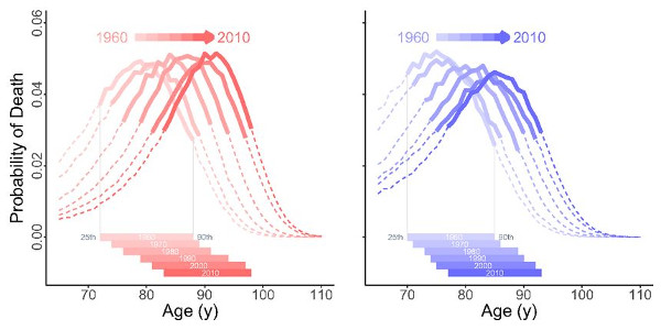
Today's open access paper provides an interesting visualization of the slow upward trend in life expectancy that has taken place over the last 60 years. A plot of the distribution of human mortality by age over the last third of life results in a wave-like curve, peaking at around 90 years of age. But those are today's numbers. In the 1960s, the curve had much the same shape, but the peak was at 80 years of age. Life was shorter, but the distribution of mortality at the end of life was much the same.

This is the case despite large changes in the causes of death over this span of decades. Mortality risk due to heart disease has diminished greatly, for example, thanks to the advent of statins and similar treatments. The slow march of medicine - meaning control of infection and improved health throughout life, not just incrementally better ways to treat age-related disease - has resulted in life expectancy at birth increasing by two years every decade. Remaining life expectancy at 65 has increased by about a year with every passing decade.

These trends are now a matter of history, and will not continue as they have. The advent of senolytic therapies to selectively remove senescent cells, one of the causes of aging, will cause an upward leap in life expectancy at 65. Other rejuvenation therapies that arrive in the decades ahead will result in further gains. The era of slow, incidental increases in life span is over. The era of deliberately engineered longevity has started, but it will most likely take two decades or more for the results of present clinical development to start to appear in population-wide demographic data. There is a lot of work left to accomplished not just in development but also for distribution of rejuvenation therapies: senolytics already exist, but next to no-one is using them, for example. This must change.

Advancing front of old-age human survival

We conclude that an advancing old-age front characterizes old-age human survival in 20 developed countries. The long-term speed of the advancing front is ≃0.12 year per calendar year, about 3 years per human generation. The location of the survival front is the 25th percentile of mortality. Thus, the front implies that, e.g., age 68 years today is equivalent, in terms of mortality, to age 65 years a generation ago. Our findings echo aspects of an earlier proposal that mortality hazards have, over the years, shifted rigidly to older ages. Our analysis of percentiles makes no assumptions about the pattern of mortality at young or old ages and focuses on older deaths. However, our finding of a shifting front in the percentiles of death at old age is consistent with some patterns of shifts in old-age mortality hazards.

Our findings provide no support for an impending limit to human lifespan, certainly not at an age that affects the movement of the survival front (between the 25th and 90th percentiles). To the extent that we can rely on the long-term speeds of percentiles above the 90th, the oldest deaths are being compressed in some countries but definitely not in others. Here again we find no support for an approaching limit to human lifespan. Nor do our results suggest that endowments, biological or other, are a principal determinant of old-age survival. The advancing survival front that we find suggests that the effects of inequality on mortality may be much smaller among old-aged adults than among younger adults.

Our analyses use period life tables, not cohorts, and suggest that continued mortality improvement depends largely on period processes such as economic growth, investment and advances in health science research and practice, and increases in the age of transition to disability. Our results also constrain biological arguments about the causes of death, especially the plasticity of death rates in response to environmental factors. Our moving survival front is consistent with a plateau in mortality rates, but implies that the location and possibly the level of the plateau change over time.

Early Onset of Menopause Correlates with Shorter Life Expectancy

Aging is a phenomenon affecting all organs and systems throughout the body, driven by rising levels of molecular damage. The variation in aging between individuals is largely determined by variations in the overall burden of such damage, the compound interest of small differences arising from lifestyle choices and happenstance such as infection in the first half of life. Thus for any given individual, manifestations and measures of aging tend to be fairly well correlated. That doesn't necessarily tell us anything about causation. So in this study, in which the researchers look at two very high level manifestations of aging, menopause and life span, there is probably no direct thread of causation at all. These are downstream manifestations of the summed effects of every cause of aging.

It is a well-accepted fact in the medical community that both type 2 diabetes and early onset of natural menopause may be associated with early death. Emerging evidence shows an association between age at menopause and diabetes, with studies reporting almost a two-fold increased risk of type 2 diabetes with early onset of menopause. To date, however, there are no other known studies that have quantified (calculated the number of years lived with and without diabetes) the combined association of early menopause and type 2 diabetes with life expectancy.

In this study involving 3,650 postmenopausal women, the difference in life expectancy was compared in women experiencing early, normal, and late menopause, as well as in those with and without diabetes. Compared with late menopause (defined as menopause that occurs at age 55 years and older), the difference in life expectancy for women who experienced early menopause (defined as menopause that occurs at age 44 years or younger) was -3.5 years overall and -4.6 years in women without diabetes. Compared with age at normal menopause (defined as menopause that occurs at 45-54 years of age), the difference in life expectancy for women who experienced early menopause was -3.1 years overall and -3.3 years in women without diabetes.

The authors suggest the need for future research to examine the mechanisms behind this association to help tailor prevention and treatment strategies that improve women's health across all age categories of menopause.

Link: https://www.eurekalert.org/pub_releases/2018-10/tnam-eoo101618.php

Random Mutations in Nuclear DNA are Prevalent in Old Tissues

Evolution requires happenstance mutation in order to progress, but too much of this random mutation leads to cancer or other forms of dysfunction sufficient to reduce reproductive fitness. The result is our present balance: enough mutation to make cancer a major cause of death, to ensure that mitochondria contribute to aging via mutation of mitochondrial DNA, and to produce some level of general dysfunction in aged tissues due to the accumulated mutational burden. The research noted here is one representative example of a range of research that seeks to quantify the degree to which our cells exhibit random mutational damage as we age. You might compare it with another set of results published recently on competition between mutations in skin, and how this paradoxically manages to suppress cancer risk.

Every person accumulates genetic changes, or mutations, throughout their lifetime. These mutations in normal tissue, called somatic mutations, are key to understanding the first steps to cancer and likely contribute towards ageing, but are uncharted territory due to technical limitations. For the first time, scientists have uncovered that on average, healthy cells in the oesophagus carry at least several hundred mutations per cell in people in their twenties, rising to over 2,000 mutations per cell later in life. Only mutations in a dozen or so genes seem to matter however, as these give the cells a competitive advantage allowing them to take over the tissue and form a dense patchwork of mutations.

The team used targeted and whole-genome sequencing to map groups of mutant cells in normal oesophageal tissue from nine individuals aged 20 to 75 years. The individuals' oesophageal tissues were considered healthy as none of the donors had a known history of oesophageal cancer, nor were taking medication for problems relating to the oesophagus. The study also casts new light on the mutations that are found in the squamous kind of oesophageal cancers. One mutated gene, TP53, which is found in almost all oesophageal cancers is already mutated in 5-10 per cent of normal cells, suggesting that cancer develops from this minority of cells.

In contrast, mutations in the NOTCH1 gene, known to control cell division, were found in nearly half of all cells of normal oesophagus by middle age, being several times more common in normal tissue than cancer. This observation suggests that researchers need to reconsider the role of some genes recurrently mutated in cancer in the light of mutations in normal tissue, and raises the possibility that the NOTCH1 mutation may even protect cells against cancer development. "This study shows that some genetic changes linked to cancer are present in surprisingly large numbers of normal cells. We still have a long way to go to fully understand the implications of these new findings, but as cancer researchers, we can't underestimate the importance of studying healthy tissue."

Link: https://www.sanger.ac.uk/news/view/mutant-cells-colonise-our-tissues-over-our-lifetime

Support the Development of Rejuvenation Therapies: Become a SENS Patron and We Will Match Your Donations

The SENS Research Foundation year end fundraiser has started. From now until the end of 2018, every new monthly donor will have the next year of their charitable donations to the SENS Research Foundation matched from our $54,000 challenge fund. The fund sponsors, Josh Triplett, Christophe and Dominique Cornuejols, and Fight Aging! challenge you to fund the development of rejuvenation therapies: sign up as a recurring donor, and we will match your donations for the next year.

Collectively, the SENS programs provide a path to comprehensive human rejuvenation. The SENS Research Foundation asks us to reimagine aging, to give our support to building a future in which being old does not have to mean being frail, sick, suffering, and diminished. This is not just theory. After years of effort, the work of our community of scientists, advocates, and philanthropists is paying off with the existence of working rejuvenation therapies that are well on their way to the clinic. The first rejuvenation therapies based on the SENS model, those that selectively destroy senescent cells, have been proven in mice and are presently undergoing heavily funded, rapidly expanding clinical development in multiple startup companies. Clinical trials in patients are underway.

So far, so good, but this is just the first step. The progress to date proves that the SENS vision of rejuvenation through damage repair is correct, but even as senescent cell clearance receives the attention and funding that it merits, a score of other equally important lines of research and development continue to languish, lacking resources. It is our job to provide those resources, the funding that can be used to bring these areas of research towards proof, widespread support, and active clinical development. The SENS Research Foundation has demonstrated its ability to make very good use of our charitable donations: you won't find a better way to change the world than this opportunity.

Reimagine Aging: the Campaign Begins Now

Thank you for your dedicated support of SENS Research Foundation's mission to end age-related disease. We know you share our passion and vision for a world with extended, healthy lifespans for all. How much human suffering would be alleviated if science and medicine could comprehensively treat the diseases of aging at their root cause?

Our donors are making this a reality. With your support, we are conducting and funding research, educating new scientists, and engaging in outreach to the public and industry partners. Research focuses on a unique damage-repair approach to treat diseases of aging. Education engages the next generation of rejuvenation biotechnology professionals. Outreach encourages and inspires the general public, policymakers, and academia to Reimagine Aging.

For more ways to donate, including how to receive tax benefits from the EU, visit our Ways to Donate page. From all of us at SRF, thank you! Your help is vital to moving our mission forward.

Chrdl1 Loss of Function Mutation Increases Synaptic Plasticity in Mice

Researchers here suggest that the protein chrdl1 plays an important role in the regulation of synaptic plasticity, the ability of the brain to generate new connections between neurons. Synaptic plasticity declines with age, and is important in cognitive function. There is thus considerable interest in ways to enhance plasticity, not just to turn back this aspect of aging, but also potentially as a form of enhancement therapy to improve memory or other aspects of the mind.

Researchers have shown that astrocytes - long-overlooked supportive cells in the brain - help to enable the brain's plasticity, a new role for astrocytes that was not previously known. The findings could point to ways to restore connections that have been lost due to aging or trauma. "To investigate this role, we used a lot of techniques in the lab to identify a signal made by astrocytes that's very important for brain maturation."

The signal turned out to be a protein astrocytes secrete called Chrdl1, which increases the number and maturity of connections between nerve cells, enabling the stabilization of neural connections and circuits once they finish developing. To further understand the role of Chrdl1, the team developed mouse models with the gene disabled by introduced mutations. These mice had a level of plasticity in their brains that was much higher than normal. Adult mice with the Chrdl1 mutation had brain plasticity that looked very much like that of young mice, whose brains are still in early stages of development.

Not much is known about the role of Chrdl1 in humans, but one study of a family with a Chrdl1 mutation showed they performed extremely well in memory tests. Other studies have shown the level of the gene encoding Chrdl1 is altered in schizophrenia and bipolar disorder, suggesting that Chrdl1 may have important roles in both health and disease. Future research by the team will dive deeper into the relationships between astrocytes and neurons and look for potential ways to use astrocytes as therapy. "We're interested in learning more about what the astrocytes are secreting into the brain environment and how those signals affect the brain. We plan to look at this relationship both early in development and in situations where those connections are lost and you want to stimulate repair, like after someone has had a stroke."

Link: https://www.salk.edu/news-release/brain-cells-called-astrocytes-have-unexpected-role-in-brain-plasticity/

Reviewing the Evidence for HSV1 to Contribute to Alzheimer's Disease

Alzheimer's disease starts with a slow rise in levels of amyloid-β present in the brain, an imbalance between dynamic processes of creation and clearance. This produces a state of mild biochemical and cognitive dysfunction that sets the stage for the later, much more destructive phase characterized by chronic inflammation, deposition of altered tau protein, and cell death. The roots of Alzheimer's must lie in the early mechanisms, in the poorly studied initial years of the condition, that cause some people to accumulate amyloid-β at a faster pace. In recent years evidence has emerged for persistent viral infection to play a role. Amyloid-β is coming to be seen as an anti-viral mechanism, and its creation and aggregation is prompted by the presence of viral particles.

Strong evidence has emerged recently for the concept that herpes simplex virus type 1 (HSV1) is a major risk for Alzheimer's disease (AD). This concept proposes that latent HSV1 in brain of carriers of the type 4 allele of the apolipoprotein E gene (APOE-ε4) is reactivated intermittently by events such as immunosuppression, peripheral infection, and inflammation, the consequent damage accumulating, and culminating eventually in the development of AD.

Population data to investigate this epidemiologically, e.g., to find if subjects treated with antivirals might be protected from developing dementia - are available in Taiwan, from the National Health Insurance Research Database, in which 99.9% of the population has been enrolled. This is being extensively mined for information on microbial infections and disease. Three publications have now appeared describing data on the development of senile dementia (SD), and the treatment of those with marked overt signs of disease caused by varicella zoster virus (VZV), or by HSV. The striking results show that the risk of SD is much greater in those who are HSV-seropositive than in seronegative subjects, and that antiviral treatment causes a dramatic decrease in number of subjects who later develop SD.

It should be stressed that these results apply only to those with severe cases of HSV1 or VZV infection, but when considered with the over 150 publications that strongly support an HSV1 role in AD, they greatly justify usage of antiherpes antivirals to treat AD.

Link: https://doi.org/10.3389/fnagi.2018.00324

Assessing the Interaction Between Telomerase Activity and Epigenetic Age in Cell Cultures

One of the more interesting aspects of the various epigenetic clocks that have been developed in recent years is that it is still largely unknown as to what exactly it is that they are assessing in our aging biochemistry. These clocks are weighted measures of epigenetic markers, such as DNA methylation, over a comparatively small number of genes. The resulting number certainly reflects chronological age, with the best clocks having a margin of error of a few years when assessed over a group of people. There is also sound evidence for it to reflect biological age, the burden of damage and dysfunction, which varies between individuals. Some people are more burdened by aging than others, and it is thought that these clocks can assess that difference.

But what underlying processes of aging are driving the results? That isn't clear at all. It is quite possible that an epigenetic clock measures the changes resulting from only a limited subset of the full range of age-related damage and dysfunction. Because aging is a global phenomenon in which all of its aspects tend to be fairly well correlated with one another, the clock nonetheless works well as a measure of overall aging. We will only find out whether or not this is the case as rejuvenation therapies start to emerge, treatments that very selectively address one and only one of the root causes of aging. Does treatment with senolytics to reduce the number of lingering senescent cells reverse the epigenetic clock measure, for example? We'll know the answer to that question in the near future, but for now those studies are still underway or pending publication.

Researchers here perform an preliminary investigation of what happens to epigenetic age in cells in which telomerase is at work. Telomerase acts to extend cell life by extending telomeres. Telomeres are the caps at the end of chromosomes, and are reduced in length with each cell division. This is a part of the countdown mechanism that leads to the Hayflick limit, preventing normal somatic cells from replicating indefinitely. Once they reach the limit, they self-destruct, or become senescent and are then destroyed by the immune system. Stem cells can replicate indefinitely because they use telomerase, and their role is to create new somatic cells with long telomeres to replace those lost over time. This split between a few privileged cells and the vast majority of limited cells is the way in which cancer risk is kept low enough for evolutionary success in higher animals.

A faction in the research and development communities are quite enthusiastic about telomerase gene therapy as a means to extend life, based on results from animal studies over the past decade or more. This most likely produces benefits through enhanced cell activity, and particularly stem cell activity, in a context in which the evolved balance of declining cell activity with age, most likely a defense against cancer, has some wiggle room. It appears possible to produce greater regeneration in later life without greatly raised risk of cancer by pressing damaged cells into undertaking more work. As is discussed by the authors of the paper below, this may also have something to do with reduced levels of senescent cells: a damaged cell that continues operating might, on average, be less immediately harmful than a lingering senescent cell, even though one would imagine this to raise cancer risk. The degree to which these and other mechanisms might contribute to the improved health and extended life observed in mice as a result of telomerase gene therapies has not yet been rigorously determined. But what does this do to epigenetic age? Running studies in cells doesn't really tell us what happens in animals; it is more a way to get a handle on the basics that can then be used to argue one position or another.

Epigenetic ageing is distinct from senescence-mediated ageing and is not prevented by telomerase expression

Ectopic expression of hTERT, the catalytic sub-unit of telomerase, which can preserve telomere length and avert senescence of some cells. It was initially thought that the functional and physical deterioration that characterise organismal ageing are a result of insufficient replenishment of cells due to telomere-mediated restriction of cellular proliferation. Senescent cells, which accumulate increasingly in tissues in function of age, were assumed to be passive and merely a consequence of the above-described processes. This notion was short-lived when senescent cells were found to secrete molecules that are detrimental to cells and tissues.

As such, it would follow that if cells were prevented from becoming senescent in the first place, ageing could be avoided. Although there are external instigators such as stress and DNA damage that can also cause cells to become senescent, replicative senescence is particular in that it is an intrinsic feature that is part of cellular proliferation and occurs even in an ideal environment. As expression of hTERT has been repeatedly demonstrated to prevent replicative senescence of many different cell types, it is reasonable to consider ectopic expression or re-activation of endogenous hTERT expression as potential means to prevent replicative senescence, delay ageing, and improve health.

The above proposition would be valid if senescent cells were indeed the only cause of ageing. Relatively recently, an apparently distinct form of ageing, called epigenetic ageing was described. This discovery stems from observations that the methylation states of some specific cytosines that precede guanines (CpGs) in the human genome changed rather reliably and strictly with age. This allowed supervised machine learning methods to be applied to DNA methylation data to generate an DNA methylation-based age estimator, which in the majority of the human population is similar with chronological age. Epigenetic age is not merely an alternative means of determining chronological age but is to some degree a measure of biological age or health; a proposition that is further supported by the impressive demonstration that acceleration of epigenetic ageing is associated with increased risk of all-cause mortality.

We recently developed a new epigenetic age estimator, referred to as skin and blood clock that is more accurate in estimating age of different cell types including fibroblasts, keratinocytes, buccal cells, blood cells, saliva and endothelial cells. Studies employing skin and blood clock and the pan-tissue epigenetic age clock revealed a startling consistency of epigenetic age across diverse tissues from the same individual, even though cellular proliferation rates and frequencies of these tissues are not the same. This suggests that the ticking of the epigenetic clock is not a reflection of proliferation frequency, which is in stark contrast to telomere length, which enumerates cellular division. It would therefore appear that the process of epigenetic ageing is distinct from that which is driven by telomere-mediated senescence.

To understand their relationship or interaction, if one indeed exists, we set out to test the impact of hTERT on epigenetic ageing. To this end we employed wild type hTERT that can prevent telomere attrition and its mutants that cannot, with some still able to nevertheless prolong cellular lifespan. Expressing these hTERT constructs in primary cells from numerous donors, ages and cell types, we observe that while hTERT expression can indeed prevent cellular senescence, it does not prevent cells from undergoing epigenetic ageing and that extension of cellular lifespan is sufficient to support continued epigenetic ageing of the cell. These simple observations provide a very important piece to the puzzle of the ageing process because it reveals the distinctiveness of epigenetic ageing from replicative senescence-mediated ageing. They provide further empirical support to the epidemiological observation that hTERT variant that is associated with longer telomeres are also associated with greater epigenetic ageing.

Mechanisms from Non-Mammalian Species Used to Regrow Sensory Hair Cells in Mice

Many other species are more regenerative than we are, and mammals in general are less regenerative than is the case for some other clades. It is too early to say whether ongoing investigations of the basis for proficient regeneration will lead to ways to safely adjust our biochemistry to perform greater feats of healing. Even simple mechanisms, if found, may be turn out to be very hard to introduce into humans. Everything in cellular biochemistry is connected to everything else; nothing can be changed in isolation. The greatest hope is that mechanisms of regeneration that are active in other species are merely dormant in mammals, but again it is too early to say with any confidence as to whether or not this is the case, despite promising signs in recent years.

Hearing impairment has long been accepted as a fact of life for the aging population - an estimated 30 million Americans suffer from some degree of hearing loss. However, scientists have long observed that other animals - namely birds, frogs, and fish - have been shown to have the ability to regenerate lost sensory hair cells. "It's funny, but mammals are the oddballs in the animal kingdom when it comes to cochlear regeneration. We're the only vertebrates that can't do it."

In 2012 researchers identified a family of receptors - called epidermal growth factor (EGF) - responsible for activating support cells in the auditory organs of birds. When triggered, these cells proliferate and foster the generation of new sensory hair cells. The researchers speculated that this signaling pathway could potentially be manipulated to produce a similar result in mammals. "In mice, the cochlea expresses EGF receptors throughout the animal's life, but they apparently never drive regeneration of hair cells. Perhaps during mammalian evolution, there have been changes in the expression of intracellular regulators of EGF receptor family signaling. Those regulators could have altered the outcome of signaling, blocking regeneration. Our research is focused on finding a way switch the pathway temporarily, in order to promote both regeneration of hair cells and their integration with nerve cells, both of which are critical for hearing."

In a new study, researchers tested the theory that signaling from the EGF family of receptors could play a role in cochlear regeneration in mammals. The researchers focused on a specific receptor called ERBB2 which is found in cochlear support cells. One set of experiments involved using a virus to target ERBB2 receptors. Another involved mice genetically modified to overexpress an activated ERBB2. A third experiment involved testing two drugs, originally developed to stimulate stem cell activity in the eyes and pancreas, that are known activate ERBB2 signaling. The researchers found that activating the ERBB2 pathway triggered a cascading series of cellular events by which cochlear support cells began to proliferate and start the process of activating other neighboring stem cells to become new sensory hair cells. Furthermore, it appears that this process not only could impact the regeneration of sensory hair cells, but also support their integration with nerve cells.

Link: https://www.urmc.rochester.edu/news/story/5448/study-points-to-possible-new-therapy-for-hearing-loss.aspx

Neurotransmitters Envisaged as Controllers of Stem Cell Activity

Adult, or somatic, stem cells support surrounding tissues by delivering a supply of daughter somatic cells, ready to replace those lost over time. This stem cell activity declines with age, and in the best studied stem cell populations this appears to be more a matter of signaling than a matter of inherent dysfunction. Stem cells react to rising levels of damage in tissues, or rather to the changes in signaling that result from that damage. Old stem cells put into a young environment perform as well as their younger counterparts. This decline with age may have evolved to limit cancer risk, but it brings the certainty of a slow decline into organ failure.

Many research groups are searching for the signals responsible for adjusting stem cell activity. The scientists here demonstrate that the autonomic nervous system makes important contributions to this signaling environment, and thus specific neurotransmitters may be a useful target for therapies to suppress or enhance stem cell function in various contexts. When it comes to aging, the function of the autonomic nervous system is known to change in later life, but more work is needed to solidify how this new research fits in to the bigger picture.

Somatic stem cells are microscopic workhorses, constantly regenerating cells throughout the body: skin and the lining of the intestine, for example. Researchers have demonstrated for the first time that stem cell proliferation is directly controlled by the autonomic nervous system (ANS). The ANS controls all of our unconscious functions: breathing, blood flow, digestion, and so forth. Its two major networks of nerve fibers run from the brain through the entire body, with neurons reaching into nearly every organ. These neurons release chemicals called neurotransmitters, which can affect target cells directly or indirectly.

When neurotransmitters bind to receptors in the membranes of certain cells, they elicit a direct response within the cell. But changes in cells can also occur when neurotransmitters induce a general state of inflammation or alter blood flow, an indirect route of action for the ANS. Scientists had suspected the ANS was involved in stem cell proliferation, but they didn't know if the relationship was direct or indirect. A direct relationship could have greater implications for drug interventions to treat medical conditions. "If you wanted to change the regeneration potential of an organ, for example, you wouldn't have to stimulate or suppress the activity of those neurons. Instead, you could just figure out what neurotransmitters are controlling proliferation and then get that chemical to those stem cells with targeted drug delivery."

To demonstrate that stem cell behavior was changing as a result of ANS stimulation, the researchers grew intestinal epithelial cells in the lab and exposed them to high levels of two neurotransmitters, norepinephrine and acetylcholine. Norepinephrine is a major neurotransmitter of the sympathetic nervous system, or "fight or flight" branch of the ANS, while acetylcholine is produced by the parasympathetic nervous system, or "rest and digest" branch. When the researchers simulated activation of either of those systems, they saw a decrease in stem cell proliferation. This suggests the body may avoid putting energy into making new cells when the fight or flight system is active.

Link: https://www.eurekalert.org/pub_releases/2018-10/uoic-isf101718.php

Anti-Amyloid CPHPC Therapy Used in a Clinical Trial for Alzheimer's Disease

CPHPC, now called miridesap, is a cautionary tale of what all too often happens to promising approaches in the field of medical development, once they advance to the point of expensive clinical trials and the requirement for partners with deep pockets to fund those trials. Miridesap was one of the earlier methodologies demonstrated to clear out transthyretin amyloid from tissues. This form of amyloid appears to be an important contribution to risk of cardiovascular disease, as well as a factor in osteoarthritis, and the evidence suggests it is the majority cause of death in supercentenarians. Its accumulation in old tissues is a form of damage, one of the root causes of aging. Ways to remove transthyretin amyloid should be pursued aggressively, but so far most of the effort in the research community has focused on the inherited form of transthyretin amyloidosis, using therapies that are not all that helpful for the age-related form of amyloidosis.

The first attempt to develop miridesap with a major pharmaceutical concern failed in the 1990s and early 2000s. The company founded to develop miridesap, Pentraxin Therapeutics, then partnered with Glaxosmithkline, GSK, at which point it took something like nine years to get to the point of running a small trial in 2015. That trial was successful, but thereafter GSK discontinued the work. The problem is less that initiatives sometimes fail, and more that (a) major pharmaceutical entities do not have the right incentives operating in order to carry out development programs rapidly and reliably, and (b) their ownership of intellectual property rights prevents anyone else from trying variants on the same approach, even when very little is being done, or the research is entirely halted. While in principal it is possible to obtain rights to a moribund program, in practice that is far from easy, and too expensive for most of the people who would consider trying it. This might all be seen as a symptom of excessive regulatory costs. Either way, research and development languishes.

Miridesap has a third act, however, one that has been in the works for a few years now. Those involved are attempting to use it as a way to remove amyloid-β in Alzheimer's disease, and are organizing a trial that is now recruiting. It will be interesting to see whether this works well enough to make it competitive. It is certainly less harsh on patients in comparison to the immunotherapies that make up the majority of attempts to treat Alzheimer's disease. Perhaps if this works, then the rights for use against transthyretin amyloid can be wrested from GSK, or GSK might be convinced to proceed again with that line of development.

NIHR-backed trial to test miridesap in Alzheimer's

Mark Pepys, who has been working on amyloidosis for 43 years, discovered way back in the 1980s that SAP, a normal, nonfibrous circulating plasma glycoprotein, is involved in the formation of amyloid deposits. He went on to show it is always present as a minor component of human amyloid deposits of all types, and that it prevents amyloid fibrils from being cleared via opsonization and phagocytosis. Despite attracting the interest of big pharma, attempts to translate those insights to the clinic have been slow to bear fruit.

A collaboration with Roche Holding AG that started in 1993 led to the discovery of miridesap (then known as CPHPC). When tested in the rare disease systemic amyloidosis, miridesap removed SAP from the blood, but could not shift large deposits of amyloid from organs. Amyloidosis patients treated with miridesap remained stable, but the deposits did not disappear. After Roche handed back rights in 2008, Pepys formed a collaboration with Glaxosmithkline to develop miridesap in combination with an anti-SAP antibody for treating amyloidosis. The rationale was to remove SAP from the blood and then use the antibody to target SAP in amyloid, activating the complement system to clear the deposits. That played out in a phase I study published in 2015, but a phase II, 30-patient study of the combination therapy recently was suspended by GSK.

Meanwhile, Pepys has been pursuing development of miridesap as a monotherapy in Alzheimer's disease. His basis for thinking miridesap can remove amyloid from the brain when it was not effective in removing it from other organs, is related to the much lower level of SAP that needs to be sponged up. SAP is generated and catabolized only in the liver and is not expressed in the brain. In a mouse model of Alzheimer's that is genetically engineered to generate human SAP, depleting SAP in the bloodstream removed all detectable SAP from amyloid in the brain.

The study is funded with $6.2 million in grants from NIHR. GSK has no commercial interest, but has assisted with the logistics of setting up the Despiad (Depletion of serum amyloid P component in Alzheimer's disease) trial. Patients in the trial will be required to inject miridesap three times a day over 12 months and to undergo a wide range of tests, including PET scans, lumbar punctures, and cognitive assessments. Pepys hopes the 100-patient, double-blind, placebo-controlled Despiad trial, will show the reduction in SAP levels translates into clinical benefit.

Exercise and Calorie Restriction Lower Blood Pressure In Part via β-Hydroxybutyrate

Researchers here describe one very thin slice of the sweeping metabolic changes produced by exercise and calorie restriction. Both interventions act to reduce blood pressure, most likely through numerous distinct mechanisms. One of those mechanisms involves raised levels of β-hydroxybutyrate, an effect that can in principle be mimicked or enhanced via carefully designed therapies. The raised blood pressure that occurs with age is one of the more destructive changes that take place with aging; it is in effect a way to translate accumulating damage and dysfunction at the cellular level into a physical bludgeon that destroys delicate structures throughout the body. Blood pressure is so influential in aging that current pharmacological methods that force a lowered blood pressure result in sizable reductions in disease incidence and mortality even though they fail to address the underlying damage of aging in any way.

Hypertension is a modifiable risk factor for cardiovascular disease and exercise is widely recommended for hypertensive patients as a lifestyle modification because of the well-documented beneficial effect of exercise on lowering blood pressure (BP). Similarly, calorie restriction, although not widely recommended for patients, is also documented to lower hypertension. Interestingly, both exercise and calorie-restriction are associated with increased circulating levels of ketone bodies such as β-hydroxybutyrate (βOHB). βOHB is produced predominantly in the liver, transported to other tissues, and traditionally recognized as a vital alternative metabolic fuel during times of starvation. However, contemporary evidence indicates that apart from serving as energy fuels, ketone bodies such as βOHB block inflammasome-mediated inflammatory diseases and thereby play a prominent role in maintaining physiological homeostasis.

In contrast to exercise and calorie-restriction, consumption of high salt promotes hypertension. Studies on the effects of dietary salt have focused mainly on organs and tissues relevant to BP regulation such as kidney, vasculature, heart, and brain. A recent report suggests that a reduction in salt intake serves as an additional interventional approach for reducing the risk for developing metabolic syndrome, of which, hypertension is one of the hallmark features. Taken together, these studies point to an intriguing possibility that a high salt diet induced a deleterious effect on hypertension and could mechanistically represent the opposite scenario to that of the protective effects of exercise and calorie-restriction on hypertension by altering the levels of metabolites such as ketone bodies.

Here, we examined this possibility, first by an untargeted mass spectrometry-based plasma metabolomics study and discovered altered ketogenesis and over-activation of renal Nlrp3 as a key mechanistic link between high salt and hypertension. These results indicated that a high salt diet has mechanistically opposite effects of exercise and calorie-restriction on BP. Next, we demonstrated that nutritional intervention with 1,3-butanediol, a precursor of the endogenous ketone body, βOHB, reversed the adverse effects of high salt induced renal Nlrp3-mediated inflammation, fibrosis, and hypertension. Based on these observations in the Dahl S rat, which is a salt-sensitive pre-clinical model of hypertension, we propose dietary intervention with 1,3-butanediol as an intriguing strategy for the clinical management of salt-sensitive hypertension.

Link: https://doi.org/10.1016/j.celrep.2018.09.058

Any and All Trend Projection Models Showing Slow, Incremental Future Increases in Longevity are Simply Wrong

This open access paper is an example of a model of future life expectancy that projects existing trends, with a little variation in here and there based on whether or not public health measures related to smoking and diet prove to be more successful or less successful. It predicts an average global increase in life expectancy of 4 to 5 years by 2040. In recent years I would have said that this is probably incorrect. I think we are at the point now in the development of rejuvenation therapies at which I can say that it is definitely incorrect. Any study that fails to consider progress in the treatment of aging as a medical condition is disconnected from reality.

Twenty years from now senolytic drugs will be used by a sizable percentage of the world's population, and will cost cents per dose. They will dramatically reduce the suffering and death resulting from inflammatory age-related diseases by removing some fraction of lingering senescent cells from old tissues. The first such therapies already exist today, are easily available, and some cost a few hundred dollars per dose or less. It isn't hard to see that the use of senolytics will spread like wildfire just as soon as the first clinical trials report their results over the course of 2019. Further consider that this is just one branch of rejuvenation biotechnology. Numerous other branches are under development today, and will certainly be clinically available by the late 2020s. The historical trend in life expectancy will be smashed; life expectancy will jump upward quite dramatically.

This was the first study to forecast a comprehensive set of cause-specific and all-cause mortality and associated indicators using a framework that allows for exploring different scenarios for many risk factors and other independent drivers. In our reference scenario, life expectancy was forecasted to continue increasing globally, and 116 of 195 countries and territories were projected to have significant advances in life expectancy by 2040. Gains were projected to be faster among many low-to-middle SDI countries, indicating that inequalities in life expectancy could narrow by 2040.

As shown by the better health scenarios, greater progress might be possible, yet for some drivers such as high body-mass index (BMI), their toll will rise in the absence of intervention. We forecasted global life expectancy to increase by 4.4 years for men and 4.4 years for women by 2040, but based on better and worse health scenarios, trajectories could range from a gain of 7.8 years to a non-significant loss of 0.4 years for men, and an increase of 7.2 years to essentially no change (0.1 years) for women.

In 2040, Japan, Singapore, Spain, and Switzerland had a forecasted life expectancy exceeding 85 years for both sexes, and 59 countries including China were projected to surpass a life expectancy of 80 years by 2040. At the same time, Central African Republic, Lesotho, Somalia, and Zimbabwe had projected life expectancies below 65 years in 2040, indicating global disparities in survival are likely to persist if current trends hold.

Taken together, our forecasts point to a world where most populations are living longer and many health improvements are likely to occur if current trajectories hold; at the same time, such gains are not without potential important social consequences, particularly if long-term planning and policy design are not fully considered today.

An important finding is that in the reference scenario, we forecasted slower progress in 2040 than that achieved in the past; however, in the better health scenario, global life expectancy improvements exceeded gains that occurred from 1990-2016. This forecasted slowdown in the reference scenario is rooted in a combination of several factors. First, some risks were projected to worsen in the future, most notably high BMI. Second, past progress on other leading risk factors for premature mortality, namely tobacco and ambient particulate matter air pollution, was highly variable and thus such heterogeneity was projected through 2040. Third, several countries that have already achieved higher levels of life expectancy have also had stagnated gains.

Link: https://doi.org/10.1016/S0140-6736(18)31694-5

Infection as the Link Between Cellular Senescence, Autophagy, and Immunosenescence

This open access paper reviews the interactions between cellular senescence, autophagy, and immunosenescence, with chronic infection as a mediating mechanism. Given the present state of knowledge and biotechnology, it is challenging enough to look at any two aspects of the aging body and consider how they might interact in isolation, but this can only ever be a thin slice of the bigger picture. All systems and states in our biochemistry interact with one another in some way, directly or indirectly, and examining ever larger sets of relationships between greater numbers of systems and states is the path to greater understanding of aging as a phenomenon. It is also somewhat beyond present capabilities, a complex, challenging endeavor for the scientists of future decades, which is why bypassing the need for this sort of understanding is highly desirable when working towards therapies to treat aging. We cannot afford to wait for a near complete knowledge of the progression of aging.

The state of cellular senescence, in which replication is shut down, can be a reaction to damage. It is one of the ways in which cancer risk is sufficiently minimized to allow higher forms of multicellular life to exist. Senescent cells are unfortunately harmful to surrounding tissues, and their accumulation with age is one of the root causes of degenerative aging. Autophagy is a collection of cellular damage control processes, responsible for recycling broken and unwanted proteins or structures in the cell. Loss of autophagy to the point of excessive accumulation of molecular damage is one way for cells to become senescent, and unfortunately autophagy declines with age. Immunosenescence is the aged state of the immune system, characterized by chronic inflammation and incapacity. In later life, the immune system becomes far less effective in removing damaged cells, such as senescent cells, as well as less effective when it comes to a defense against invading pathogens.

Even when simply considering just these three line items, the potential interactions are complex and challenging to rigorously prove. The authors of this paper advance the common view that chronic infection impairs autophagy, and thus in turn generates increased numbers of senescent cells, which accelerates the progression of immunosenescence.

Chronic Infections: A Possible Scenario for Autophagy and Senescence Cross-Talk

Cellular senescence is induced as a consequence of cellular damage accumulation, with the extent of activation directly depending on a fine-tuned balance between cellular conditions generating damage and those involved in counteracting them. The autophagic pathway plays a key role in preventing cell damage accumulation, however, the aging process leads to a decrease in autophagy capacity, and therefore also its effectiveness. In this context, senescence activation shows a more preponderant protective role.

The immune system does not escape from aging effects and displays senescence characteristics in aged individuals. Immunosenescence refers to the state of dysregulated immune function that contributes to the increased susceptibility to infections, autoimmune diseases, or cancer. Aged individuals are predisposed to more severe symptoms from certain infections and they do not mount an effective immune response upon vaccination. In general, aged populations fail to generate an appropriate innate and adaptive immune response against microorganisms, thus it becomes clear that senescence is involved in this failure.

Besides the normal occurrence of immunosenescence, several pathogen microorganisms accelerate the activation of senescence and predisposal to premature immunosenescence. For instance, hosts infected with bacteria such as P. aeuruginosa, M. tuberculosis, or H. pylori, some viruses, including HCMV, or the parasite T. cruzi, show characteristics of immunosenescence. A common issue of all of these pathogens is that they are able to generate chronic infections. In each of these, regardless of the fact that the host is faced with the same antigen several times during its lifetime, the immune response is inefficient. Furthermore, data shows that this condition generates an immune exhaustion and immunosenescence seems to be the major causative factor offering the pathogens an extra advantage since their elimination by the host tends to be even less effective.

Interestingly, a common characteristic of chronic infections is the autophagy blockage that usually occurs during autophagosome maturation, representing a factor that could contribute to or accelerate immunosenescence activation since it predisposes cells to damage accumulation. Deeper exploration to elucidate whether the activation of senescence in chronic infection is a consequence of autophagy impairment produced by pathogens to avoid degradation or, alternatively, whether it is a mechanism employed by the host to diminish infection spreading when the degradation of the pathogens has been halted. This exploration is needed to further understand the infection-autophagy-senescence relationship. With the available data, we hypothesize that chronic infections induce senescence with similar characteristics of aging, i.e., increase of inflammatory state and autophagy inhibition.

Will Strength Training with Blood Flow Restriction Help Cognitive Function?

There is plenty of evidence for exercise of all sorts to improve cognitive function in later life. That outcome might be mediated via increased blood supply to the brain, which is a particularly energy-hungry organ. Or it might be mediated via improved mitochondrial function, for much the same underlying reasons relating to energy demands. Or via any one of a number of other related mechanisms that one can link to exercise. Strength training is thought helpful in yet another way, via building or retaining muscle mass that then in turn alters metabolism in favorable ways (that usually lead back to blood flow and mitochondrial function in some way).

There is some overlap between researchers interested in strength training and those interested in ischemic conditioning, a form of intermittent restriction of blood flow that appears synergistic with exercise. One can view this all from the perspective of triggering stress responses. Exercise triggers stress responses, and so does transient ischemia. The former is far more explored, and the latter is harder to undertake safely. One might also view this area of research as the preliminary exploration that leads to drug candidates somewhere down the line, ways to artificially trigger beneficial stress responses, but I think that the past few decades of work on calorie restriction have demonstrated that to be slow, expensive, and challenging.

The integrity of the musculature and the muscle strength is of great importance throughout the entire life span. Age-related decreases in muscle mass and strength are also associated with morphological losses in the brain and decreased cognitive functions. There is growing evidence with respect to positive effects of physical activity preventing and treating morphological and functional losses in muscles and the brain. In recent years, evidence has emerged emphasizing the existence of a bidirectional relationship between physical performance and brain health. The bidirectional relationship suggests that physical training may be a valuable intervention strategy to decelerate not only physical but also cognitive decline in old age. However, the exercise type (e.g., resistance training, endurance training) and exercise variables (e.g., load, duration, frequency), which would be optimal to efficiently enhance cognitive performance are largely unknown.

A promising and cost-effective physical intervention strategy which preserves and enhances both, physical performance (especially with regard to the musculature) and cognitive functions, is resistance training (also known as strength training). A relative new method in the field of resistance training is blood flow restriction training (BFR). While resistance training with BFR is widely studied in the context of muscular performance, this training strategy also induces an activation of signaling pathways associated with neuroplasticity and cognitive functions. Based on this, it seems reasonable to hypothesize that resistance training with BFR is a promising new strategy to boost the effectiveness of resistance training interventions regarding cognitive performance.

Link: https://doi.org/10.3390/jcm7100337

Calorie Restriction Produces Beneficial Changes in Gut Flora Populations

Members of the research community have in recent years exhibited a growing interest in the analysis of gut microbes in the context of metabolism and the pace of aging. Some inroads are being made into better understanding helpful versus unhelpful microbial populations and behaviors, and how exactly their activities might influence health over the long term. It is unclear as to how large this influence is. Perhaps it is in the same ballpark as exercise, but perhaps not. The usual problems arise when comparing results between species, in that short-lived species have greater plasticity of life span, their length of life more readily extended or shortened in response to changing circumstances. It should be no great surprise to find that the practice of calorie restriction, well known to slow aging in near all species tested to date, induces changes in gut microbial populations that conform to alterations that are seen as being helpful for health in other contexts.

The gut microbiota (GM) largely derives nutrients from dietary intake. In this respect, a large number of studies have been reported on the variations of the GM composition occurring according to different diets. The majority of these studies have focused on the comparison of low vs. high energy density (i.e., high fat or high sugar) diets in animals fed ad libitum (AL), showing an increase in the Firmicutes/Bacteroidetes ratio and the proliferation of pro-inflammatory Proteobacteria in the latter condition.These changes occur rapidly and can be partially restored by reverting to the control diet. Animal experimental data also agree with observational studies in humans, where similar taxonomic features were found to be changed between obese and lean individuals.

In addition, the GM composition varies rapidly and significantly in response to macronutrient changes, even when equal numbers of calories are provided. This clearly suggests that the relative abundance of the specific GM members strongly depends on the quality of nutrients they have access to. Hence, given the strong relationship among diet, GM and health, there is a growing interest in developing novel dietary strategies to modulate the composition and, possibly, the metabolic functions of the GM.

Among dietary interventions, caloric restriction (CR) is well known for the health-promoting impact on lipid metabolism and longevity. CR is generally applied without changing the macronutrient composition and solely reducing the caloric intake compared to the AL condition. As a consequence, in experimental models, caged individuals fed a CR diet consume completely their food and then fast for several hours before the next feed administration. We have recently reported that CR induces a rapid change (as early as after 3 weeks of CR) of the GM composition in young rats, that parallels a reduction of triglycerides and cholesterol levels in the blood, and that these changes are maintained up to mid age. In particular, a CR diet enabled the expansion of Lactobacillus rapidly and persistently up to adulthood. CR-induced variation of the GM composition might then play a role in helping extend lifespan and delay the onset of age-related disorders by preserving gut homeostasis. However, the precise biochemical changes the GM undergoes during CR are still undetermined, in the short and in the long term.

Here, we investigated the short- and long-term effects of CR on the rat GM using a metaproteogenomic approach. We show that a switch from ad libitum (AL) low fat diet to CR in young rats is able to induce rapid and deep changes in their GM metaproteomic profile, related to a reduction of the Firmicutes/Bacteroidetes ratio and an expansion of lactobacilli. Specifically, we observed a significant change in the expression of the microbial enzymes responsible for short-chain fatty acid biosynthesis, with CR boosting propionogenesis and limiting butyrogenesis and acetogenesis.

Link: https://doi.org/10.1038/s41598-018-33100-y

A Biotech Industry CEO on mTOR Inhibitors and the Treatment of Aging

There is a point in the life of a young biotech company at which one traditionally appoints an established figure from industry as the CEO. Running a company that is in the public eye due to clinical trials and heading in the direction of an IPO requires a whole different set of skills than were needed for early growth and technical success in development programs. It also tends to be a sign of the changing balance of influence between founders, investors, and industry partners as development programs progress. This happened earlier in the year for Navitor Pharmaceuticals, one of a number of companies working on mTOR inhibitor therapies capable of modestly slowing the aging process.

Talking up one's position is a part of the duties of an industry CEO: a good CEO is an advocate for the company, for the technology, for the industry. That is expected. I point out this commentary from the new Navitor CEO not for the expected content, but rather as an example of our present slow movement though an important tipping point in the great, many-threaded cultural conversation about aging and the prospects for treating aging as a medical condition. The message of the life science community, that aging can be slowed and reversed, is being taken up by industry and media. It is spreading broadly, and more rapidly than in past years.

In short, the goal of bringing aging under medical control is increasingly being taken seriously, finally, after more than twenty years of earnest advocacy and hard-fought, incremental progress in obtaining research funding. Now, the battle must turn to one of steering funding towards the better rather than the worse options for development. When people agree that the goal must be reached, it becomes very important to settle on the best possible strategy.

On that note, I don't think that therapies that function via inhibition of mTOR, based as they are on modulation of dysfunctional metabolism without doing much to address the causes of that dysfunction, have anywhere near as large an upside, considered in terms of additional healthy years of life, as is the case for the SENS approaches to aging. SENS rejuvenation therapies are intended to repair the underlying damage that causes aging, while mTOR inhibition and similar approaches largely adjust harmful reactions to that damage. They are beneficial to some degree, particularly now that it is possible to separate the desirable and undesirable components of the early mTOR inhibitors such as rapamycin. Still, while modest gains are better than nothing, we should be aiming for large gains.

Targeting Aging Comes Of Age

We finally are beginning to understand the biological basis of aging and age-related diseases, making the discovery of new therapies actionable for the first time. Aging and its underlying biological mechanisms are becoming recognized as a catalyst, if not the central catalyst, for a wide range of poorly treated prevalent diseases. This is a promising new area in science providing actionable insights with potential for tremendous impact on human healthspan.

I have been following the field of the biology of aging since the beginning of my career in science, more than thirty years, while working in targeted ways to find and advance new therapies in the areas of metabolic and cardiovascular disease. Recently, I became the CEO of a biotechnology company, Navitor Pharmaceuticals, that is squarely in this space and focused on leveraging new discoveries to target the activity of mTOR (mechanistic target of rapamycin). In many ways, the progress in the field has reached a tipping point and has prompted me to reflect on the advancements.

Chronic conditions of aging are the major cost drivers for healthcare. There are some shocking statistics to be found regarding the cost of chronic conditions affecting our healthcare system. The multiple chronic conditions chartbook published in 2010 by the Agency for Healthcare Research and Quality is a short and fascinating read. Almost half of all people aged 45-64, and 80% of those 65 and over, have multiple chronic conditions. 71 cents of every US healthcare dollar go to treating people with multiple chronic conditions. Just take a moment to think about that. First off, this is a huge portion of our healthcare budget. It's not cancer. It's not rare diseases. It's not cosmetic or elective procedures. It's chronic illnesses, primarily associated with aging.

Id you have one disorder that is commonly associated with aging, chances are you have another or will develop another one. Basically, it's tough to get old. We all know that. But, now science is leading us to harness some fundamental mechanisms of aging. Two core aging mechanisms appear to have emerged as being accessible to new pharmacological intervention - the mechanistic target of rapamycin, or mTOR, and cellular senescence (which is wrapped up in mTOR as well). Drug development approaches using cellular senescence are emerging, and they are fascinating and worthy of attention. It's an exciting time in the aging space, and hopefully one that yields important new medications capable of reducing the personal, societal, and financial burdens of chronic diseases.

RNA Interference of ATP Synthase Subunits Slows Aging in Nematodes

Mitochondria are the power plants of the cell, responsible for generating adenosine triphosphate (ATP), a chemical energy store molecule used to power cellular operations. The inner workings of each mitochondrion are energetic and complicated, consisting of a number of interacting protein complexes that collectively perform the work needed to manufacture ATP molecules. Mitochondrial function occupies a central position in the interaction between metabolism and aging for a number of reasons. Firstly, they generate reactive oxygen species (ROS) as a side effect of ATP production, and the flux of ROS is both damaging and a signal to the cell to step up its efforts to repair damage. A little more ROS than usual can be beneficial. Too much ROS is harmful. Secondly, some of the critical proteins in mitochondrial complexes are produced from DNA inside the mitochondria rather than in the cell nucleus, and that DNA is vulnerable to damage. Some forms of mitochondrial DNA damage can produce damaged mitochondria that cause great harm to the cell and surrounding tissue. Thirdly, cells need ATP, and reductions in ATP production have detrimental consequences over time.

There are many ways in which mitochondrial function can be altered through the removal or reduced production of a specific subunit of one of the mitochondrial protein complexes. Some such changes are disastrous, some are beneficial. Why that is the case is a complicated topic. It has a great deal to do with the balance between production of ROS and production of ATP, the needs of cells, and the reactions of cells, particularly the activation of repair and maintenance mechanisms. That balance is different in each case, and it is a slow and expensive process to run through the protein biochemistry needed to gain insight into what exactly is going on under the hood. The paper here is an example of the sort of work that takes place in this part of the field.

Mitochondria play an essential role in many important physiological processes, including aging. Mitochondrial function has been thought to gradually decline with age, while oxidative damage and mitochondrial DNA mutations accumulate. Although complete disruption of mitochondrial function is detrimental or even lethal for many eukaryotes, including humans, accumulating evidence has revealed that partial inhibition of mitochondrial function tends to increase lifespan. In C. elegans, mutations in various mitochondrial electron transport chain (ETC) genes can greatly extend lifespan; these include mutations in isp-1 and clk-1. In addition, RNAi knockdown of various mitochondrial ETC genes prolongs lifespan in yeast, worms, and fruit flies.

The effects of mitochondrial ETC genes on modulating lifespan appear to be complex. Inhibition of some ETC genes increases lifespan, whereas inhibition of others decreases or does not alter lifespan in C. elegans and Drosophila. For example, mutations in mev-1, which encodes a subunit of complex II, causes a short lifespan in worms. In addition, the underlying causes for lifespan regulation by ETC genes remain incompletely understood. For example, the roles of reactive oxygen species (ROS) production and mitochondrial function in aging and lifespan of ETC mutants can be opposite ways. One model interpreting these opposite effects is that moderate mitochondrial impairments increase lifespan until a threshold is reached, beyond which animals display wide-spread damage, shortened lifespan, or even death. Nevertheless, how mitochondrial genes modulate lifespan and whether they function in modulating lifespan in other species remain incompletely elucidated.

ATP synthase, also known as complex V of the mitochondrial respiratory chain, is the primary cellular energy-generating machinery. In mammals, ATP synthase deficiency is one of the rarer mitochondrial oxidative phosphorylation deficiencies. ATP synthase is also intimately linked to aging. In worms, genetic inhibition of the atp-2 gene, which encodes a subunit in complex V, leads to developmental delay and increased lifespan. Additionally, a genome-wide RNAi screen revealed that RNAi knockdown of subunits atp-3, atp-5, or asb-2 prolongs worm lifespan. However, the underlying mechanism for lifespan extension due to inhibition of these subunits in the ATP synthase remains unclear. As ATP synthase is highly conserved throughout evolution, understanding the role of the ATP synthase in lifespan regulation can lead to untangling of the complexity of mitochondrial ETC genes in modulating lifespan.

Link: https://doi.org/10.1038/s41598-018-32025-w

Considering the Adaptive Response Hypothesis for Calorie Restriction

The practice of calorie restriction reliably slows aging and extends life span in most species tested to date. The degree to which this happens is much reduced in longer-lived species, but a detailed understanding of why this is the case is yet to be assembled. It makes sense from an evolutionary perspective: extended health and life in response to famine helps to raise the odds of successful reproduction. Famines tend to be seasonal, and a season is a large fraction of a mouse life span, but only a tiny faction of a human life span. Thus only short-lived species evolve a sizable gain in life span in response to reduced calorie intake, even though the short-term benefits to health appear quite similar in both short-lived and long-lived mammals.

From a biochemical perspective, cellular metabolism is so complex, and calorie restriction changes so much of it, that it remains a major undertaking to try to put everything in order to understand how exactly calorie restriction works. It is clearly an adaptive response, a shift of the whole of metabolism from one state into another, a change of great complexity. Deciphering all of the details is a fascinating scientific endeavor, but one that will come to be of increasingly little relevance to the future of human longevity. Calorie restriction mimetic therapies that modestly slow aging are hard to construct, while rejuvenation therapies based on repair of the damage that causes aging will deliver far greater benefits with far lower expense.

In 1989, the anti-aging and prolongevity actions of calorie restriction (CR) were explained from the evolutionary viewpoint of organisms having evolved adaptive response systems to maximize survival during periods of food shortage. On the basis of this evolutionary viewpoint, we divided the beneficial actions of CR into two systems; "systems activated under sufficient energy resource conditions" and "systems activated under insufficient energy resource conditions". The former is activated under natural environmental conditions that grant animals free use of energy by providing a plentiful food supply. In other words, when there is grace for free use of energy, animals grow well, reproduce more, and store excess energy as triglyceride in white adipose tissue for later use, but not to such an extent that they become obese. The latter is activated under natural environmental conditions that do not permit free use of energy because of food shortages.

In other words, when there is no grace for free use of energy, animals suppress growth and reproduction and shift energy use from growth and reproduction to maintenance of biological function, but not to such an extent that they become severely starved. Adaptation to natural environmental changes is a top priority for survival in animals. On the basis of the adaptive response hypothesis, we propose a suite of mechanisms for the beneficial actions of CR. Since experimental CR conditions can mimic insufficient energy conditions, we hypothesized that CR suppresses "systems activated under sufficient energy conditions" and activates "systems activated under insufficient energy conditions", and additively induces anti-aging and prolongevity actions. The first set of systems involves GH/IGF1, FOXO, mTOR, adiponectin and BMAL1 signaling, and CR appears to suppress these anabolic reactions. The second set of systems involves SREBP-1c/mitochondria redox, SIRT and NPY signaling, and it is likely that CR activates these reactions to make optimal use of insufficient energy resources.

Studies using monkeys suggest that the beneficial actions of CR may occur in humans as well as other mammals. Ongoing CR research focuses on two themes, i.e. elucidation of the molecular mechanisms of CR, and development of CR mimetic medicines. We consider development of novel CR mimetic medicines to be difficult without an understanding of the molecular mechanisms of CR. To develop CR mimetic medicines that are applicable to humans, further studies are therefore required on the molecular mechanisms of CR, particularly in non-human primates. In this report, we propose that the molecular mechanisms of beneficial actions of CR should be classified and discussed according to whether they operate under rich or insufficient energy resource conditions. Future studies of the molecular mechanisms of the beneficial actions of CR should also consider the extent to which the signals/factors involved contribute to the anti-oxidative, anti-inflammatory, anti-tumor and other CR actions in each tissue or organ, and thereby lead to anti-aging and prolongevity.

Link: https://doi.org/10.18632/aging.101557

Year End SENS Patron Fundraiser for 2018: Challenge Fund Supporters Sought

Our community year end fundraiser for 2018 will soon be underway to support scientific programs for the development of rejuvenation therapies carried out at the non-profit SENS Research Foundation. As was the case last year, once again Fight Aging! and a few fellow travelers will assemble a challenge fund to encourage new SENS Patrons to set up subscriptions to make monthly or yearly recurring donations to the SENS Research Foundation. The first year of any such new donations will be matched dollar for dollar from the challenge fund.

We think that recurring donations are important: the more that our community supports the SENS programs by providing a regular supply of funding, the easier it becomes for the SENS Research Foundation staff to plan ahead and commit to long-term projects. In past years this initiative has been a success: our matching fund was met last year, and the new monthly donors largely stick around for the long term to continue to support SENS rejuvenation research. This year regular donor Josh Triplett is going above and beyond to put up $36,000 to encourage new SENS Patrons to make the leap. Christophe and Dominique Cornuejols are contributing $12,000, and I myself will put in $6,000. We are looking for other challenge fund donors to join us in this initiative. Do you want to make a sizable difference to the future of human health and longevity? This is how it is done.

The SENS Research Foundation uses our donations to fund a range of scientific work on the foundations of rejuvenation therapies, focused on those areas that are furthest behind or that most need unblocking in order to achieve meaningful progress. These are all programs that achieve rejuvenation through repair: validating the list of cell and tissue damage that lies at the roots of aging, and then reversing these forms of damage, one by one. It is in large part thanks to the advocacy, networking, and funding provided by the SENS Research Foundation, and by the Methuselah Foundation before it, that rejuvenation research is as far ahead as it is. When the SENS programs started, popular culture and the scientific community were opposed to any initiative aiming to produce rejuvenation via targeting the molecular damage that causes aging, despite decades of evidence to strongly support this strategy.

In recent years the naysayers have been proven clearly and categorically wrong. Clearance of senescent cells through the use of senolytic therapies has been shown to produce rejuvenation in mice. The first such treatments are in human trials, in development by multiple biotech companies, and being used by a growing number of self-experimenters worldwide. That today there is a new and rapidly growing senolytics industry, poised to deploy rejuvenation therapies that can remove some of the burden of senescent cells in older individuals, is due in large part to the network of advocacy, science, and funding centered on the SENS Research Foundation and Methuselah Foundation. Clearance of senescence cells was in the SENS proposals, front and center, from the very start. Back then, at the turn of the century, the goal of rejuvenation was widely ridiculed. Nonetheless, with persistence, persuasion, and the support of our community of everyday philanthropists, here we are today, embarking upon the construction of an industry that aims to reverse aging.

Senolytics are just the start. They are only a part of the story, and only a narrow slice of the complete human rejuvenation that remains only a possibility, rather than a certainty. Scores of other equally important and beneficial projects under the SENS umbrella of repair therapies are still comparatively neglected, or blocked by the lack of tools, or blocked by the lack of funding, or lacking strong champions in the research community. We can help to change this. We did a great deal to make that change come about for senescent cell clearance, and we can do the same for mitochondrial DNA repair, for breaking the cross-links that stiffen tissues, for clearing amyloids and other harmful metabolic wastes, and more. We shine the light that shows the way, and, given time and resources, we are successful.

Give some thought to joining us. A future in which being old does not mean being sick and diminished is a future worth bringing into existence. We can all help in some way to make this vision a reality.

Enthusiasm for Senolytic Therapies

I think it is entirely appropriate to greet the advent of senolytics with enthusiasm. These treatments are the first legitimate rejuvenation therapies to successfully target one of the root causes of aging, the accumulation of lingering senescent cells in old tissues. The first human trial data is approaching publication, but even before it arrives, the evidence to date strongly suggests that meaningful levels of rejuvenation can be achieved in old people at a very low cost. The first senolytic drugs (such as dasatinib and navitoclax) and plant extracts (such as fisetin and piperlongumine) cost very little, and remove only some senescent cells, no more than half in some tissues, and far fewer than that in others. Nonetheless, in mouse studies they reliably reduce chronic inflammation, reverse the progression of numerous conditions ranging from arthritis to Alzheimer's disease, and extend healthy life span even when applied a limited number of times in very late life.

As we get older, more and more of our the cells in our bodies become dysfunctional and enter into a state known as senescence. These senescent cells no longer divide or support the tissues and organs of which they are part; instead, they secrete a range of harmful inflammatory chemical signals, which are known as the senescence-associated secretory phenotype (SASP). Dr. Judith Campisi from the Buck Institute for Research on Aging, along with her research team, identified that senescent cells secreted the various harmful chemicals that characterize the SASP in 2008, which was when interest in senescent cells really began.

The SASP is a real problem: it increases inflammation, harms tissue repair and function, causes the immune system to malfunction, and raises the risk of developing age-related diseases such as cancer. Even worse, the SASP also encourages nearby healthy cells to become senescent, so even a very small number of senescent cells can cause big problems. Normally, senescent cells destroy themselves by a self-destruct process known as apoptosis or are cleared away by the immune system. Unfortunately, as we age, the immune system becomes weaker, and the senescent cells start to build up in the body. The accumulation of senescent cells is considered to be one of the reasons why we age and develop age-related diseases.

With these experiments, the biotechnology industry had initial proof that targeting one of the aging processes directly could improve health by delaying aging in mice; this began the search to develop therapies that target and destroy these harmful cells. This was the birth of a new class of drugs and therapies that would become known as senolytics. So far, there have been a number of drugs and naturally occurring compounds with senolytic potential and multiple mouse experiments demonstrating that the clearance of these cells can delay the onset of diseases such as cancer, heart disease, osteoporosis, arthritis, and Alzheimer's.

Interest in senolytics has seen a meteoric rise in the last couple of years, with investment money pouring in as confidence in the approach has reached new heights. There are also a number of companies developing therapies to destroy senescent cells, and it is likely that more will join them in the coming years. Leading the charge is Unity Biotechnology, which was founded in 2011 and has raised over $385 million in funding since then. Other companies are hot on its heels developing ways to seek and destroy these harmful cells. Oisin Biotechnologies, based in Seattle, is one such company. Founded in 2016, it has raised around $4 million to date and is developing a unique lipid nanoparticle-based system to deliver senolytic and cancer therapies. Cleara Biotech, based in the Netherlands, and Spain-based Senolytic Therapeutics are also busy developing senolytic therapies.

Link: https://www.leafscience.org/senolytics-target-aging/

Controlling Pattern and Structure in 3-D Bioprinting of Tendons and Ligaments

Naturally grown tissues are intricately structured, and the physical properties of tissue derive from the patterning of cells and their behavior in generating a supporting extracellular matrix. This natural complexity ensures that there is still a great deal of work to be accomplished when it comes to the 3-D bioprinting of functional tissue structures; not all tissues can be produced using the current state of the art systems, or at least not in a useful state. The work here is an example of the sort of incremental advance needed to produce tissues that are closer in form and function to those growing naturally inside bodies.

The 3-D-printing method, which took two years to research, involves taking stem cells from the patient's own body fat and printing them on a layer of hydrogel to form a tendon or ligament which would later grow in vitro in a culture before being implanted. But it's an extremely complicated process because that kind of connective tissue is made up of different cells in complex patterns. For example, cells that make up the tendon or ligament must then gradually shift to bone cells so the tissue can attach to the bone. "This technique is used in a very controlled manner to create a pattern and organizations of cells that you couldn't create with previous technologies. It allows us to very specifically put cells where we want them."

To do that, the team used a 3-D printer typically used to print antibodies for cancer screening applications. The researchers developed a special printhead for the printer that can lay down human cells in the controlled manner they require. To prove the concept, the team printed out genetically-modified cells that glow a fluorescent color so they can visualize the final product. The technology is initially designed for creating ligaments, tendons and spinal discs, but in the future it could be adapted to any type of tissue engineering application, such as the 3-D printing of whole organs, an idea researchers have been studying for years.

Link: https://unews.utah.edu/the-fine-print/

Salivary Gland Organoids Integrate and Generate Saliva When Implanted into Mice

Salivary glands are one of many small organs that we give little thought to until they fail, and then it becomes difficult to think of anything else. Just like every other tissue in the aging body, that failure becomes more likely with each passing year, with the accumulation of molecular damage and its consequences. One of the potential approaches to this general category of gradual organ failure is the generation of new organs or new functional tissue for transplantation, building tissues in bioreactors from the starting point of cells. This can in principle fix damage that is internal to an organ by replacing that organ entirely, or augment function of a failing organ with the use of tissue patches. The aged environment and its harmful influence on organ function through signaling will remain a challenge, however, until more general rejuvenation therapies are widely deployed.

Japanese researchers have been working on the tissue engineering of functional salivary glands for some years now, and the paper noted below reports on their latest success. Like most groups in the field, they are focused on discovering the necessary signals and environment that can direct cells to build a specific tissue in the same way that occurs during embryonic development. This is quite different on a tissue by tissue basis, but nonetheless progress is being made. The researchers here can build organoids, small sections of functional salivary gland tissue that are limited in size because they lack a capillary network. An important demonstration of functionality is to implant organoids into an animal and show that they integrate and perform the tasks expected of the naturally grown organ. That rarely implies complete success, as the assessed function usually isn't exactly the same, but nonetheless, it may indicate that the research program has progressed far enough to start thinking about use in human medicine.

Researchers create a functional salivary gland organoid

Salivary glands develop from an early structure called the oral ectoderm, but the actual process is not fully understood. It is known that organ development takes place through a complex process of chemical signaling and changes in gene expression, so the scientists began to unravel what the important changes were. They identified two transcription factors - Sox9 and Foxc1 - as being key to the differentiation of stem cells into salivary gland tissue, and also identified a pair of signaling chemicals - FGF7 and FGF10 - which induced cells expressing those transcription factors to differentiate into salivary gland tissue.

To create an organoid, researchers used a cocktail of chemicals that allowed the formation of the oral ectoderm. They used this cocktail to induce embryonic stem cells to form the ectoderm, and then used viral vectors to get the cells to express both Sox9 and Foxc1. Adding the two chemicals to the mix induced the cells to form tissue that genetic analysis revealed was very similar to actual developing salivary glands in the embryo.

The final step was to see if the organoid would actually function in a real animal. They implanted the organoids into actual mice without saliva glands and tested them by feeding them citric acid. When the organoids were transplanted along with mesenchymal tissue -another embryonic tissue that is important as it forms the connecting tissue that allows the glands to attach to other tissues - the implanted tissues were found to be properly connected to the nerve tissue, and in response to the stimulation secreted a substance that was remarkably similar to real saliva.

Generation of orthotopically functional salivary gland from embryonic stem cells

Organoids generated from pluripotent stem cells are used in the development of organ replacement regenerative therapy by recapitulating the process of organogenesis. These processes are strictly regulated by morphogen signalling and transcriptional networks. However, the precise transcription factors involved in the organogenesis of exocrine glands, including salivary glands, remain unknown. Here, we identify a specific combination of two transcription factors (Sox9 and Foxc1) responsible for the differentiation of mouse embryonic stem cell-derived oral ectoderm into the salivary gland rudiment in an organoid culture system.

Following orthotopic transplantation into mice whose salivary glands had been removed, the induced salivary gland rudiment not only showed a similar morphology and gene expression profile to those of the embryonic salivary gland rudiment of normal mice but also exhibited characteristics of mature salivary glands, including saliva secretion. This study suggests that exocrine glands can be induced from pluripotent stem cells for organ replacement regenerative therapy.

Evidence for Gender Longevity Differences to Arise from Mating Strategies

That females live longer than males in numerous species is a topic of some interest to evolutionary theorists and other researchers in the life sciences. There are any number of possible explanations, but that this phenomenon exists in many different species tends to favor evolutionary arguments. Something fundamental to gender as it exists in most higher species is closely tied to aging, and the result is near always females that age more slowly than males. In the research noted here, scientists report on an experiment in fly populations that suggests this longevity difference will arise quite naturally from the differing mating strategies of male and female genders, each under selection pressure to maximize their success in reproduction.

Differences in aging and the length of life between males and females are common in the animal realm. Males often have shorter lifespans than females. Researchers used fruit flies, Drosophila melanogaster, to investigate whether sexual selection lies behind sex differences in aging. They wanted to determine whether the two sexes are affected differently when they are in poorer physical condition, in other words, when they have poorer access to nutrients and energy. In particular, they were interested in the ability of the flies to reproduce, and how this ability changes when the flies age, in a process known as "reproductive aging".

Researchers had manipulated the genetic material of some of the flies, such that they had many small harmful mutations in their genes. These mutations had a negative influence throughout life, meaning that an individual with such mutations converted food to useful energy slightly less efficiently. Thus, even though all of the flies had access to the same food and could eat equal amounts, the manipulated flies were in poorer physical condition.

In order to mate with available females, the aging males were compelled to compete with young males. It turned out, as expected, that males in good physical condition were better at this than those who were in poorer condition, independently of how old they were. The reproductive aging of males, however, decreased at the same rate, independently of whether they were in good or poor physical form. Things were different for females. Early in life, there was no difference between the number of offspring produced by females in good condition, who could use the available resources better, and the number produced by mutated females, who were in poorer condition. The two groups, however, aged at different rates. As the females became older, those who were in good physical form had more offspring than their less fortunate sisters.

"The results show that sexual selection contributes to the differences between the sexes in reproductive aging. This is probably because females in good condition, with good access to nutrients, invest the extra resources into maintaining their bodies, such that they can continue to reproduce to a more advanced age. Males, in contrast, seem to invest a great deal of their resources, independent of their condition, into trying to ensure that they achieve successful mating here and now."

Link: https://liu.se/en/news-item/darfor-aldras-honor-langsammare

Discussing the Longevity Investor Network

Bill Cherman and I, cofounders of Repair Biotechnologies, were recently interviewed on the topic of the Longevity Investor Network, an initiative organized by the Life Extension Advocacy Foundation volunteers. The Network is a group of angel investors and venture capitalists of varying backgrounds, all of whom are interested in the rapidly growing longevity industry. Some want to speed the advent of therapies capable of turning back aging, some are long-time fellow travelers from our broader advocacy community, some are newly arrived, just starting to learn about the science and the potential scale of this market. It is a real mix of views and motivations.

Every month a few aging-focused startup companies are presented to the network, and the gatherings are a chance to make connections and put names to faces. To an outsider it might sometimes seem that all of the behind the scenes communication in the venture community just happens automatically, with no need for effort. Nothing could be further from the truth; communication is hard, and building professional networks is an essential part of growing any industry. This is a very helpful initiative for a period in which we are striving to connect promising lines of research to commercial development groups and venture capital.

Why, generally, do you invest in longevity companies?

Reason: It is an effective means of advancing the state of rejuvenation biotechnologies that are at a certain stage of maturity. It is at least ten times easier to raise investment funding than it is to raise philanthropic funding, but there is very little difference in the use such money is put to when comparing late-stage lab work with early-stage startup work.

Venture capital and its angel community cousin like to present themselves as bold and risk-taking, but there is nonetheless an awful lot of herd behavior taking place. Investors follow for preference. A great deal can be accomplished in terms of steering money to sensible destinations by stepping out in front of the crowd and presenting a solid rationale for investment choices, by being the first to put some money down and explaining in detail why you choose to do that. It works at the level of small angel investments, and it works at the level of Jim Mellon's Juvenescence venture.

Bill: There are mission and financial motivations. Mission-wise, no industry can have a more positive impact on humanity than the longevity industry; after all, life is man's fundamental value, and all others require it. Biotech startup investing has historically delivered distinctive results to investors; if longevity startups succeed in extending healthspan, even larger financial outcomes will follow, I believe. I particularly like early-stage preclinical companies, which are often valued in the 7-, low-8-digit range and can IPO and reach unicorn status in as early as 2-3 years.

Why do you see value in having a network of investors who share and collaborate on deals?

Reason: Rare is the deal in which a network of investors was not in some way involved in bringing it about. The present ad hoc assembly of happenstance meetings, persuasion, and passage of information is an essential part of setting up companies, even if the investment is ultimately made by just a few of those participants. Formalizing the networks helps greatly in lowering the barriers to entry for entrepreneurs (there are never enough entrepreneurs) and to finding good investment opportunities on the part of investors. AngelList, I think, has proven this quite comprehensively. The same applies at any level of investment.

Ultimately, however, this is a little different from your run-of-the-mill investment where, at the end of the day, the point is to obtain more of those funny little tokens called money. Here, the goal is more life and the medical control of aging, and, at some point, the funny little tokens become a little less important than getting the job done. That dynamic is still shaking itself out, but I think we need communities whose members recognize that doing no more than aiming at increments of net worth to enable an ever-more luxurious tomb marker at some increasingly near point in the future is obsolete thinking when it comes to life science investment.

Bill: I would note there is value to investors and entrepreneurs. Investors get a more curated deal flow and a more thorough due diligence process, while entrepreneurs, many of whom lack business experience (to their benefit, many times), get access to several people who they can bounce ideas with and who can give them some guidance on fundraising, communicating with stakeholders, etc.

What do you hope the Longevity Investor Network can grow into?

Reason: A much bigger group of investors who largely understand that the point of this exercise is to generate a world in which aging can be controlled and that funding and profit are just means to an end. In a world in which money can truly buy additional health in late life, buy time spent vigorously alive, then money is somewhat less the central focus that it is today. The point becomes living, and, in this, we all win together or we all lose together. Senolytics show the way: high-tech development at the core, and a surrounding halo of cheap, highly beneficial treatments, something that will benefit the entire world as a result of early investments in the field.

Bill: Ideally, a one-stop shop for longevity startups to quickly raise money from smart and helpful investors, so they don't have to burn months of energy with fundraising and can go back to the science as soon as possible.

Link: https://www.leafscience.org/investing-in-longevity/

A Few More Reasons Not to Become Overweight and Obese

Being obese or overweight is, for the overwhelming majority of such individuals, a choice. There is plenty of ink spilled over how hard or easy the choice of body weight is to make, but it is nonetheless a choice. Want to weigh less? Then persist in eating fewer calories in the context of a sanely balanced diet. It really is as simple as that. The only way to fail is to fail to eat fewer calories. That this is eternally a challenge, and that obesity is increasingly prevalent in an environment of cheap calories, tells us more about human nature than it does about our biology.

The present consensus on the effects of excess visceral fat tissue is that it increases incidence of near all age-related disease, shortens life expectancy, and raises overall lifetime medical expenditure. Raised levels of chronic inflammation produced by fat tissue are an important mediating mechanism in this outcome, regardless of whether they are produced by greater numbers of senescent cells in fat, immune cells infiltrating fat tissue, inappropriate interactions with cell debris, inflammatory signaling from adipose cells, or other fat-associated mechanisms.

This is a graded effect. Even more modest levels of excess fat tissue, additional weight that in this age of obesity wouldn't merit a second glance when seen on the street, produce significant increases in the risk of age-related disease and later life mortality. The more fat tissue, and the longer that fat tissue is retained, the worse the prognosis. Fat accelerates the damage and dysfunction of aging. On this topic, the publicity materials here note a couple of recent papers that reinforce the message: early life obesity leads to a shorter life expectancy, and fat tissue greatly increases chronic inflammation, exacerbating the serious downstream consequences that inflammation causes.

Being overweight or obese in your 20's will take years off your life, according to a new report

Young adults classified as obese in Australia can expect to lose up to 10 years in life expectancy, according to a new study. The model used by the researchers calculates the expected amount of weight that adults put on every year depending on their age, sex, and current weight. It also takes into account current life expectancy in Australia and higher mortality of people with excess weight. The model predicted remaining life expectancy for people in their 20s, 30s, 40, 50s and 60s in healthy, overweight, obese and severely obese weight categories. It also calculated the number of years lost over the lifetime for people with excess weight in each age group, compared to those with a healthy weight.

On average, healthy weight men and women in their 20s can expect to live another 57 and 60 years, respectively. But, if they are already in an obese weight category in early adulthood, women will lose 6 of these years and men will lose 8. If they are in a severely obese weight category, women will lose 8 years and men will lose 10. The risks of early death associated with excess weight were apparent at every age group but decreased with age. Obese women in their 40s will experience a reduction of 4.1 years, whilst obese men stand to lose 5.1 years. For individuals in their 60s, this reduction in life expectancy is estimated at 2.3 years for women and 2.7 years for men.

New study finds that inflammatory proteins in the colon increase incrementally with weight

Studies in mice have demonstrated that obesity-induced inflammation contributes to the risk of colorectal cancer, but evidence in humans has been scarce. A new study shows that two inflammatory proteins in the colon increase in parallel with increasing weight in humans. An incremental rise in these pro-inflammatory proteins (called cytokines) was observed along the entire spectrum of subjects' weights, which extended from lean to obese individuals. In participants with obesity, there was evidence that two pre-cancerous cellular pathways known to be triggered by these cytokines were also activated.

Sixteen research participants were lean, with a BMI between 18.1 and 24.9, while 26 participants with obesity had a BMI ranging from 30.0 to 45.7. The participants were between the ages of 45 and 70 years of age and were undergoing routine screening colonoscopies. Using blood samples and colonic biopsies, the researchers determined that the concentrations of two major cytokines rose in parallel with BMI. Cytokines are proteins that mediate and regulate immunity and inflammation, among other things. In addition to evidence that they can promote cancer risk in certain tissues, pro-inflammatory cytokines have been identified as actors in insulin resistance and diabetes, as well as inflammatory disorders such as arthritis.

In an effort to identify potential confounding factors, the research team determined that thirteen of the 42 study participants were also regular users of NSAIDs, such as aspirin and ibuprofen. The research team discovered that participants who took NSAIDs at least once per week, compared to those who did not, had lower levels of pro-inflammatory proteins in the colon. This pattern was consistent across the two BMI groups.

Immune System Aging and Risk of Lymphoma

Cancer is an age-related condition in large part because the immune system declines with age. One of the many important tasks undertaken by the immune system is suppression of cancer. This is achieved by destroying cancerous and potentially cancerous cells quickly, before they can establish a tumor that will go on to subvert the immune system's normal responses to errant cells. This process of cancer eradication (and tumor development when eradication fails) is enormously complex in detail, but straightforward enough to understand at the high level. How does this interaction between aging, the immune system, and cancer risk work in practice when we are talking about a cancer of the immune system, however? The evidence suggests that persistent viral infection plays a larger direct role here than is the case in most other forms of cancer, which is intriguing given that these viral infections are also likely a major cause of adaptive immune system decline with age.

Immunosenescence is a peculiar remodeling of the immune system, caused by aging, associated with a wide variety of alterations of immune functions. It is has been implicated in pathophysiology of dementia, frailty, cardiovascular diseases, and it is the cause of increased susceptibility to infectious disease, autoimmunity, and cancer. Indeed, about 55% of tumors affect subjects who are over 65 years of age. It is well known that both the innate and the adaptive immune system protect the host against carcinogenesis by a process called "immunosurveillance". By means of this process, the immune cells identify and eliminate cancerous cells before a tumor develops.

The current available data focuses on B cell Non Hodgkin Lymphomas (NHL), which represent more than 90% of lymphoid neoplasms worldwide. Between lymphoma and aging, a complex interplay can be described. B cell NHLs develop by a multistep process closely related to normal B cell counterpart that can be favored with aging. As with all other cancer types, chronological ageing is associated with the accumulation of DNA damage particularly in stem cells. Also, epigenetic abnormalities that have a role in lymphoma and leukemia development can accumulate with aging.

In addition to abnormal genetic events, also age-related impairment in cancer protection is expected to promote B cell lymphomagenesis. The phenotype called "immunosenescence" is associated with a complex dysfunction that increases sensitivity to infections. Chronic infection with Cytomegalovirus (CMV) and Epstein-Barr Virus (EBV) in the elderly caused by restricted T cell response can alter the B cell immune repertoire, leading to infection-linked diseases as well as some types of lymphoma. Also, a causal relationship between Hepatitis C Virus (HCV) and NHL has been demonstrated and the most plausible molecular mechanism is lymphoma development by continuous antigenic stimulation.

Link: https://doi.org/10.1186/s12979-018-0130-y

A Human SGLT1 Variant Reduces Glucose and Calorie Uptake, thereby Lowering Disease Risk and Mortality

Researchers here report on a gene variant associated with reduced incidence of metabolic disease, type 2 diabetes, and heart disease. The mechanism of action is a reduced uptake of glucose (and thus calories) in the gut. The estimated effect size over decades of life based on the short term data gathered is large: a reduction of a third in mortality risk. That is sizable enough for me to think that the study needs replication before taking it at face value, but it is thought-provoking nonetheless.

One thing to consider while reading this paper is that gene variants of this nature may help to pin down the plausible scope of benefits that could result from beneficial alterations to gut microbial populations. Differences in these microbial populations is a more commonplace way in which glucose uptake and many other aspects of the interaction between diet and health can differ between individuals. It is an area of increasing research interest, though of course the potential benefits pale beside those that can be realized through rejuvenation biotechnologies after the SENS model.

After ingestion, complex carbohydrates are enzymatically broken down to produce monosaccharides (glucose, galactose, and fructose), which are absorbed in the small intestine and used as substrate for the body's metabolically active tissues. The sodium/glucose co-transporter (SGLT)-1 protein is a rate-limiting factor for absorption of glucose and galactose in the small intestine, and it uses transmembrane sodium gradients to drive the cellular uptake of these molecules. Loss-of-function mutations, including missense, nonsense, and frameshift mutations, of the SGLT1 gene result in impaired cellular glucose transport and cause glucose-galactose malabsorption (GGM), a severe genetic disorder.

Functional gene variants in SGLT1 associated with altered glucose metabolism in the general population have not been described. However, in the process of identifying causal mutations for GGM, SGLT1 gene variants that are associated with subtle abnormalities of glucose absorption in vivo have been identified; the importance of these variants, which do not result in GGM, is unknown. We hypothesized that rare or low-frequency variants in SGLT1 that are predicted to be damaging, but still preserve some of the protein's function, result in lower postprandial blood glucose levels by decreasing glucose uptake in the small intestine and thereby reduce overall caloric absorption.

Among 5,687 European-American subjects (mean age 54 ± 6 years; 47% male), those who carried a haplotype of 3 missense mutations (frequency of 6.7%) had lower blood glucose and odds of impaired glucose tolerance than noncarriers. The association of the haplotype with oral glucose tolerance test results was consistent in a replication sample of 2,791 African-American subjects and an external European-Finnish population sample of 6,784 subjects. Using a Mendelian randomization approach in the index cohort, the estimated 25-year effect of a reduction of 20 mg/dl in blood glucose via SGLT1 inhibition would be reduced prevalent obesity (odds ratio 0.43), incident diabetes (hazard ratio 0.58), heart failure (hazard ratio 0.53), and death (hazard ratio: 0.66).

Link: http://dx.doi.org/10.1016/j.jacc.2018.07.061

Aging as Damage versus Aging as Evolved Program from the Viewpoint of a Programmed Aging Theorist

Today I'll point out a view of the divide between theories of programmed aging and non-programmed aging, written by one of the more prominent programmed aging theorists in our community. I think it matters deeply as to whether we are guided by the theory that aging is caused by accumulated damage, or whether we are guided by the theory that aging is caused by an evolved program that is actively selected for. Is aging a matter of damage causing epigenetic change and cell dysfunction or a matter of epigenetic change causing damage and cell dysfunction?

This is an important division in the research community. The strategies for treating aging that must be proposed, agreed upon, and funded in well in advance of any evidence of effectiveness are very different in either case, and there is no reason to believe that the strategies of the wrong camp will prove to be useful. This is because addressing root causes is a powerful way to produce sizable gains, removing many downstream problems. Addressing downstream problems, on the other hand, has very limited utility: it is much harder, the benefits are much smaller, and the root causes will continue to cause a range of other harms. One side of this debate is wrong, and their proposed therapies will largely be a waste of time and energy, producing only marginal benefits at the end of the day.

Why can't we just determine who is right and who is wrong from an inspection of what is known of aging to date? Well, arguably we can, or at least form strong opinions about it, but there is nonetheless sufficient room for debate. The majority consensus is that programmed aging is an incorrect interpretation of the evidence, but the programming aging community is thriving nonetheless. Aging is complex and poorly understood in the details of its progression, and this is because cellular metabolism is complex and poorly understood. There is a great deal of latitude to argue about which correlated metrics in aging are cause and which are effect when it comes to the inner details of cell behavior, molecular damage, tissue function, and so forth. So given the very same data and evidence as a starting point, for much of aging it is still possible for programmed aging theorists to argue that epigenetic changes are the root cause, and for the rest of the field to argue that epigenetic changes are reactions to underlying molecular damage.

This is somewhat threatening from my point of view. While most researchers don't agree with programmed aging, they do undertake research that is more in accordance with programmed aging than with the view of aging as damage. The strategy doesn't match to the vision of aging, for reasons that have a lot to do with the way in which clinical development is regulated. This is a huge problem, and it is why progress is slow and will continue to be slow. Most researchers believe that all that can be done to intervene in aging is to adjust the operation of metabolism into more resilient states - such as by mimicking the calorie restriction response, adjusting the epigenetics of cells in old tissues. They fully understand that the potential upside here is very limited. The programmed aging advocates think that this is great and exactly what we should be doing, and in that the presence of their faction is an additional hindrance. A battle must be fought into order to steer the research community towards effective strategies, those based on repair of damage, and this is already a tall order.

Where a therapy is newly demonstrated to be effective, the side that didn't predict it will adjust their theoretical framework to contain it. That is happening at the moment for senescent cell clearance, predicted by the damage repair advocates of the SENS rejuvenation research community. Programmed aging theorists will now argue that rising levels of senescent cells are a part of the aging program, in some way a consequence of changing epigenetics. Alternatively, both sides might agree that senescent cell accumulation has a lot to do with immune system aging, and then disagree entirely about why it is that the immune system fails with age. Based on progress to date, I'm not optimistic that this debate will be conclusively resolved any time soon, even as we enter the golden age of therapies based on repair of molecular damage, informed by the theoretical view that aging is at root caused by that damage.

Aubrey and Me

I've been in the field of aging research from the late 1990s, just the time when Aubrey de Grey was getting his start. Before others, Aubrey had the vision to realize that cancer, heart disease, and Alzheimer's would never be conquered without addressing their biggest risk factor: aging. From the beginning, I admired Aubrey's successes in communicating with scholars and the public, and I reached out to him. He has always been gracious and supportive of me personally, appreciating the large common ground that we share.

There is, however, one foundational issue on which we disagreed from the start. Aubrey regards aging as an accumulation of damage. Evolution has permitted the damage to accumulate at late ages because (as Medawar theorized in 1952) there is little or no selection against it, since almost no animals live long enough in the wild to die of old age. Aubrey's program is called SENS, where the E stands for "engineering." The idea is to engineer fixes to the 7 major areas where things fall apart with age.

I regard aging as a programmed process, rooted in gene expression. Just as we express growth genes when we are in the womb and ramp up the sex hormones when we reach puberty, so the process continues to a phase of self-destruction. In later life, we over-express genes for inflammation and cell suicide; we under-express genes for antioxidants, autophagy (recycling), and repair of biomolecules. I believe in an approach to anti-aging that works through the body's signaling environment. If we can shift the molecular signals in an old person to look like the profile of a young person, then the person will become young. The body is perfectly capable of doing its own repair, and needs no engineering from us.

Over the years, research findings have accumulated, and both Aubrey and I have learned a thing or two. I'm happy to say that our favored strategies are converging, even as our philosophical underpinnings continue to differ.

Aubrey now finds optimism in the existence of what he calls "cross-talk". If we engineer a fix for one kind of damage, the body may sometimes regain the ability to repair other, seemingly unrelated kinds of damage. Hence, we may not have to engineer solutions to everything-some will come for free. A dramatic example is in the benefit of senolytics. Cells become senescent over time. I see this as a programmed consequence of short telomeres; Aubrey sees it as a response to damage in the cells. But both of us were surprised and delighted to learn, a few years ago, that elimination of senescent cells in mice had 20-30% benefits for lifespan in mice. Even though only a tiny fraction of all cells become senescent, they are a major source of cytokines (signal molecules) that promote inflammation and can cause nearby cells to become senescent in a vicious circle; this apparently accounts for the great benefit that comes from eliminating them. If we find appropriately selective senolytic agents that can eliminate senescent cells without collateral damage, then the signals that up-regulate inflammation will be cut way back, and a great deal of the work needed to repair inflammatory damage is obviated.

A Problem Population of Monocytes are Found to be Senescent Cells

The Life Extension Advocacy Foundation volunteers here note an open access paper from earlier this year. The authors characterize a small, problematic population of the immune cells known as monocytes as being senescent cells, having the same character of inflammatory signaling and disruptive behavior as other types of senescent cell. This finding is one of many discoveries emerging from the great expansion of funding and interest in cellular senescence that has taken place in recent years. The accumulation of senescent cells is an important cause of aging and age-related disease, but broad recognition of this point has required a great deal of time and hard work. Now that research in this field has picked up, the consensus on a range of cell types and behaviors, those observed in age-related disease and known to be harmful, is likely to be revised in the direction of the involvement of cellular senescence.

Monocytes are immune cells that can differentiate into macrophages and are involved in the processes of both innate and adaptive immunity. There are three known types of monocytes: classical, intermediate, and nonclassical. The nonclassical ones are the most pro-inflammatory even though they express high levels of miR-146a, a microRNA that is known to limit inflammatory responses. This apparent contradiction is what led the authors of this study to discover if there is more to miR-146a than meets the eye.

Cellular senescence is a phenomenon by which normal cells stop dividing and begin secreting a highly inflammatory cocktail of chemicals known as the senescence associated secretory phenotype (SASP). In modest amounts, senescent cells have beneficial roles; however, they tend to accumulate as we age, which results in a constant, low-grade inflammation as well as a higher susceptibility to a range of age-related diseases, cancer included, in the elderly. Given that the elevated pro-inflammatory activity of nonclassical monocytes is rather reminiscent of the SASP and that they display such high levels of miR-146a, the scientists reasoned that nonclassical monocytes may well undergo senescence.

Scientists found that elderly patients display an accumulation of these cells compared to younger people. They collected samples from 30 healthy volunteers between the ages of 22 and 35 years and 30 healthy elderly people aged 55 and older. While there was no significant difference in the total percentage of any of the three monocyte types between the two groups, the researchers found out that the elderly had a higher monocyte count per volume of blood, especially nonclassical monocytes. Accordingly, the level of inflammatory cytokines in the blood of the elderly was significantly higher. This led the scientists to conclude that senescent monocytes do indeed accumulate in the blood of the elderly and may well contribute to inflammaging, which is the chronic, low-grade inflammation that is typical among older people.

The researchers suggest that nonclassical monocytes might be a viable target for treating age-related and chronic inflammatory conditions, even non-age-related ones. It may be possible to reduce the SASP secreted by nonclassical monocytes or reduce the number of circulating nonclassical monocytes.

Link: https://www.leafscience.org/type-of-human-monocytes-found-to-undergo-senescence/

The Road to Dementia Starts Early

The consensus on neurodegenerative diseases, particularly Alzheimer's disease, is coming to be that these varied age-related conditions have deep roots. People on the road to developing Alzheimer's most likely have a biochemistry that is distinguishable from their peers ten or twenty years prior to the emergence of evident symptoms, and perhaps even earlier. The open access paper noted here discusses some of the evidence that supports this viewpoint.

Along these lines, I think that we will see a sizable growth in efforts to find early biomarkers that predict later development of neurodegeneration, building on the work of recent years in which the first few comparatively non-invasive approaches have appeared in the literature. It remains unclear at this time as to the degree to which lifestyle choices matter in these considerations. While there are certainly arguments for Alzheimer's risk to be increased by being sedentary and overweight, one of the biggest questions regarding Alzheimer's is why only some people with these risk factors go on to develop the condition rather than the majority one might expect in the case of a strong causal relationship.

Alzheimer's disease (AD) accounts for around 60-80% of dementia cases, and its symptoms are projected to affect greater numbers of people every year. Insidious and irreversible memory decline is the most recognized feature of AD, beginning with initial short-term memory deficits that make learning new information difficult, but other areas of cognition such as word-finding and executive function can also decline. As a patient progresses through mild, moderate, and severe stages of AD, greater memory deficits, increased confusion, and personality and behavioral changes, among other symptoms, are frequently observed and lead to round-the-clock assistance needs with everyday activities.

The precise brain mechanism affected by neural degeneration in the earliest stages of AD is still largely hypothesized. Recent evidence suggests that various subcortical brain nuclei may show the first AD-related pathology. The transentorhinal region is thought to be the first affected site in the cerebral cortex, and in later stages of the disease, atrophy spreads throughout cerebral cortex association areas. The question of when and in what ways healthy aging diverges from the incipient AD remains poorly understood and the focus of active research, with very recent research suggesting that this divergence may be observed as early as midlife. The identification of pathological aging in midlife could be transformational. The brain is thought to be modifiable in neural and cognitive ways, so early detection and intervention could lead to improved treatment and, ultimately, prevention of Alzheimer's dementia.

Before dementia's symptoms occur, an intermediate stage of mild cognitive impairment (MCI) may occur. MCI can be a transitional stage between normal aging and dementia, but not all people who experience it will develop dementia. MCI is characterized by observable cognitive deficits that resemble, but are less severe than, those typical of different dementias. Particularly in AD, pathophysiological processes leading to the disorder may have already begun an irreversible trajectory of neurodegeneration by the stage of MCI, as several studies suggest that dementia's pathology may be present years or even decades before its clinical diagnosis. Intervention prior to the development of MCI thus may be necessary to significantly reduce dementia incidence. However, the early divergence of healthy and pathological aging remains elusive.

Associations have been found between higher risk for AD and greater midlife decline in episodic memory and executive function. Other evidence may suggest, however, that trends in visuospatial ability deficits more strongly differentiate healthy vs. pathological aging in midlife. Other cognitive domains such as attention and language abilities have not yet displayed substantial differences in middle-aged individuals of varying dementia risk.

In addition to cognitive markers, structural neuroimaging has shown diverging trends in gray matter reduction and loss of white matter integrity in healthy vs. pathological aging. Healthy aging is more strongly associated with decline in frontal regions, while middle-aged individuals more likely to develop AD have shown greater gray matter reductions and loss of white matter integrity. Additionally, midlife volumetric reductions in the fronto-striatal executive network seem to be a normal part of aging, while reductions in the medial temporo-parietal episodic memory network seem to indicate pathological aging. Finally, entorhinal cortex and hippocampal atrophy rates appear to diverge in healthy and pathological brain aging, but it is not yet known if this divergence is relevant to midlife.

Link: https://doi.org/10.3389/fnagi.2018.00275

A Biodegradable Device for Electrical Stimulation of Nerve Regrowth

Living tissue has an electromagnetic component to its operation, both at the very small scale inside cellular processes, but also at the larger scale of signaling through the nervous system. I would say that beyond a few well established lines of research and development, such as work on pacemakers or direct stimulation of nerves, the manipulation of electromagnetic fields and currents for therapeutic effect is far from being a mature area of the life sciences. If one roves the literature in search of connections between electromagnetism, regeneration, and metabolism, there are many small interesting areas of study, a few papers here and a few papers there, but nothing that approaches the breadth and funding of, say, any given field under the broad umbrella of small molecule drug development. Perhaps this indicates a comparative lack of potential. Alternatively, perhaps it indicates that modern materials science and biotechnologies are a requirement to proceed effectively, and thus the field is by necessity still young.

The most advanced lines of work in this corner of the life science community are those involving forms of direct electrical stimulation of tissues, often in attempts to mimic natural electrical currents in the body. In these places in our physiology comparatively crude approaches can achieve results that are useful enough to build into therapies. Consider pacemakers, for example, or deep brain stimulation. While modern examples are increasingly subtle and reliable, benefits nonetheless result from electrical stimulation in absence of a complete understanding of what that stimulation does to cellular metabolism. The same sort of paradigm operates for research groups working on the electrical stimulation of damaged nerves; the ability to produce benefits for patients is somewhat ahead of the understanding of what exactly is going on under the hood in terms of cellular activity and signaling. It does tend to make progress more a matter of trial and error than it might otherwise be, but progress is progress; it should all be welcomed.

Implantable, biodegradable devices speed nerve regeneration in rats

Researchers have developed an implantable, biodegradable device that delivers regular pulses of electricity to damaged peripheral nerves in rats, helping the animals regrow nerves in their legs and recover their nerve function and muscle strength more quickly. The size of a quarter, the device lasts about two weeks before being completely absorbed into the body. "We know that electrical stimulation during surgery helps, but once the surgery is over, the window for intervening is closed. With this device, we've shown that electrical stimulation given on a scheduled basis can further enhance nerve recovery. This and other platforms represent the first examples of a 'bioresorbable electronic medicine' - engineered systems that provide active, therapeutic function in a programmable, dosed format and then naturally disappear into the body, without a trace."

The researchers studied rats with injured sciatic nerves. This nerve sends signals up and down the legs and controls the hamstrings and muscles of the lower legs and feet. They used the device to provide one hour per day of electrical stimulation to the rats for one, three or six days, or no electrical stimulation at all, and then monitored their recovery for the next 10 weeks. Any electrical stimulation was better than none at all at helping the rats recover muscle mass and muscle strength. In addition, the more days of electrical stimulation the rats received, the more quickly and thoroughly they recovered nerve signaling and muscle strength. "Before we did this study, we weren't sure that longer stimulation would make a difference, and now that we know it does we can start trying to find the ideal time frame to maximize recovery. Had we delivered electrical stimulation for 12 days instead of six, would there have been more therapeutic benefit? Maybe. We're looking into that now."

Wireless bioresorbable electronic system enables sustained nonpharmacological neuroregenerative therapy

Peripheral nerve injuries represent a significant problem in public health, constituting 2-5% of all trauma cases1. For severe nerve injuries, even advanced forms of clinical intervention often lead to incomplete and unsatisfactory motor and/or sensory function. Numerous studies report the potential of pharmacological approaches (for example, growth factors, immunosuppressants) to accelerate and enhance nerve regeneration in rodent models. Unfortunately, few have had a positive impact in clinical practice.

Direct intraoperative electrical stimulation of injured nerve tissue proximal to the site of repair has been demonstrated to enhance and accelerate functional recovery, suggesting a novel nonpharmacological, bioelectric form of therapy that could complement existing surgical approaches. A significant limitation of this technique is that existing protocols are constrained to intraoperative use and limited therapeutic benefits. Herein we introduce (i) a platform for wireless, programmable electrical peripheral nerve stimulation, built with a collection of circuit elements and substrates that are entirely bioresorbable and biocompatible, and (ii) the first reported demonstration of enhanced neuroregeneration and functional recovery in rodent models as a result of multiple episodes of electrical stimulation of injured nervous tissue.

Prelamin A May Contribute to Sarcopenia in Normal Aging

Progeria is caused by a mutation in Lamin A (LMNA), a gene that codes for a vital component of cellular structure. The cells of progeria patients are misshaped and dysfunctional, leading to symptoms that appear superficially similar to highly accelerated aging. One of the outcomes of this discovery is a broadening of research into lamin proteins in normal aging; researchers have found low levels of malformed lamins and related proteins in older individuals. Evidence is accumulating for the presence of these proteins to contribute to aspects of aging, but the size of the effect is still very much in question. It may or may not be significant in comparison to, say, the harms caused by the various forms of molecular damage outlined in the SENS rejuvenation research programs. The open access paper here delves into an association between lamins and muscle cells, drawing a potential connection to the loss of muscle mass and strength that occurs with age, a condition called sarcopenia.

Biological aging involves complex dysfunctional cellular processes with unclear underlying mechanisms, including a potential involvement of alterations at the nuclear level in a wide range of tissues. Normal nuclear function requires lamin A, a protein located at the inner nuclear envelope, where it regulates nuclear integrity, architecture, and chromatin organization. Defective processing of lamin A and accumulation of its precursors, progerin and/or prelamin A, occurs during physiological aging and is also responsible for premature aging syndromes. Symptoms include growth impairment, bone and skin abnormalities, joint contractures, and muscle dysfunction. In the present study, we aimed to determine whether and how high levels of prelamin A deteriorate the function of skeletal muscle fibers.

Myofibers contain several hundred peripherally located nuclei. Each of them controls protein synthesis in a defined volume of cytoplasm termed the myonuclear domain (MND). Regular positioning of these nuclei is essential for optimal nuclear cooperation, MND size, and efficient regulation and distribution of gene products. Here, we tested the hypothesis that an accumulation of prelamin A would alter nuclear number and positioning, ultimately disrupting the ability of fibers to generate force. To test this, we used various transgenic mouse models that mimic premature aging syndromes, wherein the composition of nuclear envelope proteins is altered. We isolated and membrane-permeabilized individual muscle fibers, then ran a series of contractile and morphological analyses, including an evaluation of the 3D organization of nuclei.

Our results indicate that, in the presence of prelamin A, the abundance of nuclei and myosin content is markedly reduced within muscle fibers. This leads to a concept by which the remaining myonuclei are very distant from each other and are pushed to function beyond their maximum cytoplasmic capacity, ultimately inducing muscle fiber weakness.

Link: https://doi.org/10.1172/jci.insight.120920

Deciding How Much Life is Enough: Another Way to Sabotage Your Future Self

We are adept at sabotaging the person we will be. Time preference is a tyrannical aspect of the human condition; we aggressively and instinctively discount the value of everything in the decades ahead, even our own lives. People let their health run down through lack of maintenance, sabotaging their future selves of two decades hence. Another more subtle manifestation is the decision made on just how much life is enough life. The infrastructure of savings, retirement, life insurance, our peers and our families, our stories, our cultural myths and traditions, all are geared towards a life of a certain shape and length. We are encouraged to plan ahead with a line to be drawn at a given age, a time to wrap it up and shut things down.

In the era in which aging was set in stone, there was a lot to be said for managed expectations. Stoic acceptance of the inevitable requires a little time to work though and put in place firmly enough to carry through to that end. But that is no longer our era. Now that the first rejuvenation therapies exist and can be accessed easily, the extensive infrastructure devoted to a fixed span of life is an impediment. It steers people incorrectly. Numerous biotechnologies of rejuvenation are progressing towards the clinic, and most will be available in some form a few decades from now. Human life is no longer of a certain span - unless you yourself decide it should be by shutting yourself away from what is happening in the labs and the clinics.

When asked how long they want to live, people often say no more than ten years above their country's average lifespan. This, mind you, is in a world where aging is still inevitable; people know that they won't be in top shape during those ten extra years, and yet, perhaps hoping that they might be an exception to that rule, they still wish for that little extra time. Even when told that they will live these extra years in complete health, the most common choice is the current maximum recorded human lifespan, which is roughly 120 years.

If we assume that no rejuvenation therapies are available to extend the time you spend in youthful health, then it is somewhat understandable if you don't feel up for a very long life, because the odds are that its final decades will be increasingly miserable; however, if rejuvenation therapies were available, and you could be fully healthy for an indefinite time, why stop at 120 years? Life extension advocates have probably all had their fair share of conversations with people who insist that 80-odd years will be more than enough for them, health or no health - worse still, some don't care about preserving their health precisely because they think that 80 years is a sufficiently long time to live.

How long one wants to live is only his or her business; just like no one should have the right to force other people to live no longer than the current maximum (an imposition that would indirectly result from a hypothetical ban on life extension therapies), no one should have the right to force anyone else to live longer than 80 years, if that's what he or she wishes for whatever reason. Indeed, it's not the right to die when you see fit that's at issue here; the question is whether people who claim that 80 years are enough have seriously thought the matter through before making their minds up or are simply parroting what others typically say out of social pressure.

Link: https://www.leafscience.org/are-you-sure-eighty-years-are-enough/

The Popular Press in Better Form on Senolytic Research and Development

Research programs and investment in commercial development related to senolytic therapies are growing rapidly, particularly in the last couple of years. As today's article demonstrations, journalists in the popular press are improving when it comes to their ability to report sensibly on these developments. This has taken far too long to come to pass; it wasn't all that long ago that near every article in the media on the prospects for treating aging was some combination of nonsense, scorn, and fear-mongering.

Senolytic treatments are those that selectively destroy senescent cells in aged tissues. The accumulation of senescent cells is one of the root causes of aging; even in small numbers these errant cells cause chronic inflammation and degrade tissue function in numerous ways via the signal molecules they generate. Removing senescent cells is a form of rejuvenation, capable of reversing aspects of aging and age-related disease and extending healthy life span. The data in mice is robust, impressive, and expanding. The first human data will be published over the course of the year ahead.

Clearance of senescent cells as a way to intervene in the aging process has been recognized as a plausible goal for quite some time, and in fact was in the SENS rejuvenation research proposals from their inception around the turn of the century. Unfortunately, aging was not seen as a legitimate target for therapy at that time, and obtaining support for this line of work has required long years of advocacy and philanthropy. In a better world, in which the research community had not relinquished its duty in the matter of aging for the better part of a generation, this all could have happened two decades or more before it finally arrived.

These days there seems a certain eagerness to forget the years in which the SENS program was mocked, researchers dismissed the likely relevance of senescent cells to aging, and the talking heads of the media sneered at the idea of treating aging as a medical condition. It is now said that nothing could have happened any faster than it did, that in fact everyone was doing the right thing just as soon as they could. This is self-serving nonsense. Countless lives have been lost and continue to be lost because of entirely unnecessary delay in the matter of addressing aging and age-related disease as an urgent concern. Senolytics is just one branch of many needed approaches. Most of the others, biotechnologies that could be just as influential on the progression of aging, are still minority concerns, disregarded by the research community, the press, and the public at large. Much work remains to be accomplished.

Want to live for ever? Flush out your zombie cells

Two blown-up images of microscope slides are the same cross-sections of mouse knees from a six-month-old and an 18-month-old animal. The older mouse's image has a splattering of little yellow dots, the younger barely any. That staining indicates the presence of so-called senescent cells - "zombie cells" that are damaged and that, as a defence against cancer, have ceased to divide but are also resistant to dying. They are known to accumulate with age, as the immune system can no longer clear them. They have been identified as a cause of ageing in mice, at least partially responsible for most age-related diseases. Seeing the slides, it makes me worried about my own knees. "Tell us about it," says Pedro Beltran who heads the biology department at Unity Biotechnology, a 90 person-strong company trying to halt, slow or reverse age-associated diseases in humans by killing senescent cells. "We think about it all the time... Wait until you see your brain."

Developing therapies to kill senescent cells is a burgeoning part of the wider quest to defeat ageing and keep people healthier longer. Unity, which was founded in 2011, has received more than $385m in funding to date. Its first drug entered early clinical trials in June, aimed at treating osteoarthritis. Other startups with zombie cells in their sights include Seattle-based Oisín Biotechnologies which was founded in 2016 and has raised around $4m; Senolytic Therapeutics whose scientific development is based in Spain and which was established last September; and Cleara Biotech, formed this June backed by $3m in funding and based in the Netherlands. In addition, Scottish company CellAge, also founded in 2016, has raised about $100,000 to date, partly through a crowdfunding campaign.

"The concept is totally getting the imagination of investors because it isn't about just slowing down the clock but actually turning it back and rejuvenating people," says Aubrey de Grey, who for nearly a decade through his campaigning charity the Strategies for Engineered Negligible Senescence (Sens) Research Foundation has been urging scientists to work towards eliminating ageing and extending healthy lifespan indefinitely. "I've never seen a field grow so quickly," says Laura Niedernhofer, a researcher who studies ageing at the University of Minnesota Medical School, adding that there isn't even as yet any human data. "There is a recognition that there is potential here to go to a root cause of ageing."

To date about a dozen drugs have been reported that can mop up zombie cells. Clearance of the cells in mice has been shown to delay or alleviate everything from frailty to cardiovascular dysfunction to osteoporosis to, most recently, neurological disorders - though whether killing senescent cells extends life is complicated. Most of the benefit seen in mice seems to be in extending healthspan, the time free of frailty or disease, and as a result median lifespan. True longevity - the maximum time the animals remain alive for - remains relatively unchanged, though studies show a 36% extension of remaining lifespan in mice that were treated when they were very old.

Unity's method is based on targeting the biological pathways senescent cells use to resist the normal death of ageing cells. Inhibit the right pathway and death can be "nudged" to occur. The company's approach is to find small molecules (so called "senolytics") that can do this. Oisín is trying to do something more ambitious: killing all a person's zombie cells in one go. The idea is to load the body with nanoparticles that insert a "suicide gene" into every cell. It only triggers if a cell has a lot of a particular protein (p16) that acts as a marker of zombie cells, albeit imperfectly.

Oisín is planning to run what co-founder Gary Hudson calls a "stealth ageing trial" in people with a variety of late-stage cancers next year (there are lots of cancers for which no treatment is available so the regulatory bar to the clinic is lower). That will test a version of its anti-ageing therapeutic modified to target cancer, but it may also be possible to see - by virtue of observable age characteristics - whether the drug has had any effect on senescent cells.

If eliminating senescent cells does improve specific age-related diseases in humans, the next step will be to go broader. That's tough because regulators don't recognise ageing as a treatable condition. On the positive side, if there is an eventual treatment it wouldn't have to be taken every day. Imagine an annual or biennial therapy, starting from middle age, that sweeps away any senescent cells building up. And because you wouldn't chronically be on the drug, the risk of side-effects would be minimised.

Glycation Damage as a Hub of Aging Pathology

Glycation is a form of chemical reaction in which a sugar bonds to a protein or lipid. There are many forms of sugary molecules floating around in our metabolism, but broadly the role of glycation in aging might be divided into two portions, both of which involved what are known as advanced glycation endproducts (AGEs). In the first, short-lived AGEs produce chronic inflammation and otherwise disrupt cell function through their interaction with cell surface receptors such as RAGE and RANKL. This is a prominent feature of metabolic syndrome, type 2 diabetes, and other pathological states of metabolism. In the second, long-lived AGEs accumulate slowly over time, linking together molecules in the extracellular matrix and as a consequence altering the structural properties of tissue. This may be most important in skin and blood vessels, where it contributes to loss of elasticity, but is also apparent in cartilage and bone, where it causes loss of strength and resilience.

In the first case, the solution is to eat less and lose weight, as this can address near all of the prevalent problems related to metabolic disorders in this modern world of cheap calories and indolence. In the second case new biotechnology is required, however: our biochemistry just isn't capable of dealing with persistent AGEs and the cross-links they produce in the extracellular matrix. The most advanced of present approaches involves mining the bacterial world for species capable of breaking down persistent AGEs and extracting the relevant enzymes as the basis for a therapy. This is by no means a popular area of research, however. When it comes to AGEs, most of the scientific community is far more interested in producing pharmaceutical therapies that tinker with short-term AGE balance and consequences in type 2 diabetes. We can hope that this will change in the years ahead.

Glycation is both a physiological and pathological process which mainly affects proteins, nucleic acids, and lipids. Exogenous and endogenous glycation produces deleterious reactions that take place principally in the extracellular matrix environment or within the cell cytosol and organelles. Advanced glycation end product (AGE) formation begins by the non-enzymatic glycation of free amino groups by sugars and aldehydes which leads to a succession of rearrangements of intermediate compounds and ultimately to irreversibly bound products known as AGEs.

The accumulation of AGEs with aging has been found in many parts of the body, including the blood, blood vessel walls, retina, lens, kidney, brain, peripheral nerves, joints, and skin. The build-up of these products results in significant changes in the metabolism, appearance, and biomechanical properties of these organs. AGEs accumulate over time because kidney function decreases with age regardless of the subject having diabetes. However, aging itself is a condition that favors AGE formation and accumulation due to the age-associated increase in oxidative stress.

In addition, repair processes are less efficient. Basal glycation that occurs over a number of years contributes to aging and can lead to various pathologies by exerting deleterious effects that, while similar to those caused by diabetes, are expressed later and often to a lesser degree. In contrast, it can also be hypothesized that the dietary restriction and qualitative and quantitative changes observed in the elderly diet, may limit their consumption of exogenous AGEs.

The accumulation of AGEs during aging is especially notable in structures that contain collagen. A build-up of glycation products is correlated with increased rigidity in the arteries, tendons, and skin. AGEs play adverse proinflammatory roles in osteoporosis and the serum level of soluble RAGE could therefore have a potential diagnostic role in the monitoring of osteoporosis progression. AGEs also play a role in the aging of skeletal muscle. Muscle mass and strength decrease during the aging process, which can increase the fragility and dependence of the elderly. Glycation and oxidation, especially with respect to lipids, also affect the pathophysiological process of age-related macular degeneration and formation of cataracts, thereby disrupting the quality of vision and the visual field.

Link: https://www.ncbi.nlm.nih.gov/pmc/articles/PMC6147582/

Replacement of Aged Microglia Partially Reverses Cognitive Decline in Mice

Researchers here report on a compelling demonstration that shows the degree to which dysfunctional microglia contribute to age-related neurodegeneration. The scientists use a pharmacological approach to greatly deplete the microglial population and then allow it to recover naturally. The influx of new microglia improves many aspects of brain function, though interestingly this procedure doesn't appear to affect the inflammatory status of brain tissue. Most neurodegenerative conditions are thought to be driven to some degree by inflammation, while the data here suggests that the activities of glial cells that support neuronal function are not to be neglected.

The data also suggests that inflammation is a reaction to the state of brain tissue, rather than something that arises from intrinsic issues within glial cells. That conclusion is contradicted by other recent research in which senescent glial cells are shown to definitively contribute to the pathology of neurodegenerative disease. Perhaps the resolution of this contradiction is that senescent glial cells are resistant to depletion via the methodology used here, but that is pure speculation on my part.

Microglia are the primary immune cells of the central nervous system (CNS), where they act as responders in the event of infection or injury. Microglia "at rest" are highly dynamic cells, constantly extending and retracting their processes to sample the local environment. Beyond immune function, studies implicate microglia in maintaining tissue homeostasis and synaptic connectivity. In neurodegenerative disease or following traumatic brain injury, microglia can assume long-lasting changes in morphology, densities, gene expression, and cytokine/chemokine production. Studies have indicated that these signals, when persistent in the brain, can lead to further harm.

Microglia are critically dependent upon signaling through the colony-stimulating factor 1 receptor (CSF1R) for their survival. We identified several orally bioavailable CSF1R inhibitors that noninvasively cross the blood-brain barrier, leading to brain-wide microglial elimination within days, which continues for as long as CSF1R inhibition is present. In particular, removal of CSF1R inhibition stimulates the rapid repopulation of the entire brain with new microglial cells, effectively replacing the entire microglial tissue. This process takes approximately 14-21 days to complete; thereafter, the new microglia are virtually indistinguishable from the resident microglia.

With 28 days of repopulation, replacement of resident microglia in aged mice (24 months) improved spatial memory and restored physical microglial tissue characteristics (cell densities and morphologies) to those found in young adult animals (4 months). However, inflammation-related gene expression was not broadly altered with repopulation nor the response to immune challenges. Instead, microglial repopulation resulted in a reversal of age-related changes in neuronal gene expression, including expression of genes associated with actin cytoskeleton remodeling and synaptogenesis.

Age-related changes in hippocampal neuronal complexity were reversed with both microglial elimination and repopulation, while microglial elimination increased both neurogenesis and dendritic spine densities. These changes were accompanied by a full rescue of age-induced deficits in long-term potentiation with microglial repopulation. Thus, several key aspects of the aged brain can be reversed by acute noninvasive replacement of microglia.

Link: https://doi.org/10.1111/acel.12832

Senescent Cells in Skin Contribute to the Formation of Age Spots, and Can be Destroyed by Radiofrequency Treatment

Two quite interesting findings are presented in this open access paper. Firstly, the pigmented areas of skin called age spots are in large part generated by the presence of senescent cells and their detrimental effects on mechanisms of skin pigmentation. Secondly, one the skin treatments that has for years been touted as rejuvenating by vendors in the more dubious, unscientific end of the medical community in fact destroys a fair number of senescent cells and therefore might actually be a legitimate rejuvenation therapy, albeit limited to the skin. This is certainly a novelty, but I suppose that the research community might find more such cases as the understanding of senescent cells in aging continues to grow in detail and sophistication. There will be a certain amount of up-ending of expectations on all sides as rejuvenation therapies and their associated research communities make progress in the years to come.

A caveat on this research is that the portion using human data involves results obtained from only a few individuals, while much of the mechanistic examination in cells and tissues largely uses senescence induced in non-physiological conditions. Based on other research, cells made senescent in various non-physiological ways can differ in state significantly from those that arise naturally in the body. They are more or less vulnerable to different senolytics, for example. Still, this work is intriguing, a good start, and plausible when taken as a whole. I wouldn't be overly surprised to find it validated when a more extensive study is undertaken. One possible approach to independent confirmation is for the groups working on human trials of senolytic drugs to start paying attention to the age spots of their patients. This could be accomplished without excessive additional cost: a photographic record of hands and forearms, for example.

Senescent fibroblasts drive ageing pigmentation: ​A potential therapeutic target for senile lentigo

Pigmentation is an outcome of the interplay between melanocytes and neighbouring cells, such as keratinocytes and fibroblasts. Cutaneous ageing is an important extrinsic process that modifies the pigmentary system. Senile lentigo, also known as age spots, is one of the major changes associated with laxity and wrinkling during the ageing of skin. It is characterized by the presence of hyperpigmented spots in the elderly.

Cellular senescence is a fundamental ageing mechanism. Senescent cells and those with the related senescence-associated secretory phenotype (SASP) are known to be the main drivers of the age-related phenotype. During intrinsic and extrinsic skin ageing, the skin can contain senescent cells in epidermal and dermal compartments. Cellular senescence has been studied in dermal fibroblasts, which secrete factors that contribute to skin wrinkling. For example, the chronic secretion of matrix metalloproteinases by senescent cells is an important contributor to the degradation of collagen and other extracellular matrix components in dermal tissue. A decrease in the expression of transforming growth factor type II receptor appeared to be a critical event in age-related skin thinning. However, despite the important role exerted by neighbouring cells on the regulation of melanocyte biology, few studies have examined how senescent cells are involved in skin pigmentation, and it remains unclear whether senescent cells affect nearby epidermal melanocytes and influence ageing pigmentation.

In this study, we reveal what we believe is a novel mechanism whereby aged fibroblasts contribute to the local regulation of melanogenesis. We show that as an individual ages, pigmented skin contains an increasing proportion of senescent fibroblasts. Phenotype switching in these cells results in the loss of SDF1, and SDF1 deficiency appears to be a potent stimulus for the melanogenic processes that contribute to uneven pigmentation. These changes might be epigenetic. For example, the level of hypermethylation of the SDF1 promoter was remarkably different between hyperpigmented and perilesional skin.

The human skin, unlike other organs, undergoes photo-ageing in addition to natural ageing processes, and photo-ageing has been attributed to ageing pigmentation. Both processes are cumulative, and the most noticeable age-related changes therefore occur in the superficial layer of the skin. In the present study, we show that cellular senescence is especially likely to occur in fibroblasts located in the upper dermis of pigmented skin. Senescent fibroblasts are expected to influence melanocytes via cross-talk that can readily occur through a damaged basement membrane. We showed that senescent fibroblasts play a stimulatory role in pigmentation by upregulating the expression of the melanogenesis regulators MITF and tyrosinase in melanocytes.

Moreover, the impact of senescent fibroblasts on skin pigmentation was directly demonstrated when eliminating senescent cells with an intervention that reduced pigmentation. Microneedle fractional radiofrequency (RF) is a cosmetic therapy that induces skin rejuvenation via electromagnetic thermal injury. The microneedle RF device was chosen to manipulate only dermal cells, in which the microneedles generate thermal coagulation columns in the dermis, not in the epidermis. It was previously demonstrated that fractional laser treatment decreases the occurrence of senescent fibroblasts in aged dermis. Ten volunteers with senile lentigo were treated with RF, and skin samples were collected from 4 participants who agreed to undergo a skin biopsy before and at 6 weeks after treatment. Following RF treatment, the number of senescent fibroblasts was significantly reduced. The elimination of these cells was thought to be caused by RF-induced cell death. The elimination of senescent fibroblasts from senile lentigo was accompanied by skin lightening.

An Interview with James Peyer of Apollo Ventures

James Peyer of Apollo Ventures has a good sense of the biotechnology industry. If you are engaged in starting up a new biotechnology company, then he should be high on the list of folk to talk to while in the process of learning how it is that life science funding and development works in practice. The presently young longevity industry must initially fit into the existing life science ecosystem, even though it is destined to outgrow and eventually become enormously larger than that ecosystem. Half of humanity at any given time is the size of the market for rejuvenation therapies, vastly larger than the equivalent markets for any present medical technology intended to treat clinical disease after it emerges. Today just a handful of companies are taking the first steps in the creation of this ultimately gargantuan industry. Tomorrow comes the flood.

What turned you on to the field of anti-aging biology?

I became a scientist because I felt like we were treating the diseases of aging the wrong way. We were waiting for people to get cancer or Alzheimer's disease or something and then trying to do something about it, which felt totally backwards to me. By the time the diseases rear their heads they're at such a level of complexity that biologically, walking them backwards is an enormous - and maybe in many cases insurmountable - challenge.

We still don't know much about aging and how to stop it. Is it premature to start investing?

I think definitely not. Are we ready to administer new medicines to healthy people and help them live longer and prevent disease? The short answer is we're not there. But are new medicines that may eventually be able to do that ready to undergo clinical development for other diseases? Absolutely yes. And that's exclusively what Apollo works on.

What is your vision for Apollo?

Creating a portfolio approach to aging. There's not going to be one single pill that eliminates cancer, Alzheimer's disease, and every other disease of aging. Diseases of aging aren't caused by just one type of damage, so in the long run to make us all healthier, we're going to have to use multiple medicines targeted at the different types of damage. For example, in Alzheimer's disease we may need to both break down unwanted protein aggregates and also regulate glucose levels to really beat the disease. Cancer might need increased immune surveillance and also better DNA repair. For this reason, I think we'll see the serious benefits to healthy lifespan once we start combining multiple safe and effective therapeutics.

If you develop a drug for a rare disease, it will be very expensive. So if it also works as an anti-aging therapy, will it only be affordable to the rich?

Drug prices can always come down to match a market. Let's say our drug starts out as chronic treatment for an orphan disease. Our next trial would be to prevent Alzheimer's or early stage Parkinson's or something like this, in which you give it chronically to a large number of healthy or nearly healthy people. If it succeeds, the price point for that drug will have to drop really sharply to match the market. Something that can increase the median healthy lifespan of a population, even if it's just for a year or two years, already approaches the value of a miracle cure for cancer. Even if it's a quarter of a cure for cancer, it's still a massive deal.

Link: https://medium.com/neodotlife/this-scientist-turned-vc-wants-to-extend-life-a84b2ad31ca2

Loss of Plasticity in the Brain with Age isn't a Simple One-Dimensional Decline

Plasticity in the brain refers to the ability to generate new neurons and new connections between neurons. This is important for learning, memory, and recovery from damage. There is some question as to whether humans and mice are at all similar when it comes to the ability to generate new neurons in adulthood, but in either species overall plasticity declines with age, and this is thought to be an important contributing factor in cognitive decline. This decline isn't simple, however, as illustrated by the research results here. Like many aspects of aging, it may be more of a ragged dysregulation, a running awry of mechanisms that operated correctly in youth, rather than a matter of a process slowly and cleanly shutting down.

Neuroplasticity refers to the brain's ability to modify its connections and function in response to environmental demands, an important process in learning. Plasticity in the young brain is very strong as we learn to map our surroundings using the senses. As we grow older, plasticity decreases to stabilize what we have already learned. This stabilization is partly controlled by a neurotransmitter called gamma-Aminobutyric acid (GAB), which inhibits neuronal activity.

Researchers tested the hypothesis that plasticity stabilization processes become dysregulated as we age. They ran an experiment where rats were exposed to audio tones of a specific frequency to measure how neurons in the primary auditory cortex adapt their responses to the tones. They found that tone exposure caused neurons in older adult rats to become increasingly sensitized to the frequency, but this did not happen in younger adult rats. The effect in the older adult rats quickly disappeared after exposure, showing that plasticity was indeed dysregulated. However, by increasing the levels of the GABA neurotransmitter in another group of older rats, the exposure-induced plastic changes in the auditory cortex lasted longer.

These findings suggest the brain's ability to adapt its functional properties does not disappear as we age. Rather, they provide evidence that plasticity is, in fact, increased but dysregulated in the aged brain because of reduced GABA levels. Overall, the findings suggest that increasing GABA levels may improve the retention of learning in the aging brain. "Our work showed that the aging brain is, contrary to a widely-held notion, more plastic than the young adult brain. On the flip side, this increased plasticity meant that any changes achieved through stimulation or training were unstable: both easy to achieve and easy to reverse. However, we also showed that it is possible to reduce this instability using clinically available drugs. Researchers and clinicians may build upon this knowledge to develop rehabilitation strategies to harness the full plastic potential of the aging brain."

Link: https://www.mcgill.ca/neuro/channels/news/new-insight-aging-289791

Across Large Populations, Telomere Length Falters as a Biomarker of Aging in the Oldest Cohorts

Telomeres are the repeated DNA sequences found at the ends of chromosomes. A little of that length is lost with each cell division, and this serves as a part of the mechanism that limits the number of times a somatic cell can divide. Stem cells employ telomerase to maintain long telomeres through the asymmetric divisions needed to supply tissues with new daughter somatic cells equipped with long telomeres. This split of responsibilities between many restricted cells and a few privileged cells is the primary strategy by which multicellular organisms keep the risk of cancer low enough for evolutionary success.

Given this arrangement, average telomere length in any given tissue is a blurred measure of how fast cells divide and how frequently new cells are delivered by the supporting stem cell population. Over large populations of people, shorter telomere length tends to correlate with greater age, most likely because stem cell activity declines with age. Unfortunately, it is the case that telomere length as presently measured - in leukocytes from a blood sample - is quite dynamic in response to day to day environmental circumstance, and is thus only poorly correlated to aging for any given individual. Telomere measurement services are readily available, but there really isn't all that much that can be deduced from the result. It isn't actionable. If measured again next week or next month, or with a passing infection versus without, then the number will likely be significantly different.

Further, for every study population in which the correlation with aging is affirmed, there is another in which the telomere length data stubbornly refuses to do the expected thing. The study here produces both of these outcomes, confirming the correlation in younger people, but also finding that the relationship falters for individuals older than 80 years of age. All in all telomere length just isn't a very useful measure of aging. It is not robust enough, and its individual variability means that the numbers are next to useless when it comes to guiding medical decisions.

Telomere Length and All-Cause Mortality: A Meta-analysis

Telomere attrition has been widely reported to be associated with increased morbidity and mortality of various age-related diseases. In 2003 was reported for the first time that telomere shortening contributed to all-cause mortality based on a study of 143 unrelated Utah residents aged 60-97 years. More recently, other researchers used the largest study so far (n = 64,637) to demonstrate that short telomeres were associated with a higher risk of all-cause mortality. Although several other studies reported an association of telomere length (TL) with all-cause mortality, there is a substantial variability among the findings of these studies due to the different TL measurement techniques and the varying age, sex, and ethnicity of the study participants. To this end, we aimed to perform a meta-analysis of the association of TL with all-cause mortality, taking advantage of both previously published results from cohort studies of the general population and un-published original data from the Swedish Twin Registry (STR).

We found that shorter leukocyte TL was associated with an increased risk of all-cause mortality, although some between-study heterogeneity was observed. The magnitude of the association of TL and all-cause mortality was similar for the youngest groups (younger than 75 years and 75-80 years), but weaker for the oldest old (over 80 years). The results of our STR cohorts were similar in effect sizes compared to several earlier studies, but slightly weaker than those reported by others.

Women have on average longer telomeres and life expectancy compared to men of the same age. Our STR study further confirmed the sex difference in TL. Several plausible biological mechanisms have been proposed to explain the phenomenon. First, estrogen may stimulate the production of telomerase and may be protective against reactive oxygen species damage. In addition, estrogens have been shown to stimulate the phosphointositol 3-kinase/Akt pathway, which contributes to enhanced telomerase activity. Second, the heterogametic sex hypothesis suggests that shorter telomeres in men may arise if the unguarded X chromosome in men contains inferior telomere maintenance alleles. Third, men have a faster rate of telomere attrition than women although there is no difference of TL at birth. The longer telomeres may on the other hand be a reason for the overall lower risk of age-related diseases and consequently longer lifespan of women compared to men.

Mesenchymal Stem Cell Aging as a Contributing Cause of Osteoporosis

Bone tissue is constantly remodeled, broken down by osteoclasts and built up by osteoblasts. With age the balance of activity between these two cell populations shifts to favor osteoclasts. The result is ever weaker and more brittle bones, the condition known as osteoporosis. Numerous mechanisms may contribute to this cellular imbalance, with the signaling of senescent cells clearly implicated on the basis of recent evidence. The open access paper noted here looks another of the possible contributions, the aging of mesenchymal stem cells in the bone marrow.

Aging is a gradual process that results in a loss of tissue homeostasis, driving a progressive deterioration of tissue and organ functions mainly due to cellular damage accumulated throughout life. The human skeleton is especially affected by the passage of time: bone loss begins as early as the third decade of life, immediately after peak bone mass. In humans, bone is a highly active tissue which undergoes continuous self-regeneration throughout adulthood to maintain structural integrity in a process called bone remodeling. It has been estimated that the entire skeleton is remodeled every 10 years.

Throughout young adulthood more bone is formed than is resorbed, resulting in an increase in bone mass. Later on, throughout adulthood when the growth period is finished, the amount of resorbed bone equals that which is subsequently formed (remodeling balance). In the elderly, the amount of bone resorbed is greater than the amount of bone formed; accordingly, a decrease in bone mineral density occurs. As a consequence, bone aging is the main risk factor for primary osteoporosis, characterized by a reduction in bone mineral density, predisposing the elderly population to an increased risk of fractures.

Mesenchymal stem cells (MSCs) are non-hematopoietic stem cells which can be isolated from many tissues and have the capacity of self-renewal and to differentiate into various mesodermal cell types, such as osteoblasts, chondrocytes, and adipocytes. In bone, the process of osteogenesis is driven by a sequential cascade of biological processes initiated by the recruitment of MSCs to bone remodeling sites and subsequent proliferation. During the first steps of differentiation, MSCs proliferate and commit to actively proliferating pre-osteoblasts which do not secrete extracellular matrix (ECM). They further mature into non-proliferating osteoblasts involved in initial matrix secretion, maturation, and mineralization.

In the aging process, bone loss is caused not only by enhanced bone resorption activity but also by functional impairments of MSCs. At the cellular level, the MSC pool in the bone marrow niche shows a biased differentiation into adipogenesis at the cost of osteogenesis. This differentiation shift leads to decreased bone formation, contributing to the etiology of osteoporosis.

Link: https://doi.org/10.1186/s13287-018-0995-x

Exercise Enhances the Cellular Maintenance Processes of Autophagy

How does exercise improve health over the long term and modestly extend healthspan? One of the important mechanisms is increased autophagy, the collection of cellular maintenance processes that are provoked into action by various stresses. Heat, lack of nutrients, and the oxidative molecules generated during the hard work of exercise are all sufficient to trigger greater autophagy for some period of time, continuing even after the stress has ended. This sort of stress response is an important component of near all of the methods demonstrated to somewhat slow aging in laboratory species. Sadly it isn't anywhere near as effective at extending life span in longer-lived species such as our own. Nonetheless, the benefits of exercise are both highly reliable and essentially free. It would be foolish to skip them given that cost-benefit equation.

Researchers have found that a lack of muscle stimulus due to a surgically induced sciatic nerve injury in rats resulted in a buildup of inadequately processed proteins in muscle cells and consequently led to muscle weakness or wasting. This buildup was caused by the impairment of autophagy, the cellular machinery responsible for identifying and removing damaged proteins and toxins. The researchers demonstrated that physical exercise can keep the autophagic system primed and facilitate its activity when necessary, as in the case of muscle dysfunction due to the lack of stimulus. The degenerative processes caused by a lack of muscle stimulus were found to be delayed in rats that had been subjected to a prior regime of aerobic exercise training.

"Daily exercise sensitizes the autophagic system, facilitating the elimination of proteins and organelles that aren't functional in the muscles. Removal of these dysfunctional components is very important; when they accumulate, they become toxic and contribute to muscle cell impairment and death. Imagine the muscles working in a similar manner to a refrigerator, which needs electricity to run. If this signal ceases because you pull the plug on the fridge or block the neurons that innervate the muscles, before long, you find that the food in the fridge and the proteins in the muscles will start to spoil at different speeds according to their composition. At this point, an early warning mechanism, present in cells but not yet in fridges, activates the autophagic system, which identifies, isolates and 'incinerates' the defective material, preventing propagation of the damage. However, if the muscle does not receive the right electric signal for long periods, the early warning mechanism stops working properly, and this contributes to cell collapse."

Link: http://agencia.fapesp.br/physical-exercise-improves-the-elimination-of-toxic-proteins-from-muscles/28788/

Animal Data Shows Fisetin to be a Surprisingly Effective Senolytic

It is exciting to see animal data arrive for some of the potentially senolytic compounds that may turn out to destroy enough senescent cells in mammals to be worth using as first generation rejuvenation therapies. As a reminder, the accumulation of senescent cells is one of the causes of aging; countless cells become senescent every day in our bodies, but near all are destroyed. A tiny fraction linger to cause significant harm through the inflammatory signal molecules that they secrete. If these errant cells can be removed, then inflammatory diseases and numerous aspects of aging can be turned back to some degree. The results in mice stand head and shoulders above all of the other approaches to aging in terms of reliability and breadth of benefits.

Some senolytic compounds have been tested in animals, but a larger body of candidate senolytic drugs are presently only accompanied by cell study data. The ability to selectively destroy senescent cells in a petri dish does little more than indicate potential; there is a significant rate of failure in medical research and development for compounds with promising cell data, and any number of reasons as to why they may not work well enough in tissues or otherwise turn out to be infeasible for use in animals and humans. Fisetin was one such senolytic candidate with cell study data only, and I had not viewed it as a likely prospect. It is a flavonoid, and the one other well-known possibly senolytic flavonoid turned out not to be useful on its own - though it is helpful as a part of a combination treatment.

Given that, results from the recent animal study of fisetin noted here greatly exceed expectations, surprisingly so. Fisetin appears about as effective in mice as any of the current top senolytics, such as the chemotherapeutics dasatinib and navitoclax. Per the data in the open access paper below, dosing with fisetin destroys 25-50% of senescent cells depending on organ and method of measurement. The dose level is large in absolute terms, as one might expect for a flavonoid. For aged mice and a one-time treatment, the researchers used 100 mg/kg daily for five days. The usual approach to scale up estimated doses from mouse studies to initial human trials leads to 500 mg per day for five days for a 60kg human.

Given the wealth of new results emerging these days, it seems to me that people focused on self-experimentation, open human trials, and investigative mouse studies in this field should be moving to focus on combination therapies. Consider a combination of fisetin, dasatinib, quercetin, piperlongumine, and FOXO4-DRI - multiple different mechanisms to provoke apoptosis that are all hitting senescent cells at the same time. The goal would be to see if it is possible to engineer a significantly higher level of clearance of senescent cells than any of these senolytics can achieve on their own. This seems like a plausible goal, and may turn out to present meaningful competition to efforts such as those of Oisin Biotechnologies and other groups developing more sophisticated senolytic therapies that should have high rates of clearance.

Researchers Have Discovered How to Slow Aging

As people age, they accumulate damaged cells. When the cells get to a certain level of damage they go through an aging process of their own, called cellular senescence. The cells also release inflammatory factors that tell the immune system to clear those damaged cells. A younger person's immune system is healthy and is able to clear the damaged cells. But as people age, they aren't cleared as effectively. Thus they begin to accumulate, cause low-level inflammation and release enzymes that can degrade the tissue.

Researchers found a natural product, called fisetin, reduces the level of these damaged cells in the body. They found this by treating mice towards the end of life with this compound and see improvement in health and lifespan. "These results suggest that we can extend the period of health, termed healthspan, even towards the end of life. But there are still many questions to address, including the right dosage, for example." One question they can now answer, however, is why haven't they done this before? There were always key limitations when it came to figuring out how a drug will act on different tissues, different cells in an aging body. Researchers didn't have a way to identify if a treatment was actually attacking the particular cells that are senescent, until now.

Fisetin is a senotherapeutic that extends health and lifespan

A panel of flavonoid polyphenols was screened for senolytic activity using senescent murine and human fibroblasts, driven by oxidative and genotoxic stress, respectively. The top senotherapeutic flavonoid was tested in mice modeling a progeroid syndrome carrying a p16INK4a-luciferase reporter and aged wild-type mice to determine the effects of fisetin on senescence markers, age-related histopathology, disease markers, health span and lifespan. Human adipose tissue explants were used to determine if results translated.

Of the 10 flavonoids tested, fisetin was the most potent senolytic. Acute or intermittent treatment of progeroid and old mice with fisetin reduced senescence markers in multiple tissues, consistent with a hit-and-run senolytic mechanism. Fisetin reduced senescence in a subset of cells in murine and human adipose tissue, demonstrating cell-type specificity. Administration of fisetin to wild-type mice late in life restored tissue homeostasis, reduced age-related pathology, and extended median and maximum lifespan.

Winners Announced for the Longevity Film Competition

The winners of the recent Longevity Film Competition have been announced, and their videos can be watched at the competition website. Congratulations are due to the contestants. It is a pleasure to see that our community of advocacy and support for rejuvenation research has grown in recent years to the point at which a short contest of this nature can produce a variety of quality entries. We have come a long way since the turn of the century, and our early struggles to find funding and fellow travelers on the road to an end to aging are but a memory now. Popular culture is already forgetting just how opposed people were to the idea of extending healthy life spans, now that the first rejuvenation therapies have been shown to work in animal studies. There is a long way yet to go, but with greater funding and greater popular support, we are moving much faster now.

The Longevity Film Competition is an initiative by the Healthy Life Extension Society, the SENS Research Foundation, and the International Longevity Alliance. The promoters of the competition invited filmmakers everywhere to produce short films advocating for healthy life extension, with a focus on dispelling four usual misconceptions and concerns around the concept of life extension: the false dichotomy between aging and age-related diseases, the Tithonus error, the appeal to nature fallacy, and the fear of inequality of access to rejuvenation biotechnologies.

The competition is now over; the deadline for submissions was September 15, and fittingly, the winners have been announced today, October 1, in occasion of Longevity Day. "I want to say that this was a big challenge. The creators have used very different techniques and tools, which made most of the videos in the shortlist very hard to compare. Each video has its own advantages, and I can't help but congratulate every team on their personal success in delivering the message! This year's shortlist is a wonderful collection of perfectly unique stories."

Link: https://www.leafscience.org/the-winners-of-the-longevity-film-competition/

Life Biosciences: David Sinclair Aims to be a Major Player in the Present Generation of Commercial Longevity Science

This article, unfortunately paywalled, is interesting to note as a mark of the now increasingly energetic expansion of commercial efforts in longevity science. David Sinclair has been building a private equity company to work in many areas relevant to this present generation of commercial longevity science; while I'm not sold on his primary research interests as the basis for meaningful treatments for aging, he is diversifying considerably here, including into senolytics, the clearance of senescent cells demonstrated to produce rejuvenation in animal studies. This sort of approach to business mixes aspects of investing and running a company; it allows a fair degree of flexibility if well run. For someone with comparatively easy access to large amounts of capital, it is a sensible choice. The obvious other example in our field is Juvenescence, Jim Mellon's vehicle. We should expect to see many more entities of this nature arise as the message spreads that the first rejuvenation therapies actually work, and that treating aging as a medical condition is a viable near term goal.

Life Biosciences LLC, the longevity startup founded by Harvard researcher David Sinclair and funded by WeWork's Adam Neumann, is ramping up an expansion of its bid to become the world's largest company dedicated to antiaging drugs. Launched publicly in April, the company has six subsidiaries across four continents. Adding to that, it just acquired Lua Technologies Inc., a health-care communications company, to power Life Biosciences' research collaboration platform. To continue global expansion efforts, Life Biosciences is also looking to raise up to $25 million in new financing, according to a regulatory filing.

Some of the most heavily funded longevity startups like Unity Biotechnology, now public, focus on just one or a few aging-related diseases like osteoarthritis and vision loss. Life Biosciences is aiming for an all-encompassing gambit: to own all the best research, drug-development pipelines, intellectual property, and financing opportunities for the entire sector.

For the past three years, the company has operated quietly amid a surge of activity from other venture-backed longevity startups. In the past year, Life Biosciences' workforce has grown to 90 employees, including the hiring of several veteran pharmaceutical and IT executives into key leadership positions. "Our thesis was to have a land grab of the best people before we let ourselves be known and have competition. We have achieved that now." Life Biosciences has secured several prominent aging and longevity researchers including Dr. Nir Barzilai.

Life Biosciences' portfolio covers a range of longevity research and therapeutics including drugs to target metabolic diseases like diabetes, the use of stem cells to aid in senescent, or so-called "zombie" cell removal, and compounds to prolong life for pets. Two of Life Biosciences' current companies, Senolytic Therapeutics Inc. and Jumpstart Fertility Inc., were acquired at a very early stage while the other four were formed in-house.

Link: https://www.wsj.com/articles/longevity-specialist-life-biosciences-steps-up-expansion-gambit-1538391600

Addition of Macrophages Enables Lab Grown Muscle to Regenerate

A good amount of evidence has been assembled by the scientific community to demonstrate that the innate immune cells called macrophages play a central role in tissue regeneration. Regeneration is an intricate dance of signaling between numerous cell types and cell states: stem cells, somatic cells, immune cells, and others. Macrophages supply necessary signals that help to guide regenerative processes. They are also responsible for destroying the temporary population of senescent cells that arises in wounds, cells that also deliver signals that promote regenerative activity. Senescent cells are useful in the short term, but if they linger they become disruptive and harmful.

One of the lines of evidence for the importance of macrophages in healing involves comparisons with species capable of highly proficient regeneration. In salamanders, regeneration of organs is dependent on the presence of macrophage signaling. Similarly, African spiny mice exhibit an unusually comprehensive regenerative capacity for mammals, and here again that is due to their macrophages.

Much of the investigative work on macrophages and regeneration has focused on muscle tissue, and the materials noted here today continue that theme. Researchers have been able to engineer small sections of functional muscle tissue for a number of years, with the inability to reliably produce capillary networks being the primary roadblock to the creation of large muscle sections for transplantation. Blood and nutrients can only perfuse through a few millimeters of solid tissue. These small organoids may be functional when it comes to the core capabilities of muscle tissue, but they are lacking when it comes to regenerative capacity. One logical approach to fixing this problem is to incorporate macrophages into the mix of cells, and judging from the results here, this works fairly well once the initial hurdles are overcome.

Macrophages enable regeneration of lab-grown adult muscle tissue

In 2014, researchers debuted the world's first self-healing, lab-grown skeletal muscle. The milestone was achieved by taking samples of muscle from rats just two days old, removing the cells, and "planting" them into a lab-made environment perfectly tailored to help them grow. For potential applications with human cells, muscle samples would be mostly taken from adult donors rather than newborns. There's just one problem - lab-made adult muscle tissues do not have the same regenerative potential as newborn tissue. "I spent a year exploring methods to engineer muscle tissues from adult rat samples that would self-heal after injury. Adding various drugs and growth factors known to help muscle repair had little effect, so I started to consider adding a supporting cell population that could react to injury and stimulate muscle regeneration. That's how I came up with macrophages, immune cells required for muscle's ability to self-repair in our bodies."

After a muscle injury, one class of macrophages shows up on the scene to clear the wreckage left behind, increase inflammation and stimulate other parts of the immune system. One of the cells they recruit is a second kind of macrophage, dubbed M2, that decreases inflammation and encourages tissue repair. While these anti-inflammatory macrophages had been used in muscle-healing therapies before, they had never been integrated into a platform aimed at growing complex muscle tissues outside of the body. "When we damaged the adult-derived engineered muscle with a toxin, we saw no functional recovery and muscle fibers would not build back. But after we added the macrophages in the muscle, we had a wow moment. The muscle grew back over 15 days and contracted almost like it did before injury. It was really remarkable."

The discovery may lead to a new line of research for potential regenerative therapies. According to a popular theory, fetal and newborn tissues are much better at healing than adult tissues at least in part because of an initial supply of tissue-resident macrophages that are similar to M2 macrophages. As individuals age, this original macrophage supply is replaced by less regenerative and more inflammatory macrophages coming from bone marrow and blood. "We believe that the macrophages in our engineered muscle system may behave more like the muscle-resident macrophages people are born with. We are currently working to understand if this is indeed the case. One could then envision 'training' macrophages to be better healers in a system like ours or augmenting them by genetic modifications and then implanting them into damaged sites in patients."

Incorporation of macrophages into engineered skeletal muscle enables enhanced muscle regeneration

Adult skeletal muscle has a robust capacity for self-repair, owing to synergies between muscle satellite cells and the immune system. In vitro models of muscle self-repair would facilitate the basic understanding of muscle regeneration and the screening of therapies for muscle disease. Here, we show that the incorporation of macrophages into muscle tissues engineered from adult-rat myogenic cells enables near-complete structural and functional repair after cardiotoxic injury in vitro.

First, we show that-in contrast with injured neonatal-derived engineered muscle-adult-derived engineered muscle fails to properly self-repair after injury, even when treated with pro-regenerative cytokines. We then show that rat bone-marrow-derived macrophages or human blood-derived macrophages resident within the in vitro engineered tissues stimulate muscle satellite cell-mediated myogenesis while significantly limiting myofibre apoptosis and degeneration. Moreover, bone-marrow-derived macrophages within engineered tissues implanted in a mouse model augmented blood vessel ingrowth, cell survival, muscle regeneration, and contractile function.

Idiopathic Pulmonary Fibrosis Patients Exhibit Greater Levels of Senescence in Bone Marrow Stem Cells

Idiopathic pulmonary fibrosis (IPF) appears to be significantly driven by the presence of senescent cells in the lungs. Other forms of fibrosis in other organs have been similarly linked to senescent cells. Increased cellular senescence is a feature of aging, and indeed is one of the root causes of aging. These cells secrete a potent mix of signals that induce inflammation, damage tissue structures, and change the behavior of nearby cells for the worse. In this context the results presented here are intriguing; the authors of this open access paper find that IPF patients have more senescent bone marrow stem cells.

There are a few ways to think about this. The first is that aging is a global phenomenon of accumulating molecular damage throughout the body, and people with enough damage to be predisposed to clinical levels of lung fibrosis are going to exhibit more pronounced measures of aging everywhere else as well. The second is that stem cells are negatively affected by high levels of inflammation, inflammatory signaling can spread widely by following the circulatory system, and the inflammatory conditions of IPF in lung tissues may thus be harming stem cell populations throughout the body. Lastly, one could argue causation in the other direction, as the researchers do here, suggesting that senescence of stem cells in bone marrow is a contributing factor to the development of IPF.

Idiopathic pulmonary fibrosis (IPF) is a chronic interstitial lung disease characterized by a progressive and irreversible loss of lung function though accumulation of scar tissue. Aging is considered the main risk factor for IPF. Along with others, we have demonstrated that there is an increase in markers of cell senescence in lung fibroblasts from IPF patients. Additionally, we have shown that, in animal models of lung injury, aged bone marrow-derived mesenchymal stem cells (B-MSCs) have decreased protective activity. This is in contrast to what we had previously described in young animal models of pulmonary fibrosis, where infusion of B-MSCs isolated from normal young donors in the initial stages of the injury results in a decrease in collagen deposition in the lung.

Therefore, we aimed to determine the differences in the biological and functional characteristics of B-MSCs from healthy individuals and IPF patients within the same age range. Characterization of IPF B-MSCs shows an increase in cell senescence linked to an upsurge of senescence-associated secretory phenotypes (SASPs) promoting a proinflammatory milieu and increasing deposition of components from the extracellular matrix. Our data suggest that extrapulmonary alterations in B-MSCs from IPF patients might contribute to the pathogenesis of the disease.

The consequences of having senescent B-MSCs are not completely understood, but the decrease in their ability to respond to normal activation and the risk of having a negative impact on the local niche by inducing inflammation and senescence in the neighboring cells suggests a new link between B-MSC and the onset of the disease.

Link: https://doi.org/10.1186/s13287-018-0970-6

Dementia Correlates with a History of Hypertension

Hypertension, raised blood pressure, is an important mediating mechanism in aging. It is caused by forms of low-level biochemical damage in and around the cells of blood vessel walls, and produces structural damage to organs and the cardiovascular system, leading to dysfunction and death. Hypertension is sufficiently harmful in and of itself that present methods of reducing blood pressure can reduce risk of mortality and clinical age-related disease, even given significant side-effects, and even given that none of these methods address the root causes of hypertension. They override reactions to damage rather than repairing damage. Repair of that damage, once implemented, should prove far more effective.

One of the ways in which hypertension damages organs is through an increased pace of rupture in capillaries and other forms of small-scale structural damage. This is particularly important in the brain, as it has only a very limited capacity to heal injuries of this nature. Cognitive decline driven by hypertension is in part a progression of tiny, unnoticed strokes, each destroying the function of a minuscule portion of the brain. Over time that adds up, and thus we might expect to observe correlations between hypertension and dementia. Nothing is simple in human data, of course, as even straightforward relationships can be challenging to extract from the very noisy data.

Hypertension is a highly prevalent condition, occurring in one-third of the world's adults and in two-thirds of adults over 65 years of age. Both hypertension and dementia are age-related comorbidities which may induce considerable disabilities. Some epidemiological studies showed that hypertension is an important risk factor of dementia, which was evident from the positive relationship between blood pressure at midlife and the subsequently higher risk of cognitive impairment or dementia late in life; however, some other studies provided contradictory evidence that low blood pressure was a risk factor for dementia and cognitive decline.

We, therefore, intend to explore the association between blood pressure and cognition. Data were drawn from 3,327 participants at the baseline of Shanghai Aging Study. History of hypertension was inquired and confirmed from participants' medical records. Systolic blood pressure (SBP) and diastolic blood pressure (DBP) were measured in the early morning. Participants were diagnosed with "cognitive normal," "mild cognitive impairment (MCI)," or "dementia" by neurologists. Multivariate logistic regression was used to evaluate the association between history of hypertension, duration of hypertension, SBP, DBP, or classification of blood pressure and cognitive function.

Our study indicated that history of hypertension, duration of hypertension, and high blood pressure were positively associated with dementia. A significantly higher proportion of hypertension [76.5%] was found in participants with dementia than in those with MCI [59.3%] and cognitive normal [51.1%]. Participants with dementia had significantly higher SBP [157.6 mmHg] than those with MCI [149.0 mmHg] and cognitive normal [143.7 mmHg]. After adjusting for sex, age, education, living alone, body mass index, anxiety, depression, heart disease, diabetes, and stroke, the likelihood of having dementia was positively associated with history of hypertension (odds ratio = 2.10), duration of hypertension (odds ratio = 1.02 per increment year), higher SBP (odds ratio = 1.14 per increment of 10 mmHg), higher DBP (odds ratio = 1.22 per increment of 10 mmHg), moderate hypertension (odds ratio = 2.09), or severe hypertension (odds ratio = 2.45).

Link: https://doi.org/10.3389/fneur.2018.00664

Commentary on Recent Research into Mitochondrial DNA and Aging

Today I'll point out a commentary on recent research in which a method of degrading mitochondrial function was shown to produce aspects of accelerated aging in mice. The commentary is somewhat more approachable than the paper it comments on. The challenge here is the same as in any form of research in which something vital is broken in animal biochemistry, and wherein the result looks a lot like a faster pace of aging. These forms of artificial breakage are almost never relevant to the understanding of normal aging; they create an entirely different state of metabolism and decline.

It is true that normal aging is a process of damage accumulation and reactions to that damage. But it is a specific mix of damage of specific types. That damage has the downstream consequence of loss of cell and tissue function, which in turn leads to the visible, well-known symptoms of aging and age-related disease. Near any form of significant damage and breakage in biochemistry will also lead to loss of cell and tissue function, however, even if it doesn't normally occur in the wild. Very high levels of unrepaired nuclear DNA damage, far greater than exist in normal animals, produce conditions that look a lot like accelerated aging. Consider Hutchinson-Gilford progeria syndrome as a natural example. But this doesn't tell us much about normal aging despite the fact that lower levels of nuclear DNA damage are a feature of normal aging.

In the research referenced in the commentary here, mitochondrial DNA is removed from cells, leaving them with an abnormally low count of genome copies in the mitochondrial population. The result looks a lot like accelerated aging. Mitochondria are the power plants of the cell, responsible for producing the chemical energy store molecules used to power all cellular processes. Progressive loss of function in mitochondria is implicated in aging and many age-related diseases, but just as in the case of raised levels of nuclear DNA damage, it isn't at all clear that artificial breakage of mitochondria tells us anything useful about the mitochondrial contribution to normal aging. It definitely tells us what happens when you break things, but any other insights are tenuous and highly dependent on the details.

Mitochondrial DNA keeps you young

Ageing is characterized by a decline in mitochondrial function, including a reduction in TCA cycle enzymes, a decrease in the respiratory capacity, and an increase in reactive oxygen species (ROS) production, in both animal models and humans. These alterations can lead to DNA mutations, cell death, inflammation, and a reduction in stem cell function, contributing to tissue degeneration. The increase in mitochondrial DNA mutations observed in aged mitochondria from both mouse models and humans is the proposed driving force.

Mitochondrial DNA (mtDNA) is replicated by a dedicated mitochondrial DNA polymerase (DNA pol γ), whose proofreading activity has been ablated to generate a mouse model, i.e., the so-called "mitochondrial mutator mouse", able to introduce random mutations in mtDNA. This model displays a strong ageing phenotype, including hair loss, graying, and kyphosis, along with reduced mitochondrial respiratory complex activity and increased oxidative stress.

Researchers have recently described a novel transgenic mouse with an inducible depletion of mtDNA, i.e., the mtDNA-depleter mouse. This model carries an aspartate to alanine conversion at position 1135 of POLG1 that behaves as a dominant negative for DNA pol γ, whose expression is under the control of a Tet-responsive promoter. Doxycycline administration leads to the induction of mutant DNA pol γ that blocks mtDNA replication. As mtDNA is removed by mitophagy for recycling, the activation of the transgene leads to a reduction of more than 60% in the total mtDNA content after 2 months. As mtDNA codes the core subunits of mitochondrial respiratory complexes, a significant impairment was observed in their activity.

At the macroscopic level, the mtDNA-depleter mouse shows expected accelerated ageing, including weight loss and kyphosis, but ageing of the skin was particularly severe and characterized by hair loss, wrinkles, and pigmentation, while at the histological level, this mouse displayed hyperplastic and hyperkeratotic epidermis, degeneration of hair follicles and extensive inflammatory infiltrates. Although the model requires extensive additional characterization, histological sections of other tested tissues (considered to have a high demand for mitochondrial activity), including the liver, brain and myocardium, do not display major alterations.

How mtDNA depletion affects ageing is a rather interesting question. The extended inflammatory infiltrates suggest that mitochondria could produce ROS as ROS can act as signaling molecules for inflammasome activation; unfortunately, the author did not report measurements of oxidative stress, but cells depleted of mtDNA are usually characterized by diminished oxygen consumption and ROS production, suggesting that oxidative stress should not mediate the ageing phenotype observed here. However, the following two major consequences were observed in a cell model of mtDNA depletion using the same strategy as that used in the depleter mouse: (1) a significant rearrangement of histone acetylation due to indirect alterations in the citrate levels, and (2) a reduction in cell proliferation due to a reduction in the membrane potential and destabilization of Hif1a. While the type of epigenetic rearrangement that occurs during ageing is unclear, Hif1a depletion has been shown to lead to an accelerated aged skin phenotype in mice.

Another extremely interesting point in this study is the recovery of the phenotype. Halting doxycycline exposure led to a surprising and almost complete recovery of the mtDNA content and skin phenotype after one month. The recovery of the mtDNA content is expected since the original mtDNA was not completely exhausted. The recovery of the skin phenotype is more intriguing. The mutator mouse model provided important insight into how mitochondria can induce an ageing phenotype by affecting haematopoietic and neural stem cell self-renewal capacities. We speculate that mtDNA depletion affects epidermal stem cell function, leading to skin ageing. Although it has long been thought that stem cells do not rely on mitochondrial function (at least for ATP production), additional observations in adult stem cells from other tissues suggest that mitochondria can be fundamental for stem cell self-renewal. However, progenitor cells, which have an established dependency on mitochondrial respiration in many models, could be more sensitive to mtDNA depletion and therefore responsible for the rapid recovery.

There are No Mesenchymal Stem Cells

A growing number of researchers are arguing that the term "mesenchymal stem cell" has broadened to the point of uselessness, and now serves to obscure significant differences in cell populations. This is a similar situation to that of the long-running discussion regarding very small embryonic-like stem cells, another term of art that probably lumps together a broad selection of quite different cell types. Since mesenchymal stem cells, whatever they might be in each individual case, are now widely used in therapy it seems a little more pressing to resolve questions of cell identity here, however. To what degree are varied results from treatments an outcome of failing to adequately categorize cell phenotypes and sources? Mesenchymal stem cell transplantation is a reliable way to reduce chronic inflammation, but any other outcome, such as some degree of tissue regeneration, is by no means assured.

Various populations of cells in the adult human body have been the subject of controversy since the early 2000s. Contradictory findings about these haphazardly termed 'mesenchymal stem cells', including their origins, developmental potential, biological functions and possible therapeutic uses, have prompted biologists, clinicians and scientific societies to recommend that the term be revised or abandoned. Last year, even the author of the paper that first used the term mesenchymal stem cells (MSCs) called for a name change.

Tissue-specific stem cells, which have a limited ability to turn into other cell types, are the norm in most of the adult body. Several studies indicate that the variety of cells currently dropped into the MSC bucket will turn out to be various tissue-specific cell types, including stem cells. Yet the name persists despite the evidence pointing to this, and almost two decades after questions about the validity of MSCs were first raised. A literature search indicates that, over the past 5 years, more than 3,000 research articles referring to MSCs have been published every year.

In our view, the wildly varying reports have helped MSCs to acquire a near-magical, all-things-to-all-people quality in the media and in the public mind - hype that has been easy to exploit. MSCs have become the go-to cell type for many unproven stem-cell interventions. The confusion must be cleared up. What is needed is a coordinated global effort to improve understanding of the biology of the cells currently termed MSCs, and a commitment from researchers, journal editors, and others to use more precise labels. We must develop standardized analyses of gene expression, including on a cell-by-cell basis, and rigorous assays to establish the precise products of cell differentiation in various tissues. Such efforts could put an end to lingering questions about MSC identity and function, once and for all.

Link: https://doi.org/10.1038/d41586-018-06756-9

Skin is Surprisingly Resilient to Cancer

Human skin has evolved a greater resilience to cancer than other tissue types. It is an outcome that makes a certain amount of sense, given that skin is exposed to the additional mutational burden caused by solar radiation. Researchers here investigate some of the mechanisms involved in this cancer resistance, and suggest that the level of mutational damage is high enough that potentially cancerous mutations are continually being outcompeted by other potentially cancerous mutations. It is rare for any one mutant lineage to dominate sufficiently to generate skin cancer. The goal in this sort of investigation is to find something that could potentially serve as the basis for a cancer treatment. While this is fascinating, I don't immediately see the potential for any practical use of these findings.

Non-melanoma skin cancer in humans includes two main types: basal cell skin cancer and squamous cell skin cancer, both of which develop in areas of the skin that have been exposed to the sun. Basal cell skin cancer is the most common type of skin cancer, whereas squamous cell skin cancer is generally faster growing. However, every person who has been exposed to sunlight carries many mutated cells in their skin, and only very few of these may develop into tumours. The reasons for this are not well understood.

For the first time, researchers have shown that mutated cells in the skin grow to form clones that compete against each other. Many mutant clones are lost from the tissue in this competition, which resembles the selection of species that occurs in evolution. Meanwhile, the skin tissue is resilient and functions normally while being taken over by competing mutant cells.

Scientists used mice to model the mutated cells seen in human skin. Researchers focused on the p53 gene, a key driver in non-melanoma skin cancers. The team created a genetic 'switch', which when turned on, replaced p53 with the identical gene including the equivalent of a single letter base change. This changed the p53 protein and gave mutant cells an advantage over their neighbours. The mutated cells grew rapidly, spread and took over the skin tissue, which became thicker in appearance. However, after six months the skin returned to normal and there was no visual difference between normal skin and mutant skin.

The team then investigated the role of sun exposure on skin cell mutations. Researchers shone very low doses of ultraviolet light (below sunburn level) onto mice with mutated p53. The mutated cells grew much faster, reaching the level of growth seen at six months in non-UV radiated clones in only a few weeks. However, despite the faster growth, cancer did still not form after nine months of exposure. "In humans, we see a patchwork of mutated skin cells that can expand enormously to cover several millimetres of tissue. But why doesn't this always form cancer? Our bodies are the scene of an evolutionary battlefield. Competing mutants continually fight for space in our skin, where only the fittest survive. We did not observe a single mutant colony of skin cells take over enough to cause cancer, even after exposure to ultraviolet light. Exposure to sunlight continually created new mutations that outcompeted the p53 mutations."

Link: https://www.sanger.ac.uk/news/view/skin-battlefield-mutations